Review
Nosema ceranae
in
Apis mellifera: a 12 years
postdetection
perspective
Raquel Martı´n-Hernandez ,1,2*
Carolina Bartolome ,3Nor Chejanovsky ,4 Yves Le Conte ,5Anne Dalmon ,5
Claudia Dussaubat ,5Pilar Garcı´a-Palencia ,6 Aranzazu Meana ,6M. Alice Pinto ,7
Victoria Soroker ,4and Mariano Higes 1 1Laboratorio de Patologı´a Apı´cola. Centro de
Investigacion Apı´cola y Agroambiental de Marchamalo, (CIAPA-IRIAF), Consejerı´a de Agricultura de la Junta de Comunidades de Castilla-La Mancha, Marchamalo, Spain.
2Instituto de Recursos Humanos para la Ciencia y la
Tecnologı´a (INCRECYT-FEDER), Fundacion Parque Cientı´fico y Tecnologico de Castilla – La Mancha, Spain. 3Medicina Xenomica, CIMUS, Universidade de Santiago
de Compostela. Xenomica Comparada de Parasitos Humanos, IDIS, 15782 Santiago de Compostela, Galicia, Spain.
4Agricultural Research Organization, The Volcani Center,
Rishon LeZion, Israel.
5INRA, UR 406 Abeilles et Environnement,
F-84000 Avignon, France.
6Facultad de Veterinaria, Universidad Complutense de
Madrid, Spain.
7Mountain Research Centre (CIMO), Polytechnic
Institute of Braganc¸a, 5300-253 Braganc¸a, Portugal.
Summary
Nosema ceranaeis a hot topic in honey bee health as reflected by numerous papers published every year. This review presents an update of the knowledge generated in the last 12 years in the field of N. ceranae research, addressing the routes of transmission, population structure and genetic
diversity. This includes description of how the infection modifies the honey bee’s metabolism, the immune response and other vital functions. The effects on individual honey bees will have a direct impact on the colony by leading to losses in the adult’s population. The absence of clear clinical signs could keep the infection unnoticed by the beekeeper for long periods. The influence of the environmental conditions, beekeeping practices, bee genetics and the interaction with pesticides and other pathogens will have a direct influence on the prognosis of the disease. This review is approached from the point of view of the Mediterranean countries where the professional beekeeping has a high representation and where this pathogen is reported as an important threat.
Introduction
Two different microsporidia affect the honey bee (Apis mellifera L.) causing nosemosis: the historical well known Nosema apis, responsible for nosemosis type A andNosema ceranae, responsible for nosemosis type C (Higes et al., 2010a). Both microsporidia are obligate intracellular eukaryotic parasites, nowadays classified as fungi (Adl et al., 2005). These species differ in spore morphology (Ptaszynska et al., 2014), genome size (Cornman et al., 2009; Chen et al., 2013; Pelin et al., 2015), ability to adapt to temperature, both in terms of spore production (Higes et al., 2010b) and survival (Higeset al., 2007; Martı´n-Hernandezet al., 2007), and effects on the host (Martı´n-Hernandezet al., 2011; van der Zee et al., 2014). Their pathological effects in the field are also different. Nosemosis type A is character-ised by the presence of faecal spots inside and outside the hive, weak crawling bees, reduced honey yield, increased winter mortality and a slow build-up in spring (Fries, 1993). Conversely, nosemosis type C has been associated with reduced honey production, weakness and increased colony mortality (Higes et al., 2008a,a; Paxton, 2010; Botı´as et al., 2013), in most of cases in Received 21 November, 2017; revised: 7 March, 2018; accepted 11
March, 2018. *For correspondence. E-mail rmhernandez@jccm.es;
the absence of other signs associated with nosemosis type A. Recently, a new species ofNosema, phylogenet-ically related to N. apis and named Nosema neumanni was identified in Uganda (Chemurotet al., 2017). So far, no specific clinical signs have been associated to this new microsporidia and, therefore, this species has not been yet reported to any disease.
N. apiswas initially identified in Australia, North Amer-ica and Europe, but it has now been reported on every continent (Furgala and Mussen, 1990). There is consid-erable variation in the prevalence in the different coun-tries, probably related to the scale and time of sampling. For example, Farrar (1947) found high prevalence in queens analysed in ‘package bees’ and Doull (1961) observed thatN. apiswas present in all hives at all sam-pling dates in Southern Australia. Colony surveys of the past century show that the prevalence ofN. apistended to be higher in the later years, which is most likely due to the improvements in monitoring over time. Concerning N. ceranae, it was first described in the Asian honey bee (Apis cerana) in the 1990s (Fries et al., 1996) and later detected almost simultaneously in honey bees in Europe and Asia (Higeset al., 2006; Huanget al., 2007) and later in honey bees worldwide becoming a globally distributed pathogen (Higes et al., 2006; 2010a; Huang et al., 2007; Fries, 2010; Mediciet al., 2012). Currently, N. ceranae is considered a pathogen causing important colony losses, especially given its sharply enlarged geo-graphical range in recent years (Klee et al., 2007; Martı´n-Hernandezet al., 2007). RegardingN. neumanii, no information about its distribution or prevalence has been reported so far.
In the last decade, detection ofN. ceranaeinfection in honey bees has increased worldwide and most specifi-cally in Southern European countries (Stevanovic et al., 2011). By contrast, in northern European countries, N. apis is still predominant over N. ceranae(Forsgren and Fries, 2013; Blazyte-_ Cere skiene_ et al., 2016). The first description of N. ceranae (Fries et al., 1996) did not include information about its impact on Asian honey bee health. In the last years, the knowledge of this parasite has increased exponentially. As an example, in 2010 there were 83 published papers focusing in this micro-sporidia species and currently there are more than 400 (Source: Scopus). However, despite this progress, it is still a challenge for scientists working in the fields of api-culture and insect pathology to carry out research on Nosemafor several reasons:
(i) The range and prevalence of N. ceranae has increased significantly in the past decade, with dif-ferent consequences in Northern and Southern temperate areas;
(ii) Nosema species can only be confirmed using molecular tools;
(iii) The clinical signs ofN. apis and N. ceranae infec-tion are distinct;
(iv) N. ceranae infection is detectable in both healthy and declining honey bee colonies, and thus, its overall contribution to honey bee losses is debatable;
(v) The impact of the newly describedN. neumannion colony health is still unknown, as is its potential effect on honey bees or its geographical distribution.
The aim of this review is to provide a state-of-the-art in the main field ofN. ceranaeresearch, focusing on its routes of transmission, its effect on the prevalence ofN. apis, its population structure and genetic diversity, and its effect on honey bees at both the individual and col-ony levels.
First detection and dispersion of an emergent parasite in honey bees
N. ceranaewas first detected inA. ceranaat the end of the XXst Century (Frieset al., 1996), and then inA. mel-lifera in Taiwan and Spain in the early XXIst Century (Higeset al., 2006; Huang et al., 2007). After some ini-tial doubts,N. ceranaeis now considered a predominant infective agent of A. melliferathat is related to high col-ony losses in the Mediterranean countries (Higes et al., 2008a; Bacandritsos et al., 2010; Hatjina et al., 2011; Sorokeret al., 2011; Lodesaniet al., 2014). Indeed, the detection of N. ceranae in Spain did not occur by chance but rather was a response to the demands of professional beekeepers. In 2004, there was a high number of requests for pathogen analysis to the Official Honey Bee Laboratory at Marchamalo (Spain) due to colony losses, with well-experienced beekeepers report-ing only empty hives or very weak colonies. The preva-lence ofN. ceranae in those colonies was close to 90% almost all year round, from 2004 to 2006 (Martı´n-Hernandezet al., 2007).
The original host of N. ceranae is unknown but it is generally presumed to beA. cerana, from which it was first isolated in 1996 (Frieset al., 1996). However, recent analyses of historical samples detected N. ceranae in the AsianA. ceranaandA. dorsata, in workers from Tai-wan, as early as 1968, and in A. mellifera, in workers from the USA (Traver and Fell, 2015) and Brazil (Teix-eira et al., 2013), as early as 1975 and 1979 respec-tively. After its initial detection in 2005 (Higes et al., 2006; Huang et al., 2007), in 2007 N. ceranae was reported in the USA, Brazil, China, Vietnam and eight other EU countries (Klee et al., 2007; Paxton et al.,
2007). More recently, the pandemia was verified as the pathogen crossed geographic boundaries, being detected in honey bee colonies of numerous countries such as Canada (Williams et al., 2008), Australia, (Gierschet al., 2009), Uruguay, (Invernizziet al., 2009), Japan (Yoshiyama and Kimura, 2011), Chile (Martinez et al., 2012), Jordan (Haddad, 2014) or Saudi Arabia (Ansariet al., 2017).
A clear difference with respect to N. apis is that N. ceranaeis present in different pollinators, such as bum-ble bee species (Tabum-ble 1), which can also spread the infection back to commercial honey bees (Li et al., 2012). For example, the richness of parasites in wild bumble bees increases in the proximity of commercially reared honey bees, which seems to be related to a spill over of infectious diseases from domestic livestock to wild populations (Graystock et al., 2014). N. ceranae infections in commercial bumble bees were found to reduce their survival and also to produce a sublethal effect on the sucrose response threshold (Graystock et al., 2013), which might represent a threat to these important pollinators. The recent detection of this para-site in solitary bee species confirms the wide dispersion of the parasite in wild bees (Ravoet et al., 2014). Indeed, it appears that the international movement of honey bee queens, colonies and products can intensify the spread of this pathogen.
The situation in Spain was recently replicated in Iran (Nabian et al., 2011), where an increasing number of bee samples were sent to the laboratories from colonies with no clear clinical signs, although the most beekeep-ers noticed rapid dying off of colonies in winter. The analysis of those samples allowed the first detection of N. ceranaein Iran. Although Africa is considered to be virtually N. ceranae-free (Strauss et al., 2013; Muli et al., 2014), this parasite has been reported inA. melli-fera intermissafrom Algeria (Higeset al., 2009b) and in A. mellifera adansonii from Benin (Cornelissen et al., 2011). However, in the nearby Ghana, neither N. apis nor N. ceranae were detected (Llorens-Picher et al., 2018). Migratory bee eating birds like Merops apiaster may play an important role in the spread of this patho-gen across continents (e.g., from Northern Africa to Southern Europe). These birds can regurgitate pellets that contain infective spores over the hives after eating infected honey bee foragers (Higeset al., 2008b), since apiaries are usually stop-over sites on migratory path-ways of these birds used year after year (Valeraet al., 2017).
How isNosematransmitted?
Nosema is transmitted through the ingestion of spores via contaminated water or food, through the exchange of
food between bees or when they perform their cleaning duties. The median infective dose for N. apishas been described to be 94.3 spores per bee (Fries, 1988) whereas forN. ceranaeit was established in 149 spores per bee, although the minimum dose capable of causing a detectable infection was 1.28 spores (McGowanet al., 2016). When the spores enter to the bee’s ventriculus, they extrude a polar filament through which the sporo-plasm is transferred into the epithelial cells of the host. Once the parasite multiplies and develops within the host-cell cytoplasm, the spores can be led into the gut lumen, where they may be excreted or they may infect additional epithelial cells (Fig. 1). The presence of empty spores inside the parasitized epithelium was considered evidence that autoinfection is a common feature in the life cycle of these pathogens (Fries et al., 1996; Higes et al., 2007; 2009a), causing extensive and even total destruction of the ventricular epithelial layer. Indeed, although it was thought thatN. ceranaewas only able to infect adult bees, it was also found in prepupae of A. mellifera under laboratory conditions (Eiri et al., 2015) and in drone pupae from naturally infected apiaries (Tra-ver and Fell, 2011) demonstrating the infectivity of this microsporidium in bee breeding and displaying a range of pathological problems in the subsequent adults (Ben-Vau and Nieh, 2017).
The large increase in the detection of N. ceranae worldwide is in part due to the specificity of the molecu-lar techniques that enable N. ceranae to be differenti-ated from N. apis, as well as to the more intense commercial exchange between beekeepers over recent years. In Japan, for example, tens of thousands of mated queens are imported every year (Yoshiyama and Kimura, 2011) and there is an increasing use of honey bees as pollinators for greenhouse crops that have led to an increase in the abundance and prevalence of N. ceranaeinA. mellifera(Zhuet al., 2014).
Once introduced into a country, the migratory move-ments between different climatic regions related to honey harvesting and associated to beekeeping practi-ces (e.g., migrations) enhance the potential for contact between apiaries. Thus, new colonies can easily be infected, for example, through the sharing of food resources, and even through the robbery of sick hives. Royal jelly, pollen and honey may also be sources of spores (Cox-Foster et al., 2007; Higes et al., 2008c; Giersch et al., 2009). The recent report that Nosema parasites can be transmitted via insemination as a sec-ondary mode of transmission (Peng et al., 2015; Rob-erts et al., 2015) is striking and it means that infection by this parasite should be considered in mating stations. This probably also occurs in bumble bees, where N. bombi spores have previously been reported in the semen of males (Otti and Schmid-Hempel, 2007) and
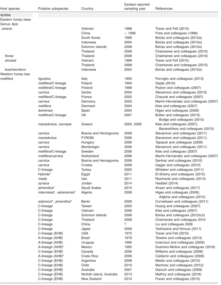
Table 1.World distribution ofNosema ceranaeacross hosts of the Apidae and Vespidae families.
Host species Putative subspecies Country
Earliest reported
sampling year References
Apidae
Eastern honey bees
GenusApis
cerana Vietnam 1968 Traver and Fell (2015)
China <1996 Fries and colleagues (1996)
South Korea 1996 Botı´as and colleagues (2012a)
Indonesia 2004 Botı´as and colleagues (2012a)
Solomon Islands 2008 Botı´as and colleagues (2012a)
Thailand 2008 Chaimanee and colleagues (2010)
florea Thailand 2008 Chaimanee and colleagues (2010) dorsata Vietnam 1968 Traver and Fell (2015)
Thailand 2008 Chaimanee and colleagues (2010)
koschevnikovi Indonesia 2004 Botı´as and colleagues (2012a) Western honey bee
mellifera ligustica Italy 1993 Ferroglio and colleagues (2013) mellifera/C-lineage Poland 1994 Gajda (2016)
mellifera/C-lineage Finland 1998 Paxton and colleagues (2007) carnica Serbia 2000 Stevanovic and colleagues (2010) mellifera/C-lineage France 2002 Chauzat and colleagues (2007) carnica Germany 2003 Martı´n-Hernandez and colleagues (2007) mellifera Denmark 2004 Klee and colleagues (2007)
iberiensis Spain 2004 Higes and colleagues (2006) mellifera/C-lineage UK 2007 Bollan and colleagues (2013),
Budge and colleagues (2015) macedonica,cecropia Greece 2005, 2009 Klee and colleagues (2007),
Bacandritsos and colleagues (2010) carnica Bosnia and Herzegovina 2006 Stevanovic and colleagues (2011) macedonica FYROM 2006 Stevanovic and colleagues (2011) carnica Hungary 2006 Tapaszti and colleagues (2009) carnica Montenegro 2006 Stevanovic and colleagues (2011) mellifera/C-lineage Sweden 2006 Klee and colleagues (2007)
mellifera/carnica Switzerland 2006 Martı´n-Hernandez and colleagues (2007) carnica Bosnia and Herzegovina 2008 Santrac and colleagues (2010)
carnica Croatia 2009 Gajger and colleagues (2010)
C-lineage Turkey 2005 Whitaker and colleagues (2011)
Hybrids* Egypt 2011 El-Shemy and colleagues (2012)
meda Iran 2011 Razmaraii and colleagues (2013)
syriaca Jordan 2014 Haddad (2014)
jemenitica* Saudi Arabia 2015 Ansari and colleagues (2017) intermissa*,sahariensis* Algeria 2008 Higes and colleagues (2009),
Adjlane and colleagues (2015) adansoni*,jemenitica* Benin 2009 Cornelissen and colleagues (2011)
C-lineage Taiwan 2005 Huang and colleagues (2007)
C-lineage Vietnam 2006 Klee and colleagues (2007)
C-lineage Solomon Islands 2008 Botı´as and colleagues (2012a,b)
C-lineage Thailand 2008 Chaimanee and colleagues 2010
C-lineage China ? Liu and colleagues 2008
C-lineage Japan 2009 Yoshiyama and Kimura (2011)
C-lineage (EHB) USA 1975 Traver and Fell (2015)
A-lineage (AHB) Brazil 1979 Teixeira and colleagues (2013)
A-lineage (AHB) Uruguay 1990 Invernizzi and colleagues (2009)
A-lineage (AHB)* Mexico 1995 Guerrero-Molina and colleagues (2016)
C-lineage (EHB) Canada 2006 Williams and colleagues (2008)
A-lineage (AHB)* Costa Rica 2006 Calderon and colleagues (2008)
C-lineage (EHB) Argentina 2008 Medici and colleagues (2012)
C-lineage (EHB) Chile 2010 Martinez and colleagues (2012)
C-lineage (EHB) Australia 2007 Giersch and colleagues (2009)
C-lineage (EHB) Norfolk Island, Australia 2013 Malfroy and colleagues (2016)
C-lineage (EHB) New Zealand 2010 Frazer and colleagues (2015)
where N. ceranae is now a common parasite in some areas.
How the spread ofN. ceranaeis affecting the prevalence ofN. apis?
The increasing worldwide prevalence of N. ceranae in the past decade, particularly in Mediterranean countries like Spain, Italy, Israel, Greece or Turkey (Klee et al., 2007; Higes et al., 2008a; Soroker et al 2011; Hatjina et al., 2011; Oguz et al., 2017), coupled with the absence of N. apis in several surveys, has led to the
hypothesis that N. ceranae might be displacing N. apis (Klee et al., 2007, Traver and Fell, 2011). The seasonal pattern typical of N. apis infection was well known: (i) low levels of infection during the hot summer months; (ii) a short peak in the autumn; (iii) a slow rise in the num-ber of infections during the winter; (iv) and a peak in the spring, with the level of infection rapidly increasing when foraging is limited by humid and cold climatic conditions (Fries, 1993). Accordingly,N. apislevels tend to drop off during the summer due to natural controlled mecha-nisms within the colony itself (Bailey, 1955). Long term studies showed that this pattern was evident forN. apis,
Table 1.cont.
Host species Putative subspecies Country
Earliest reported
sampling year References
Stingless bees
GenusMelipona
fasciculata Brazil 2015 Porrini and colleagues (2017) quadrifasciata anthidioides Brazil 2015 Porrini and colleagues (2017) marginata Brazil 2015 Porrini and colleagues (2017) rufiventris Brazil 2015 Porrini and colleagues (2017) mandacaia Brazil 2015 Porrini and colleagues (2017)
GenusTetragonisca
fiebrigi Argentina 2014 Porrini and colleagues (2017)
GenusScaptotrigona
jujuyensis Argentina 2015 Porrini and colleagues (2017) Solitary bees
GenusOsmia
bicornis Belgium 2012 Ravoet and colleagues (2014) cornuta Belgium 2012 Ravoet and colleagues (2014)
GenusAndrena
ventralis Belgium 2012 Ravoet and colleagues (2014)
GenusHeriades
truncorum Belgium 2012 Ravoet and colleagues (2014) Bumble bees
GenusBombus
atratus Argentina <2008 Plischuk and colleagues (2009)
Uruguay 2010 Arbulo and colleagues (2015)
Colombia 2013 Gamboa and colleagues (2015)
morio Argentina <2008 Plischuk and colleagues (2009)
bellicosus Argentina <2008 Plischuk and colleagues (2009)
Uruguay 2010 Arbulo and colleagues (2015)
waltoni China 2008 Li and colleagues (2012) remotus China 2008 Li and colleagues (2012) impetuosus China 2008 Li and colleagues (2012) sibiricus China 2008 Li and colleagues (2012) brasiliensis Argentina 2015 Plischuk and Lange (2016) hortorum UK <2013 Graystock and colleagues (2013)
hypnorum UK <2013 Graystock and colleagues (2013)
lapidarius UK <2013 Graystock and colleagues (2013)
lucorum UK <2013 Graystock and colleagues (2013)
pascuorum UK <2013 Graystock and colleagues (2013)
pratorum UK <2013 Graystock and colleagues (2013)
terrestris UK <2013 Graystock and colleagues (2013)
Vespidae
Polybia scutellaris Argentina 2010 Porrini and colleagues (2017)
Apis melliferasubspecies or evolutionary lineage are mostly predicted from the known native and introduced distributional ranges. Subspecies marked with an asterisk were mentioned in the reference whereas those marked in bold were identified either morphometrically or molecularly.
even though N. ceranae was present throughout the year (Higes et al., 2008a; Martı´n-Hernandez et al., 2012). However, the levels ofN. ceranae(percentage of bees infected) vary over time with very high levels from the end of summer up to spring and the maximum dur-ing winter in Spain (Higes et al., 2008a). These profile can be influenced by some undetermined factors since the higher levels were reported in summer in Canada (Copley et al., 2012), in March in Serbia (Stevanovic et al., 2013) or in spring (reflecting the development over winter) in Germany (Gisder et al., 2017). Also, N. ceranae can multiply at higher temperatures, displaying a greater biotic potential thanN. apis(Martı´n-Hernandez et al., 2009; Higes et al., 2010b; Gisder et al., 2017). Indeed, their spores are tolerant to temperatures as high as 608C and they can survive desiccation (Fenoyet al., 2009; Martı´n-Hernandezet al., 2009). By contrast, cold has a negative effect on N. ceranaewhose spores are sensitive to low temperatures and freezing (Fries, 2010; Gisderet al., 2010; Sanchez Colladoet al., 2014).
A first study of the within-host competition effect betweenN. apisand N. ceranaedid not show any clear competitive advantage for any of them (Forsgren and Fries 2010). However, a later study identified a priority effect when N. ceranae was the first infection (Natso-poulouet al., 2016). Apparently both environmental vari-ables and interspecies competition are important elements of mathematical models that help explain the differential prevalence ofNosemaspp. in distinct climatic
regions. Although such models can overestimate preva-lence, the predictions derived from them are consistent with field data obtained across Europe. Hence, they reveal a transition zone in the relative prevalence of the two species, withN. ceranaepredominating overN. apis in Southern regions (e.g., Spain) and vice versa (e.g., Sweden). Accordingly, the apparent global advantage of N. ceranae appears not to be due to differences in spore production or infectivity (as shown by Milbrath et al., 2015). The replacement ofN. apisbyN. ceranae is unlikely to occur due to a competitive advantage for within-host spore production (Martı´n-Hernandez et al., 2012; Gisderet al., 2017).
Genetic diversity ofN. ceranae
Many studies assessing the intraspecific variability inN. ceranae rely on the analysis of the ribosomal DNA (rDNA) (Huang et al., 2008; Sagastume et al., 2011; 2014; Suwannapong et al., 2011; Roudel et al., 2013), which is organized into ribosomal units (Huang et al., 2007; Huanget al., 2008) present as multiple copies in the genome (Cornman et al., 2009). Although intrage-nomic rDNA diversity is usually low as a result of con-certed evolution (Eickbush and Eickbush, 2007), rDNA markers show extensive sequence heterogeneity in N. ceranae (Sagastume et al., 2011; 2014) and in other Nosema species (Gatehouse and Malone, 1998; 1999; Tay et al., 2005; O’Mahony et al., 2007). Indeed, the
Fig. 1.Nosema ceranaeandNosema apislife cycle in honey bees. The spores ingested by the bees get to the ventricular lumen. There, spores extrude the polar filament and the sporoplasm is transferred into the epithelial cells. The sporoplasm matures into a Meront and a Mer-ogonic phase starts that comprises binary division of binucleate stages (the number of divisions is still undetermined). Lately, electrondense material is deposited in the outer face of the plasma membrane, which indicates the sporogonial phase. This phase involves the division of the
sporonts (Nosemaspp. have been described as bisporous) and then sporonts and daughter cells mature into spores. The first generation of
spores will be primary spores which can re-infect the same cell or infect neighbor cells. The second generation (after secondary meronts) will lead to environmental spores with the spore wall thicker than the primary spores (Huang and Solter, 2013). All parasitic stages develop in direct contact with the host cell cytoplasm and all phases are dyplokariotic.
A. Scheme of theNosemacell-cycle inside the host cell.
B. TEM image taken from a honey bee infected byN. ceranae. The ventricular cells can be seen with different parasitic stages.
average nucleotide diversity (p, Nei, 1987) in these
regions ranges from 0.14%–0.45% for the small subunit (SSU; Sagastume et al., 2011; Roudel et al., 2013) to 2.59% for the Intergenic Spacer (IGS; Sagastumeet al., 2011).
The analysis of single copy genes (Chaimaneeet al., 2011; Hatjinaet al., 2011; Roudelet al., 2013; G omez-Morachoet al., 2014; 2015a; 2015b; van der Zeeet al., 2014), which are better suited than multicopy markers for estimating the levels of intraspecific diversity, reveal high diversity within isolate variability in N. ceranae, regardless of whether isolates are obtained from a sin-gle bee or from homogenized pools of individuals (Hat-jina et al., 2011; Roudel et al., 2013; Gomez-Moracho et al., 2014; 2015b; Pelinet al., 2015), with an average pairwise diversity at synonymous sites of about 1% (Gomez-Morachoet al., 2015a). Most of the variation in N. ceranae is contributed by low frequency mutations (Hatjina et al., 2011; Roudel et al., 2013; van der Zee et al., 2014; Gomez-Morachoet al., 2014; 2015b; Pelin et al., 2015), especially in the parasite populations obtained from A. mellifera (Gomez-Moracho et al., 2015a), which is largely compatible with the recent expansion ofN. ceranaein this new host (Roudelet al., 2013; Gomez-Morachoet al., 2015a; Pelinet al., 2015).
The finding of multiple haplotypes within isolates can be explained by (i) the presence of several strains co-infecting honey bee colonies (Hatjina et al., 2011; Gomez-Moracho et al., 2014; 2015a), (ii) the existence of a diplokaryon with two diploid nuclei (Roudel et al., 2013; Pelinet al., 2015) or (iii) the combination of both, as these causes are not mutually exclusive. In any case, the occurrence of infections in which many different hap-lotypes co-exist, is a key factor in maintaining the genetic diversity ofN. ceranaein its new hosts (G omez-Morachoet al., 2015a).
Another important source of genetic variation is the existence of recombination. Although a clonal mode of reproduction has been proposed forN. ceranae on the basis of the detection of high levels of linkage disequilib-rium and heterozygosity (Pelinet al., 2015), the lack of genetic exchange between nuclei would make their sequences evolve independently and become more divergent over time. This scenario contrasts with the lack of structure observed in the haplotypes obtained from single bees (Roudel et al., 2013; Gomez-Moracho et al., 2015a), which rather suggests the existence of genetic flow between nuclei. The presence of sex-related loci and genes involved in meiotic recombination (Leeet al., 2010) point to the existence of cryptic sexual stages in the life cycle ofN. ceranae; however, it is still unknown if the recombinant haplotypes (Sagastume et al., 2011; Roudel et al., 2013; van der Zee et al., 2014; Gomez-Morachoet al., 2014; 2015a; 2015b) are
generated during meiosis or during mitosis, as the out-comes of these processes are difficult to distinguish (Weedall and Hall, 2015). At any rate, the weak, yet sig-nificant genetic exchange detected in N. ceranae (Gomez-Morachoet al., 2015b) has important evolution-ary implications, not only because it allows deleterious mutations to be eliminated more efficiently but also because recombination provides a better capacity to adapt to new environments or hosts than a clonal mode of reproduction (Barton, 2010).
How areN. ceranaepopulations structured?
Recent analyses suggest that, despite sharing alleles, there is moderate but significant differentiation among the N. ceranae haplotypes found in different Apis spe-cies (Chaimanee et al., 2011; Gomez-Moracho et al., 2015a). In N. ceranae populations from A. mellifera most of the variation occurs within honey bee colonies, which show no genetic differentiation and shared alleles regardless of their geographic origins (Roudel et al., 2013; Gomez-Morachoet al., 2014; 2015a; van der Zee et al., 2014; Pelinet al., 2015). In line with these obser-vations, the analysis of the genomes of eightN. ceranae isolates from distant locations revealed that more than 98% of the polymorphism detected was shared among at least two of the isolates studied (Pelin et al., 2015), confirming that population structuring in A. mellifera, if any, is still at an extremely initial stage.
In contrast, when it comes to N. apis, a considerable fraction of the genetic variance (between 20% and 34%) corresponds to differences between isolates obtained from distinct A. melliferalineages (Maside et al., 2015). Indeed, isolates collected from honey bees of lineage A (which can be found in Africa and the Iberian Peninsula) exhibit different haplotypes from those obtained from lin-eages C or M (which involveA. melliferasubspecies dis-tributed across South Eastern Europe or Western and Northern Europe respectively); the existence of this pop-ulation structure suggest a far older relationship between A. mellifera and N. apis than that between the former andN. ceranae.
What are the major effects ofN. ceranaeon honey bees?
Since the first report of N. ceranaeinfection inA. melli-fera, there has been some controversy about the conse-quences of such infection. However, in recent years most studies have confirmed that N. ceranae has a pathogenic effect in this host, expressed at least in a shortening of the workers’ lifespan in controlled (cage) experiment (e.g., Mayack and Naug, 2009; Alaux et al., 2010; Martı´n-Hernandez et al., 2011; Dussaubat et al., 2012; Goblirschet al., 2013; Schwarz and Evans, 2013;
Aufauvre et al., 2014; Basualdo et al., 2014; Roberts and Hughes, 2014; 2015; Williams et al., 2014; Doublet et al., 2015aHuanget al., 2015); only few papers failing to report this effect (Milbrath et al., 2013; Retschnig et al., 2014; Garridoet al., 2016).
The effect ofN. ceranaeinfection on honey bees’ fitness
The lesions caused by the infection in the bee ventriculi were described in depth some years ago (Higes et al., 2007; Garcia-Palencia et al., 2010). Here we focus on the studies that enlightened many of the effects that this microsporidia has on the physiology ofA. mellifera.
Changes in metabolism. The effect of N. ceranae infec-tion on the host’s gene expression has recently been thoroughly addressed, confirming the effect of this microsporidia in the infected host (Szumowski and Troe-mel, 2015). Since N. ceranae invades the ventriculus (midgut) of honey bees, most studies have focused on this organ. Honey bees consume nectar and pollen as sources of carbon and nitrogen respectively, and both require extensive processing in the gut (Kunieda et al., 2006) by enzymes that metabolize carbohydrates and lipids to breakdown the food and to release stored energy and to synthesize the organism’s primary energy stores (reviewed by Klowden, 2002). Modifications to carbohydrate metabolism have frequently been reported in N. ceranae infected bees suggesting a manipulative activity of the pathogen to ensure the availability of nutrients for its own benefit. In terms of gene expres-sion, this manipulation is apparently reflected in the up-regulation of thea-glucosidase gene and of three genes
involved in trehalose transport (the major carbohydrate energy storage molecule in insects; Dussaubat et al., 2012) observed, as well as the down-regulation of the trehalase and the glucose-methanol-choline oxidoreduc-tase three encoding genes (Aufauvre et al., 2014). These alterations to the expression of genes involved in sugar metabolism were also confirmed in a proteomic study where four proteins involved in energy supply were found to be less abundant in the midgut ofN. cera-nae-infected bees (Vidau et al., 2014). Similarly, gas chromatography–mass spectrometry highlighted a decrease in the majority of carbohydrates and amino acids implicated in various biochemical pathways, such as fructose, L-proline, sorbitol and glycerol (Aliferis et al., 2012).
The alterations of the carbohydrate metabolism reflect the nutritional and energetic stress experienced by N. ceranaeinfected honey bees (Mayack and Naug, 2010; Aliferiset al., 2012; Vidauet al., 2014). Interestingly, this has not been observed in Nosematolerant honey bees (Kurze et al., 2016). Energetic stress has been
described in infected foragers that were hungrier than uninfected bees (Mayack and Naug, 2009), consuming more sugar (Alauxet al., 2010; Martı´n-Hernandezet al., 2011; Vidau et al., 2011). These infected workers appear to be unable to utilize the excess carbohydrates consumed probably because the most of them are used by the pathogen to complete its life-cycle. Moreover, the energetically stressed bees have been reported to expe-rience higher mortality during foraging (Mayack and Naug, 2013). Hence, the mechanisms controlling the mobilization of energy reserves appear to be disturbed and there is poor carbohydrate homeostasis in their hae-molymph (Aliferis et al., 2012). The stronger sugar demand and higher consumption could be a host response to the infection, directly related to the depen-dence of microsporidia on host energy. However, the intestinal lesions caused byN. ceranaeproliferation may decrease the digestive capacity of honey bees and gen-erate signs of starvation, such as impoverishment of hypopharyngeal protein secretions in nurse bees (Vidau et al., 2014). It should also be noted that a higher sugar consumption is not always observed in such studies (Aufauvre et al., 2012; 2014), suggesting that other unknown factors could influence this parameter. Addi-tionally, Li and colleagues (2018) reported that bees infected by N. ceranae show an accelerated lipid loss, suggesting lipids may be used also as a fuel for increased metabolic demands due to the infections.
All these modifications alter the feeding behaviour of infected honey bees and their transition to become for-agers (Mayack and Naug, 2009). In fact, the inhibition of fatty acid synthesis and also the starvation can lead bees to begin foraging earlier in life (Schulzet al., 1998; Toth, 2005), and energy stressed bees in a colony first altering their activity and then their foraging rate (Mayack and Naug, 2013). Among N. ceranae infected bees, the weaker capacity to fly, probably due to the lower trehalose levels, should also be taken into account (Mayack and Naug, 2010). However, it may be that these behavioural alterations are related to the infection itself, since the altered regulation of highly conserved neurohormonal pathways (such as the octopamine path-way) on N. ceranae infection was caused by the patho-genesis itself and not indirectly by energetic stress (Mayacket al., 2015).
Changes in other vital functions. Other important meta-bolic pathways for honey bee physiology are also altered by N. ceranae infection. For example, oxidative stress has been reported due to the over-expression of genes related to the generation of antioxidant enzymes and increased glutathione-S-transferase activity (Vidauet al., 2011; Dussaubatet al., 2012), although the appearance of this detoxifying enzyme could be influenced by diet
(Di Pasquale et al., 2013). This oxidative response to infection (observed by both transcriptomic and proteomic approaches), and the higher energetic demand, strongly suggests that a negative impact on infected honey bee development may cause a reduction in lifespan (Vidau et al., 2014). Additionally, the stress response observed inN. ceranae infected bees appears to be derived from the modification of transcriptional profiles in the brain and from changes in the cuticular hydrocarbon profiles (McDonnellet al., 2013; Aufauvreet al., 2014) that are similar to those produced by the miteVarroa destructor (McDonnell et al., 2013). The enhanced impact of the infection over time, also seen in response to insecticides (see below), suggests a growing disturbance of the honey bee transcriptome that might reflect the failure of recovery from stress and could explain the higher mor-tality rates observed (Aufauvreet al., 2014).
Conversely, N. ceranaeinfection was reported to pre-vent the apoptosis of epithelial cells in the bees’ pre- ventric-uli (Higeset al., 2013; Martı´n-Hernandezet al., 2017) to avoid the host innate response to the infection. A capac-ity to inhibit genes involved in cell signalling and in the self-renewal of intestinal cells (Dussaubat et al., 2012; Huang et al., 2016) and an up-regulation of genes belonging to the IAP family (inhibitors of apoptosis genes; Martı´n-Hernandez et al., 2017) has also been reported. As microsporidia can modulate such pro-cesses in cell cultures (Del Aguilaet al., 2006), this may be a mechanism used by the parasite to favour its devel-opment. Indeed, there are differences in the transcrip-tion of an anti-apoptotic gene (inhibitor of apoptosis protein-2) betweenNosema-tolerant and Nosema -sensi-tive bees (Kurzeet al., 2015).
Effects on bee immune response. Microsporidia have been seen to modify the host’s immune response. For example, Nosema bombycis induces transcriptional changes in 34 out of 70 Bombyx mori immune genes, even inducing the down regulation of the serine protease cascade in the melanization pathway, and up-regulating lysozyme and lectins (reviewed in Szumoski and Troe-mel, 2015). This effect is especially noteworthy given that several studies have addressed how infection byN. ceranaemight affect immunity at the social (colony) and individual (bee) level. However, while all of these studies reported effects on immunity at the individual level, other controversial results have also been described. In this regard, one of the first studies on A. mellifera infected with N. ceranae showed a down-regulation of some immune-related genes likeabaecin,hymenoptaecin, glu-cose dehydrogenase (GLD) and vitellogenin (Vg), sug-gesting that N. ceranae infection suppresses immune defence mechanisms in honey bees (Antunez et al., 2009). Later studies also detected host
immunosuppression, reporting a down-regulation of defensin, abaecin, apidecin (Chaimanee et al., 2012), hymenoptaecin(Chaimaneeet al., 2012; Aufauvreet al., 2014), serine protease 40, catalase (Aufauvre et al., 2014), basket (GB16401) and u-shaped (GB16457) genes (involved in Drosophila immune responses; Dus-saubat et al., 2012). However, these changes were associated with the over-expression of other immune related genes, such as ROS (reactive oxygen species) and glutathione peroxidase like 2. Another study also reported a down-regulation of antimicrobial peptides (Badaouiet al., 2017), suppression of Toll and Imd path-ways and of the expression of Pattern Recognition Receptors-related genes, this last was persistent and intensified in time (Liet al., 2018). By contrast, Schwarz and Evans (2013) reported a complex immune response mounted by bees againstN. ceranae infection, which is also dynamic over time. In this work, ingestion of N. ceranaespores was seen to rapidly enhance Toll and Imd signalling, and to increase the expression of the cel-lular recognition molecule Dscam and AMP Defensin 2. Moreover, infection caused a diverse and extended effector immune response viaabaecin,apidaecin, hyme-noptaecin, defensin 1 and 2, mainly observed 7 days post-infection. These same genes and some other related to microbial recognition proteins as peptidogly-can recognition proteins and Gram-negative binding pro-teins were also upregulated in a field assay, both in nurse and foragers infected, mainly these latter (Liet al., 2017).
However, in a different study neither haemocyte num-ber nor phenoloxidase (PO)-activity were apparently affected by infection (Alaux et al., 2010), although the longevity ofNosemainfected bees was linked to the lat-ter (Di Pasqualeet al., 2013). Intermediate results were reported when the immune response was compared between ‘Nosema-resistant selected’ and ‘unselected’ drones, with stronger gene expression in the infected group than in the uninfected controls from day one to five post infection, although the expression of genes from the innate immune system was weaker in the unse-lected strain (Huang et al., 2012). Many factors can influence immune responses, such as the different doses of infection (which vary considerably between studies), the duration of the assays, the tissues in which gene expression was studied (whole bees, abdomen, ventriculi, etc.), or the age of the bees at infection and during the study. In this regard, it is known that the hae-mocyte number is dramatically lower in early adult life in all the bee castes, and that the dynamics of PO activity is sex and caste specific (Schmid et al., 2008). In fact, the immune response of N. ceranae infected queens changes as they age, with the expression ofapidaecin, eater and vitellogenin varying in queens inoculated at
different ages (Chaimaneeet al., 2014). Also, the quality and diversity of the pollen supplementing bees’ food influences their immunity, as observed through the gen-eral activity of Glutathione-S transferase, alkaline phos-phatase and PO (Di Pasquale et al., 2013). However, little is known about the protective role of immune related peptides after microsporidia infection. Indeed, only the increased expression of aubergine has been linked to resistance (or protection) in Nosema resistant honey bees (Huanget al., 2014a,b).
In terms of other molecules related to the honey bees’ immune response, as well as to other physiological func-tions, some alterations to Vg and Juvenile Hormone (JH) have been also described in N. ceranae infected bees. Vg fulfils several functions in workers, such as participating in the synthesis of royal jelly (Amdamet al., 2003), promoting immunity, stress resilience and longev-ity (Amdam et al., 2004), and it also proposed to regu-lates behavioural development along with JH (Robinson and Vargo, 1997; Nelson et al., 2007). Adult workers that were infected as larvae with N. ceranae spores showed significantly higher Vg titers and lower total hae-molymph protein titers than uninfected controls (BenVau and Nieh, 2017). Furthermore, these honey bees infected at the larval stage also had some modification in their sting, which developed a more queen-like sting morphology (BenVau and Nieh, 2017). Moreover, the expression of Vg has been found to be higher in bees collected from colonies with low levels of N. ceranae infection than in bees from colonies with high levels and it has been suggested to be associated with colony resistance toN. ceranae(Antunezet al., 2013).
In addition to modifying the expression of the Vg gene, N. ceranae infection can also disrupt the physio-logical regulation of the age-specific behaviour of infected workers by increasing the level of JH (III) in the haemolymph of infected bees (Ares et al., 2012), as JH is a promotor of foraging. Indeed, the atypical transcrip-tion of Vg and JH is the inverse of what would be expected for healthy, uninfected bees (Goblirsch et al., 2013). Similarly, infection ofB. moribyN. bombycis pro-vokes the differential expression of many genes involved in the synthesis and metabolism of JH, which probably leads to the described increase in JH (Ma et al., 2013). This alteration in the Vg/JH equilibrium has been pro-posed to be responsible for the effects on early foraging and the shortened lifespan of N. ceranae infected worker bees (Goblirsch et al., 2013). Also, N. ceranae alters the metabolism of bees increasing EO (ethyl ole-ate) levels (Dussaubatet al., 2010), a primer pheromone which regulates worker behavioural maturation (i.e., inhibits the transition from inside-nest tasks performed by nurse bees) to foraging tasks performed by old bees
(Leoncini et al., 2004). All these effects fit with the fact that infested bees forage earlier.
Altogether, these alterations reflect the effect of this Microsporidia on immunity at the individual bee and col-ony level, such that colonies may become more suscep-tible to other infectious diseases or the effects of pesticides (see below).
Factors related withN. ceranaeinfection
Honey bees live in an environment where they might be exposed to different factors, such as pathogens and pesticides, which may interact with one another.
Interactions betweenN. ceranaeand other pathogens of honey bees. The pathogen status of a colony is seasonal-dependent and gaining insights on the interac-tions between pathogens in the field can become com-plex. In this regard, several experimental approaches that aimed to study the interaction between Nosema and other viruses or parasites contributed to the analysis.
One of the more prevalent viruses of honey bees worldwide is the Deformed wing virus (DWV), named after the main sign of infection observed in adults. Costa and colleagues (2011) reported that varroa-free emerg-ing adults from a DWV-positive colony fed withN. cera-nae spores showed significantly lower DWV loads in their midgut than their Nosema-free counterparts. This difference did not held for other tissues, suggesting that DWV andN. ceranaemay compete for host cells or spe-cific cell functions in the honey bee midgut. However, in a later field study in Hawaii, no correlation between DWV loads andN. ceranaespore counts were observed (Martinet al., 2013). A recent survey suggested that the DWV load may negatively impact establishment of Nosema spp., as Nosema-free honey bees had signifi-cantly higher DWV loads than Nosema-infected honey bees (Traynoret al., 2016). The order of infection seems to be important; priorN. ceranaeinfection inhibited sub-sequent DWV infection but this was not reciprocal, sug-gesting asymmetry in the competitive interaction between these pathogens (Doublet et al., 2015b). In another experiment, N. ceranae fed to emerging bees from a DWV-infected colony appeared to accelerate DWV replication at early stages of viral infection in a dose-dependent manner, but not once DWV titers reached a plateau (Zheng et al., 2015). However, the discrepancies among these studies could be attributed to differences in the methodologies applied to determine the viral load, such as the analysis of a specific tissue or the whole bee. Finally, the food could also influence the infection since pollen supply was able to increase theN. ceranaeimpact on DWV replication (Zheng et al., 2015) and a negative correlation between N. ceranae spore
loads and DWV-B (formerly Varroa destructor virus-1) titers was stronger in protein-fed bees than in sugar fed bees, showing that nutrition seems to play an important role also on virus infections in insects (Tritschler et al., 2017).
The relationship between N. ceranaeinfection and V. destructor, an important vector of DWV, is also unclear. Indeed, although one study found a positive correlation betweenNosemaand varroa in commercial apiaries pre-treated against both (Little et al., 2016), others did not observe any correlation between these two pathogens but signalled an emerging genotype B of DWV as linked to overwinter worker losses (Natsopoulouet al., 2017). In this respect, the infection by N. ceranae has been related to a reduced efficacy of varroa treatments (Botı´aset al., 2012b).
Regarding other viruses, in a 6 year survey Traynor and colleagues (2016) observed a strong positive corre-lation between Lake Sinai virus-2 (LSV-2) and Nosema spp. with intensity peaks opposite to varroa-peak infesta-tion and its associated viruses DWV and ABPV (Acute bee paralysis virus). They proposed different hypotheses to explain this positive correlation, including (i) different seasonal life histories of parasites and pathogens, (ii) a double-repression relationship between ABPV/DWV/var-roa, which would compete for host resources, with DWV and ABPV outcompeting LSV-2 and (iii) a direct link between LSV-2 and Nosema that inhibit the replication of DWV and ABPV. Another study did not found signifi-cant association betweenN. ceranaeand the DNA virus, the Apis mellifera fillamentous virus, AmFV (Hartmann et al., 2015).
The Black queen cell virus (BQCV), another very fre-quent virus infecting bees, induced elevated mortality of adults when fed simultaneously withN. ceranae, but this effect was not reflected in a significant increase in the load of one pathogen over the other in the bee midgut (Doubletet al., 2015a). However, the detection of patho-gen loads was performed 13 days post infection while significant differences in bee mortality started earlier at 9 days post infection.
Experimental simultaneous co-infections of winter honey bee workers with Chronic bee paralysis virus (CBPV) and N. ceranae showed differences in CBPV replication but not in honey bee mortality, depending on the inoculation method of the virus, per oz or per cuti-cula(Toplak et al., 2013). Thus, when adult bees were simultaneously inoculated with CBPV and N. ceranae per oz, 50% of the bees had Ct (Cycle threshold) values of CBPV lower than the initial virus inoculum, which con-trasted with 71.7% of the bees with lower Ct values than the initial virus inoculum obtained when they were infected with CBPV-only per oz. Infection with N. cera-nae per ozand CBPVper cuticula resulted in 71.2% of
the bees with Ct values lower than the original virus inoculum, while infection of CBPV alone per cuticula resulted in 12% of the bees with Ct values lower than the inoculum. These results suggest a synergistic effect of N. ceranae on CBPV replication when the virus is inoculated per cuticula and an antagonistic effect when it isper oz. The percentage ofN. ceranaespores inper oz- and per cuticula CBPV-infected dead bees were 71.1% and 58. 2% respectively, compared to 54.3% in N. ceranae-only infected bees (Toplaket al., 2013).
Regarding to other gut pathogens, in the past few years, special attention has been paid to co-infection of N. ceranae with N. apis. While there is no evidence of host competitive advantage for N. ceranae (Forsgren and Fries, 2010), infection intensity and honey bee mor-tality appear to be significantly greater for N. ceranae than for N. apis or for their mixed infections (Williams et al., 2014). Indeed, while the mortality caused by N. ceranae was similar to that of N. apis, reduced spore intensity was observed. Moreover, the host competition was evident between the two microsporidia and the order of infection had an important influence (Natsopou-louet al., 2015). The first parasite to infect significantly inhibited the growth of the second, althoughN. ceranae provoked stronger inhibition. It was recently reported that mixed infection by Nosema species negatively affected honey bee survival more than a single species infection; yet no competitive advantage for N. ceranae was observed even when both species coinfected the host simultaneously (Milbrathet al., 2015). There is also some controversy to whetherN. ceranaeis more patho-genic thanN. apis, as this seems to vary greatly among different studies (Huanget al., 2015). Nevertheless, the damage to colonies is more closely related to the preva-lence under natural conditions than the pathogen’s spe-cific effects, which has been shown to be influenced by multiple factors.
Finally, co-infection of N. ceranaewith the trypanoso-matid Crithidia mellificae was found to alter the reper-toire of systemic antimicrobial peptides as well as dampening the cellular immune response of honey bees (Schwarz and Evans, 2013).
On one hand, the emerging picture from laboratory studies is that the temporal sequence of infection, route of infection, dose of the pathogen and the impact of the infecting agent on host immunity (and metabolic resour-ces of the host) are important factors determining the outcome of the co-infection, since N. ceranae appears to have an important effect on the ability of a virus to infect the bee’s midgut cells. On the other hand, each of these pathogens, the microsporidia and the virus, can weaken the bee’s immune defences facilitating the repli-cation of the co-infecting partner. Consequently, these complex interactions between N. ceranae and other
pathogens will require further study to be fully understood.
Interactions betweenN. ceranaeand pesticides. Interac-tions between pesticides and N. ceranae can be expected, as both have the potential to disturb similar metabolic functions related to immunity, energetic resources and antioxidant responses (Di Prisco et al., 2013). Several studies indicate that a detrimental inter-action occurs when honey bees are exposed to both pesticides and N. ceranae(Pettiset al., 2013). A syner-gistic effect betweenN. ceranaeand neonicotinoids, first observed under laboratory conditions, causes a signifi-cantly higher bee mortality along with a reduction in glu-cose oxidase activity, which is involved in social immunity through the sterilization of the colony and brood food (Alauxet al., 2010). This synergism between neonicotinoids and N. ceranae in adult bees has been confirmed by others (Vidau et al., 2011; Aufauvre et al., 2012; Doublet et al., 2014) and was also observed in field studies demonstrating an indirect effect of imidaclo-prid onN. ceranaegrowth, even when honey bees were exposed to levels below those considered harmful (Pet-tis et al., 2012). In contrast with those observations, Gregorc and colleagues (2016) did not detect an effect of a neonicotinoid on N. ceranae growth in laboratory conditions but a minor synergistic toxic effect on the honey bee midgut tissue compared to that of both stres-sors separately. Similar synergism was reported between N. ceranae and fipronil (Vidau et al., 2011; Aufauvre et al., 2012), yet such interaction was later questioned, despite the observation that N. ceranae -insecticide combinations significantly enhanced honey bee mortality (Aufauvre et al., 2014). Particularly, in new-born queens exposed to both N. ceranae spores and a neonicotinoid under laboratory conditions, and introduced later in small mating hives in the field, co-exposure had similar effects to individual co-exposure to each stressor, rapidly compromising queens’ survival and physiology (Dussaubat et al., 2016). When using proboscis extension response (PER) only slightly impair-ment of learning in honey bees infected withN. ceranae and no interaction with a neonicotinoid pesticide was observed (Piiroine and Goulson, 2016). Regarding other pesticides, Pettis and colleagues (2013) reported that bees consuming pollen contaminated with fungicides (as chlorothalonil or pyraclostrobin) and acaricides (as 2,4 Dimethylphenyl formamide, an amitraz metabolite, bifen-thrin or fluvalinate) have a large increased risk of N. ceranae infection. Conversely, Garrido and colleagues (2016) found no interactive effects between sublethal doses of tau-fluvalinate or coumaphos and N. ceranae on nurse bee mortality and adults, and N. ceranae development was not affected by the acaricides.
More research is needed to understand in which envi-ronmental context honey bees are more susceptible to both stressors and which interaction effects can become visible and compromise colony survival.
Other interacting factors.Many other factors could influ-ence the development and the course of N. ceranae infection. The age of an individual when exposed to a parasite can have a significant effect on its survival, immune-competence and the intensity of infection (Rob-erts and Hughes, 2014). Foragers have previously been reported to be the most intensely infected individuals in a colony (Higeset al., 2008a; Meanaet al., 2010; Smart and Sheppard, 2012; Li et al., 2017). However, older worker bees appear to survive better than younger indi-viduals when challenged withN. ceranae, although older bees develop more intense infections and have lower levels of prophenoloxidase, a marker of immunity response, which is negatively correlated with the inten-sity of infection. This facet has important epidemiological consequences since more strongly infected bees survive longer, facilitating pathogen transmission (Roberts and Hughes, 2014). Similarly, the queen becomes less sus-ceptible to N. ceranae infection as she ages, such that the time spent in the mating nuclei is also epidemiologi-cally important (Chaimanee et al., 2014). In fact, until recently only adult bees were thought to be susceptible toN. ceranaeinfection, yet larvae and pupae have been shown to develop infection, and this infection confirmed histologically in tissues as early as prepupal stages diminished adult longevity (Eiriet al., 2015).
All A. mellifera castes (workers, queen and drones) are susceptible to N. ceranae infection (Higes et al., 2008a), although drones appear to suffer higher mortal-ity than workers and surviving bees have a lower body mass, suggesting sex-specific differences in honey bee susceptibility to N. ceranae (Retschnig et al., 2014). In fact, drones have been regarded as intracolonial ‘super-spreaders’, whereby transmission is enhanced when drones rather than workers are the infected individuals. Moreover, the survival of susceptible individuals (work-ers) maintained with infected drones was generally sub-stantially worse than when they were kept with infected workers (Roberts and Hughes, 2015).
Another factor that must be considered is the dose of infection. It is known that virulence, in the sense of increased mortality rates in infected individuals, increases with the dose of inoculum (Ebert, 1999). Usu-ally, the more parasites infect an individual, the stronger the effects on host fecundity and survival (Anderson and May, 1978; Keymer, 1982; Ebert et al., 2000). Higher doses of viral, bacterial and fungal pathogens increase mortality rates and reduce the survival time of infected insects (e.g., van Beek et al., 1988; 2000; Hochberg,
1991; Arthurs and Thomas, 2001; Brunneret al., 2005). Some of these dosage effects are purely statistical, such that at higher doses the probability of a successful and potentially lethal infection increases, yet this is also the case in terms of the internal dynamics of infection. Consequently, the strong variability in longevity reported in distinct laboratory studies could be influenced by this parameter, as it also varies greatly in these studies.
Diet may also affect tolerance to N. ceranae, since nutritional quality and the diversity of pollen nutrition can shape bee health (Porrini et al., 2011; Di Pasquale et al., 2013; Jack et al., 2016; Tritschler et al., 2017). Indeed, pollen nutrition improves the survival of healthy and N. ceranae infected bees, and pollen quality (reflected in protein content and antioxidant activity) strongly influences the effects of infection on bees (Di Pasquale et al., 2013). The source of dietary protein also seems to be important. Infected or uninfected bees fed with a non-natural protein diet as a pollen substitute had lower protein titres in the haemolymph than those fed with bee-bread, and their survival was also worse (Basualdo et al., 2014). Also, the mortality of bees infected withN. ceranae and fed in the laboratory with only sucrose syrup, supplemented or not with aminoa-cids and vitamins, was higher than when the bees were fed with pollen (Porriniet al., 2011).
Does host variation influence virulence?
The host range of N. ceranae is increasingly larger denoting a low specificity and a potentially high capacity of adaption to novel hosts. Despite the high number of host taxa,N. ceranaehas been restricted to the Apidae. However, this situation changed recently with detection of N. ceranaein the Vespidae wasp Polybia scutellaris, suggesting that the pathogen is even capable of travers-ing the family barrier (Porriniet al., 2017).
Since first discovery in A. cerana(Frieset al., 1996), N. ceranaewas subsequently found in other Asian bee species, including A. florea, A. dorsata (Chaimanee et al., 2010) andA. koschevnikovi(Botı´aset al., 2012a). Following the out of Asia host shift (Higeset al., 2006; Huanget al., 2007), N. ceranae has spread worldwide and jumped across numerous social and solitary bee species and genera within the Apidae, including Bombus, Osmia, Andrena, Melipona, Tetragonisca and Scaptotrigona (Table 1). The notable host range and corresponding global distribution, represent a wide vari-ety of climates, from Mediterranean in Spain (Higes et al., 2006), temperate in Canada (Williams et al., 2008), tropical in Mexico (Guerrero-Molinaet al., 2016), to hot arid in Saudi Arabia (Ansariet al., 2017).
Despite the long list of host taxa that have been shown positive to N. ceranae (Table 1), experimental
infections have been limited toA. cerana(Suwannapong et al., 2011), A. florea (Suwannapong et al., 2010), Bombus spp. (Graystock et al., 2013) and, to a great extent, toA. mellifera(Higes et al., 2007; Paxtonet al., 2007; Mayack and Naug, 2009; Forsgren and Fries 2010; Chaimanee et al., 2013; Williams et al., 2014; Huang et al., 2015; Milbrath et al., 2015; Natsopoulou et al., 2015), with higher virulence displayed by the two latter species. WhetherN. ceranae is virulent to all Api-dae and whether it is equally virulent to every A. melli-fera subspecies is unclear. What is clear is that the current N. ceranae geographical range covers a large portion of theA. melliferadiversity with possibly 14 sub-species of the four major evolutionary lineages (C, M, A and the Middle Eastern O; Ruttner, 1988), as putative hosts. It must be noted, however, that most artificial infection studies and population surveys did not identify, or even mentioned, the subspecies or lineage (only three studies provided morphometric or molecular identi-fication; Table 1), and there is a possibility that commer-cial stock of C-lineage ancestry (either A. mellifera carnica, A. mellifera ligustica and even buckfast) was the main strain under analysis in many regions.
Infection experiments in A. mellifera have produced inconsistent results onN. ceranaevirulence, with mortal-ity rates of caged bees ranging from 10% in France (Vidauet al., 2011) to as high as 100% in Spain (Higes et al., 2007) 8–10 days post-infection. Several hypothe-ses (discussed herein) have been evoked to account for the differences across infection experiments and world-wide N. ceranae surveys, among which is the genetic variability of the host (Paxton et al., 2007; Martı´n-Hernandezet al., 2011; Dussaubatet al., 2013a; Bran-chiccela et al., 2017). Predicting from studies focusing on a wide range of host-pathogen systems, which have shown that disease virulence varies with host genotype (de Roode et al., 2004, and references therein), it is possible that different subspecies vary in their ability to counter infection. Yet, to our knowledge, there is only one cage artificial infection experiment that compared the susceptibility toN. ceranae of molecularly identified honey bees of unspecified subspecies but belonging to lineages C and O (Fontbonne et al., 2013). This study suggests that genetic variation among individual bees or colonies within lineage, and not between lineages, is a better predictor of host response to N. ceranae. In fact, inter-colony variation in susceptibility to nosemosis has long been recognized and used in Denmark in a breed-ing programme that selected for low infection rates (Traynor and Traynor, 2008).
In contrast with the cage study of Fontbonne and col-leagues (2013), field experiments of natural infection found differential levels ofN. ceranae between molecu-larly identified Russian (M-lineage) and Italian bees
lineage) in the USA (Bourgeois et al., 2012), and between Africanized (A-lineage) and Italian bees in Uru-guay (Mendozaet al., 2014), with Italian bees seemingly more susceptible in both cases. Furthermore, the results of Bourgeois and colleagues (2012) provide the first evi-dence for genetic variation in resistance toN. ceranaein the Russian stock.
However, not all studies offer support for bee strain contributing to differential virulence. In another field experiment in the USA, queen genetic origin did not seem to influence N. ceranae infection levels (Villa et al., 2013). However, the authors did not identify the colonies and, inferring from queen origin (commercial colonies from Northern USA, Canada and Australia), they were most likely of C-lineage ancestry. A similar finding was reported for Spain in aN. ceranaesurvey of colonies identified for mitochondrial DNA (Jara et al., 2012). In this study, variation in pathogen prevalence was not linked to A and M-lineage mitotypes.
While it remains to be demonstrated that variation among A. mellifera subspecies influences virulence of N. ceranae, it has been repeatedly shown that increased genetic diversity within a colony improves its resistance to a diverse array of diseases (Palmer and Oldroyd, 2003; Tarpy, 2003; Tarpy and Seeley, 2006; Seeley and Tarpy, 2007; Desai and Currie, 2015), supporting one of the hypothesis related with the evolution of polyandry (Hamilton, 1987). This hypothesis was recently tested in honey bee colonies headed by queens artificially insemi-nated with one or 12 drones and proved true forN. cera-nae, with significantly higher prevalence levels detected in genetically similar colonies as compared to genetically diverse colonies (Desai and Currie, 2015). Interestingly, no significant differences between the two types of colo-nies were found for N. apis (Desai and Currie, 2015), which agrees with a previous study from Woyciechoski and Krol (2001). In conclusion, while all these studies represent first (but not comparable) attempts to address the role of honey bee variability in N. ceranaevirulence, carefully designed cage and field infection assays with genetically characterized host and pathogen are required for a better understanding of their interaction.
CanN. ceranaekill a colony?
Accurate data on nosemosis type C as a main cause of colony mortality is difficult to find in the literature, princi-pally because of the absence of clear clinical signs (Higes et al., 2010c). However, there are some reports where nosemosis (without specifying the species) is associated with colony losses. A survey carried out in 2014–2015 in the USA, reported that 5% of the bee-keepers attributed toNosemadisease a 53.9% of colony mortality (CI 95%: 50.0–57.8) (Seitz et al., 2016).
Another survey in Europe, developed in 2012–2013 showed 21.77% (CI 95%: 14.14–31.14) winter mortaligy in colonies suffering nosemosis (based on clinical signs for N. apis) versus a 12.64% (CI 95%: 6.84–20.78) in colonies without the infection, although significant regional differences in colony losses were observed (Chauzatet al., 2016).
The first evidence of a relationship between N. cera-nae infection and colony loss was recorded in Spain (Higes et al., 2006; 2008a; 2009a; Botı´as et al., 2013; Cepero et al., 2014; Meana et al. 2017). Subsequently, a similar link between this pathogen and honey bee col-ony weakness/loss was proposed in other countries with comparable climatic conditions such as Greece (Hatjina et al., 2011), Israel (Soroker et al., 2011), South-East, North and Western-coast of USA, (Villa et al., 2013; Bekele et al., 2015), Central Chile (Bravo et al., 2014), Italy (Lodesaniet al., 2014; Cavigliet al., 2016) and Jor-dan (Adjlane and Haddad, 2016). On the contrary, it seems that colder climates like Germany, Balkan coun-tries, Switzerland and Northern Greece do not fulfill the specific conditions (climatic and/or beekeeping practi-ces) for N. ceranae to compromise colony survival (Gisder et al., 2010; Hedtke et al., 2011; Stevanovic et al., 2011; 2013; Dainat et al., 2012; Francis et al., 2014). This may well reflect the ability of N. ceranae spores to better resist to high temperatures and desicca-tion than to low temperatures (Fenoy et al., 2009; Sanchez Colladoet al., 2014) and its ability to complete the life cycle more efficiently at high temperatures (Mar-tı´n-Hernandezet al., 2009; Higeset al., 2010b). Thus, in warmer areas the infection byN. ceranaemight cause a chronic stress on honey bee colonies will be more intense, ultimately favouring colony death (Higes et al., 2008a,b; Maiolino et al., 2014) as predicted by recent models (Bettiet al., 2014; Perryet al., 2015, see below).
Is disruption of age polyethism byN. ceranaelinked to colony mortality?
Considering the studies carried out under field condi-tions, most researchers agree thatN. ceranaeproduces alterations in temporal polyethism. Studies over long periods demonstrated that N. ceranae can trigger pre-mature foraging activity and shorten the lifespan of infected worker bees (Dussaubat et al., 2013b; Gob-lirschet al., 2013). Indeed,N. ceranaeinfection appears to accelerate honey bee behavioural development (Higeset al., 2008a) and it disrupts the basic underpin-nings of temporal polyethism as workers may become less flexible in their response to colony demands, lead-ing to colony decline. It has also been shown that infected bees take longer foraging trips and that they spend less time in the hive between successive trips,