Universidade de Lisboa
Faculdade de Medicina Dentária
Effect of Chlorhexidine Loading on Surface Properties of
Acrylic Reline Resins after Chemical Ageing
Ana Lúcia Afonso Madeira
Orientadores:
Professora Doutora Maria Cristina Bettencourt Neves Professora Doutora Ana Francisca Bettencourt
Dissertação
Mestrado Integrado em Medicina Dentária
2019
iii
Agradecimentos
A realização desta dissertação assinala o final de uma etapa muito importante da minha vida. Muitas foram as pessoas que me acompanharam durante este percurso académico, a quem não posso deixar de agradecer.
À minha orientadora, Professora Doutora Maria Cristina Bettencourt Neves, Professora Auxiliar com Agregação do departamento de Prostodontia Removível da Faculdade de Medicina Dentária da Universidade de Lisboa, um agradecimento pela oportunidade de conhecer o mundo da investigação laboratorial e por me permitir dar continuidade a um projeto que lhe é tão querido. Obrigada pela constante disponibilidade e confiança transmitida, assim como pela exigência e transmissão de saberes que a distinguem como docente.
À minha co-orientadora, Professora Doutora Ana Francisca Bettencourt, Professora Auxiliar do Departamento de Ciências Toxicológicas e Bromatológicas da Faculdade de Farmácia da Universidade de Lisboa, pela disponibilidade das instalações e de todos os recursos necessários durante a execução prática deste trabalho. Agradeço a sua ajuda sincera e dedicada, preocupação, interesse e auxílio teórico e prático. Foi um prazer conhecê-la.
À Professora Doutora Sofia Arantes e Oliveira, Professora Associada, regente da unidade curricular de Odontogeriatria e docente de Biomateriais Dentários, pela disponibilização das instalações e equipamentos do Laboratório de Biomateriais da Faculdade de Medicina Dentária, assim como pelas sugestões e auxílio prestado.
Ao Professor Doutor Jaime Portugal, Professor Catedrático e Regente das unidades curriculares de Biomateriais da Faculdade de Medicina Dentária da Universidade de Lisboa, pelas sugestões, auxílio prestado e pelos conhecimentos transmitidos na área de Biomateriais. À Dra. Joana Costa, um agradecimento especial pela enorme paciência, compreensão e preocupação ao longo de todo o percurso. Todas as dicas, ensinamentos e tempo despendido a ajudar-me na minha luta contra o tempo são guardados com carinho, assim como a simpatia.
Ao Sr. Tomás, Técnico de Prótese Dentária, por toda a ajuda e dicas transmitidas. A todos os docentes e funcionários da Faculdade de Medicina Dentária da Universidade de Lisboa que, direta ou indiretamente, contribuíram para a minha formação pessoal e profissional.
Ao meu colega, amigo e companhia de todas as horas de laboratório, Luís Nepomuceno, foi um prazer não só partilhar contigo este projeto como todos os anos de curso. Obrigada pela
iv paciência, amizade, músicas motivacionais, passeios a Farmácia, por ficares até tarde… Obrigada!
À minha dupla e eterna amiga Joana Martins, a que mais me aturou, um agradecimento especial e de coração cheio, por todo o apoio e amizade, mesmo nos momentos mais difíceis e cansativos. Obrigada por teres sempre a palavra certa e reconfortante a dizer, pela aprendizagem, por tornares a clínica mais fácil e alegre e por assumires este “compromisso” comigo, será impensável não continuar assim por largos (e largos…) anos.
Ao grupinho de todas as horas, de sempre e para sempre, Anita, Nepo, Inês Craveiro e Super, foram uma marcante parte deste meu percurso que não irei esquecer e que farei questão de levar daqui em diante. Obrigada pelo companheirismo, partilha e amizade!!!!
À minha Cá, a primeira amiga com que me cruzei na Faculdade e que me acompanhará para o resto da vida. Obrigada por seres única, por só tu me contares coisas random e super divertidas, e por te manteres pertinho, és uma força da natureza!
Agradeço ainda a todos os meus colegas deste percurso de 5 duros e longos anos, pelo convívio e cumplicidade diária.
À minha madrinha e amiga, Sara Tomé, por todo o apoio e motivação transmitida, nunca será possível agradecer o suficiente.
Ao meu namorado e melhor amigo, Tiago, obrigada por todo o amor, carinho, e apoio incondicional. Obrigada pela paciência infindável para as minhas crises, planos abortados e, acima de tudo, por estares a meu lado, sem ti era impossível cumprir esta etapa. Amo-te!
Por último, mas sempre em primeiro, agradeço aos meus pais, por TUDO. Pelo apoio incondicional, compreensão e incentivo ao longo de toda a minha vida, por serem os lutadores que são e nunca desistirem… Se aqui cheguei, a vocês devo! Esta vitória é também vossa, por todo o empenho para que eu fosse mais e melhor, para que fosse feliz. Amo-vos!
À minha querida irmã, Vânia, de quem tenho muito orgulho, que tanto me ensinou e mostrou que com empenho e paciência é possível. Por me ter dado o exemplo a seguir, por todo o apoio, por ser a mana mais velha e por me ter dado o presente mais especial, o meu Gonçalinho que consegue alegrar até os meus dias mais stressantes.
v
Resumo
Devido a processos fisiológicos decorrentes da perda de peças dentárias, como a reabsorção óssea contínua e progressiva do rebordo alveolar, ocorre inevitavelmente desadaptação da prótese dentária com perda de retenção e estabilidade. A readaptação desta aos tecidos pode ser conseguida através de rebasamento com resinas acrílicas, um procedimento que pode ser realizado pelo método direto (diretamente na cavidade oral) ou indireto (por intermédio de procedimentos laboratoriais). As resinas acrílicas são constituídas por polímeros obtidos através de uma reação de polimerização, durante a qual o monómero é convertido, mas não na sua totalidade. O monómero residual não só pode ter efeitos citotóxicos nos tecidos biológicos,como efeitos inconvenientes na estrutura da resina, possibilitando a formação de porosidades. A porosidade permite a colonização de Candida albicans, devido à aderência deste agente à resina acrílica, sendo este considerado o primeiro passo da patogénese da Estomatite Protética.
A Estomatite Protética é uma condição crónica observada em 45-70% dos utilizadores de prótese removível. Em geral, manifesta-se como uma inflamação difusa na mucosa do palato, delimitada pela região de contacto com a prótese, e pode ser provocada por vários fatores, entre os quais uma higiene oral insatisfatória, baixo pH salivar e uso contínuo da prótese. A terapia com antifúngicos tópicos e sistémicos tem sido considerada como a opção mais frequente, mas depende da adesão do paciente ao tratamento e não erradica os microrganismos presentes na prótese removível. Assim, a Clorexidina (CHX) surge como um agente antimicrobiano de elevada substantividade, com capacidade de suprimir a aderência de Candida albicans na prótese e na mucosa, através da sua ação anti-biofilme. Para garantir a disponibilidade da dose terapêutica na área pretendida, é sugerido um sistema de libertação de CHX que passa pela sua incorporação em resinas de rebasamento. Estudos microbiológicos prévios evidenciaram uma atividade antifúngica ideal com uma concentração de 2,5% na resina Kooliner (K) e 5% nas resinas Ufi Gel Hard (UG) e Probase Cold (PC), no entanto é importante avaliar o comprometimento desta incorporação nas propriedades físicas e mecânicas destes biomateriais dentários. A literatura existente estuda a influência da incorporação de CHX nas resinas de rebasamento sujeitas a envelhecimento térmico, contudo não contempla a submissão a processos de biodegradação química.
Como tal, o objetivo deste estudo foi avaliar o efeito da incorporação de uma concentração específica de CHX na energia de superfície, na resistência adesiva à microtracção
vi e no tipo de falhas obtidas com a sua fratura, de três resinas acrílicas de rebasamento, após serem sujeitas a um processo de envelhecimento químico.
Para o teste de energia de superfície, quarenta e dois espécimes (25×16×1 mm) das três resinas acrílicas de rebasamento (n=7) foram elaborados com recurso a moldes de aço (125×25×1 mm), para as quais foram realizados dois grupos: controlo (sem incorporação de CHX) e experimental, com a incorporação das seguintes concentrações de CHX - Kooliner 2,5%, Ufi Gel Hard 5% e Probase Cold 5%. Os espécimes foram imersos em saliva artificial num rácio 1g/5mL e incubados a 37ºC com agitação de 300 rpm, respeitando ciclos alternados de 6h em pH=3 e 18h em pH=7 até perfazer um total de 28 dias. Posteriormente foram testados com recurso a um tensiómetro de Kruss, imergindo cada espécime em água e 1,2-propilenoglicol. Os ângulos de contacto foram obtidos através da técnica da placa de Wilhelmy para cada líquido e usados para determinação da energia de superfície (γ) pelo método de Wu. No caso da resistência adesiva à microtração (μTBS), primeiramente foram elaborados trinta e seis espécimes (n=6) com forma quadrangular (10×10×10 mm) de resina termopolimerizável de base de prótese (Probase Hot) e submetidos a 2500 ciclos de termociclagem (alternadamente submersos a 5 e 55ºC durante 20 segundos). Em seguida procedeu-se ao rebasamento de todos os espécimes com as resinas em estudo incorporadas com as concentrações de CHX específicas (Kooliner – 0% e 2,5%; Ufi Gel Hard – 0% e 5%; Probase Cold – 0% e 5%). Os cubos rebasados foram sujeitos à máquina de corte Isomet por forma a obter cinco palitos uniformes (1mm2) de cada um, sendo estes posteriormente submetidos ao processo de envelhecimento químico. Seguidamente, os espécimes foram sujeitos a uma máquina de testes universal Instron e efetuou-se o teste de resistência adesiva à microtração, com uma carga de célula de 1kN e uma velocidade de 1mm/min, até ocorrer fratura. As superfícies previamente aderidas foram observadas num estereomicroscópio e classificadas consoante o tipo de falha: adesiva, coesiva ou mista.
A unidade experimental considerada para efeitos estatísticos na γ e na μTBS foi o cubo, enquanto na avaliação do tipo de falha foi considerado o palito. Assim, no primeiro caso a normalidade foi testada pelo teste de normalidade Shapiro-Wilk e os resultados foram analisados estatisticamente com recurso a testes não paramétricos de acordo com testes de
Kruskal-Wallis e correções de Mann-Whitney. No segundo caso, os testes qui-quadrado e o teste
exato de Fisher foram aplicados. Considerou-se nível de significância de 5% em todos os testes. No que diz respeito à energia de superfície, não foram encontradas diferenças estatisticamente significativas em qualquer um dos grupos de cada material estudado. A única
vii diferença significativa diz respeito ao aumento da componente dispersiva do grupo 5% da Probase Cold em relação ao controlo, no entanto sem diferenças na energia de superfície total. Quanto à resistência adesiva, não se verificaram diferenças estatisticamente significativas nos grupos experimentais de Kooliner e Ufi Gel Hard quando comparados com o controlo. Em contraste, o grupo Probase Cold com 5% de CHX apresentou valores inferiores comparativamente com o grupo controlo (p=0,004).
Na análise do tipo de falha, não foram observadas diferenças significativas entre os grupos experimental e controlo de cada material. Observou-se que o tipo de falha predominante no estudo foi adesiva (79,4%). Em ambos os grupos Kooliner, 90% das falhas foram adesivas e nenhuma falha coesiva foi observada. No que respeita Ufi Gel Hard, foi observada uma diminuição das falhas adesivas e um aumento das falhas coesivas com a incorporação de 5% CHX, sendo que neste grupo a falha coesiva foi predominante (43,3%). No caso da Probase Cold, em ambos os grupos 90% das falhas foram adesivas, no entanto verificou-se uma eliminação das falhas coesivas e um aumento das falhas mistas com a incorporação de 5% de CHX.
Os resultados obtidos podem ser explicados pela diferença de método de polimerização e pela composição inerente a cada resina de rebasamento, influenciando a formação de mais ou menos porosidades na superfície da resina e interferindo no grau de difusão do monómero na resina da base da prótese. Apesar de importantes noções poderem ser retiradas deste estudo, o processo multifatorial inerente à cavidade oral deverá ser recriado em necessários futuros estudos, sendo sugerida a simulação de forças mastigatórias repetidas até a ocorrência de fratura e a observação das falhas obtidas com microscopia electrónica de varrimento.
Em conclusão, a incorporação das referidas concentrações de clorexidina, após um processo de envelhecimento químico não afeta a energia de superfície total dos três materiais estudados nem a resistência adesiva à microtração nos grupos de Kooliner e Ufi Gel Hard. No entanto, parece influenciar negativamente a resistência adesiva à microtração nos espécimes de Probase Cold com incorporação de 5% de CHX. O tipo de falha apresentado após fratura não foi influenciado pela incorporação de CHX nas três resinas acrílicas de rebasamento em estudo.
Palavras-chave: Estomatite protética; Resinas acrílicas; Clorexidina; Tensão superficial; Resistência à tração.
viii
Abstract
Denture stomatitis is a chronic condition for which a release system of Chlorhexidine (CHX) loaded on resins has been suggested as a promising treatment.
The purpose of the present study was to evaluate the effect of loading three acrylic reline resins, with a specific concentration of CHX, in the surface free energy, microtensile bond strength and type of bonding failure after a chemical ageing procedure, compared to a control group (0% of CHX).
Surface free energy (γ) was evaluated by immersing specimens of acrylic reline resins loaded with specific percentages of CHX (n=7) into water and 1,2-propanediol. Contact angles were obtained by the Wilhelmy plate technique and used to estimate the γ values through the Wu method.
Microtensile bond strength (μTBS) test was conducted testing sticks obtained from each specimen of denture base resin linked to a reline resin loaded with specific concentration of CHX (n=6) in a Instron universal machine, with 1kN load cell and crosshead speed of 1mm/min. Afterwards, the failure mode was assessed with a stereomicroscope and classified as adhesive, cohesive or mixed.
Data from γ and μTBS was submitted to the nonparametric tests according to the Kruskal-Wallis and Mann-Whitney tests, while failure mode data was submitted to the chi-square and the Fisher’s exact tests, considering the 5% level of significance (α=0.05).
No statistical differences were observed in the γ between groups in all three reline resins, as well as in the μTBS between experimental K and UG. However, 5% CHX PC group presented lower μTBS values than the control. For all three reline resins, no statistical significant differences were found between the type of failures observed and CHX loading.
In conclusion, after a chemical ageing procedure, loading PC with 5% CHX seems to negatively influence bond strength, without other undesirable effects in the studied properties.
Keywords: Denture stomatitis; Acrylic resins; Chlorhexidine; Surface tension; Tensile Strength.
ix
Table of Contents
Agradecimentos ... iii Resumo ... v Abstract ... viii Table of Contents... ixList of tables and figures ... x
List of abbreviations ... xi
1. Introduction ... 1
2. Objectives ... 4
3. Materials and Methods ... 6
3.1. Materials ... 6
3.2. Surface Free Energy ... 8
Preparation of the specimens ... 8
Chemical ageing procedure ... 8
Surface free energy assessment ... 10
3.3. Microtensile Bond Strenght ... 11
Preparation of denture base specimens ... 11
Relining procedure ... 12
Preparation of specimens for microtensile bond strength assessment ... 12
Chemical ageing procedure ... 13
Microtensile bond strength assessment... 13
Failure mode assessment ... 15
3.4. Statistical Analysis ... 16
4. Results ... 17
4.1. Surface Free Energy ... 17
4.2. Microtensile Bond Strenght ... 19
5. Discussion ... 22 6. Conclusions ... 28 7. References ... 29 Appendices ... 37 Appendix 1 – Tables ... 37 Appendix 2 – Figures ... 41
Appendix 3 – Experimental Data ... 44
x
List of tables and figures
Page
Table 3.1 Materials used in the study. 6
Table 3.2 CHX Concentration and number of specimens for each material. 7
Table 4.1 Total surface free energy data, as well as the dispersive and polar
components, by reline resin. 17
Table 4.2 Microtensile bond strength data by reline resin (n=6). 19
Page
Figure 3.1.1 Chlorhexidine diacetate monohydrate 7
Figure 3.2.1 Preparation of the specimens: a) Compression of the resin through metal
mold compression; b) After the cure is complete. 8 Figure 3.2.2 Incubation of the specimens: a) in graduated falcon tubes with artificial
saliva; b) in incubator at 37ºC. 9
Figure 3.2.3 Sequence of a chemical ageing cycle. 10
Figure 3.3.1 Thermocycling machine. 11
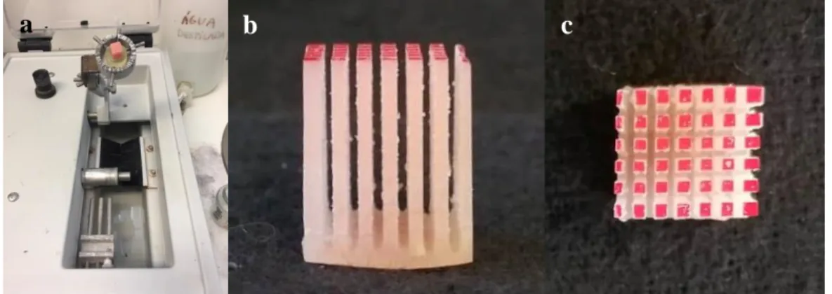
Figure 3.3.2 Putty elastomer mold used for relining procedure. 12 Figure 3.3.3 Preparation of specimens: a) Position on Isomet cutting machine; b) and c)
After section in X and Y axis to obtain sticks 13 Figure 3.3.4 μTBS specimens in graduated falcon tubes with artificial saliva; 13 Figure 3.3.5 Positioning the stick in the Geraldeli’s Jig with the interface centered, using
the stereomicroscope 14
Figure 3.3.6 Sticks fixed to Geraldeli’s Jig with cyanoacrylate glue and placed at Instron
universal testing machine. 14
Figure 3.3.7 Measurement of stick’s bonding area with a digital micrometer. 15 Figure 3.3.8 Stereomicroscope’s images of the three types of failures. 15 Figure 4.2.1 Box plot of microtensile bond strength (MPa) of Kooliner. 19 Figure 4.2.2 Box plot of microtensile bond strength (MPa) of Ufi Gel Hard. 20 Figure 4.2.3 Box plot of microtensile bond strength (MPa) of Probase Cold. 20 Figure 4.2.4 Percentage of Failure according to the acrylic reline resin and proportion of
xi
List of abbreviations
1,6-HDMA 1,6-hexanedioldimethacrylate CHX Chlorhexidine HEMA 2-hidroxyethylmethacrylate IBMA Isobutylmethacrylate IR Interquartile RangeISO/TS International Organization for Standardization/Technical Specification
K Kooliner L Liquid M Mean m Median Max Maximum Min Minimum min Minutes MMA Methylmethacrylate P Powder PC Probase Cold PEMA Polyethylmethacrylate PMMA Polymethylmethacrylate SD Standard deviation
UG Ufi Gel Hard
γ Surface free energy
γd Dispersive component of surface free energy
γp Polar component of surface free energy
1
1. Introduction
Over the last years, the adult population has been experiencing an improvement in oral health, leading to a decrease of edentulism. However, the demographic trend for an elderly population leads to a still significant number of patients needing treatment with complete or partial dentures, which is believed to rise steadily for the next two decades.(1-3) This rehabilitation allows the reestablishment of function, vertical dimension, improves aesthetics and speech, as well as decreases psychological consequences.(2,4)
Due to physiologic progression of residual ridge resorption after tooth loss, adaptation of the denture base is affected, resulting in loss of retention, comfort, trauma in the underlying mucosa and consequent rejection of the denture.(5-6) The denture and the ridges should be examined periodically to detect these changes and if a situation like this is presented, a relining procedure should be done.(7-8) With this procedure, retention and stability improve significantly and an effective distribution of the masticatory load in the denture is achieved. This is a time-saving, convenient and relatively inexpensive prosthodontic treatment when compared to the cost and time-consuming of making new dentures.(9-12)
The relining procedure can be carried out with chairside relining materials, which means that the relining is directly performed inside the mouth of the patient, or laboratory relining materials, used in the indirect method.(5,7) Acrylic resins for relining procedures, alike to the denture base, consist of polymeric biomaterials composed by chains of monomers, where a maximum conversion of monomer is necessary.(13-14) The residual monomers can be trapped on the polymer matrix, affecting the mechanical and physical properties of the biomaterial, and can be diffused into the surrounding medium causing undesirable biological reactions, including local chemical irritation, hypersensitivity, ulceration, systemic allergic reactions and development or oral diseases, like denture stomatitis.(15-17)
Denture stomatitis is a chronic condition observed in 45-70% of denture wearers and manifests as a diffuse inflammation of the palatal mucosa that is delimited by the borders of the denture, usually asymptomatic.(2,18-21) It is considered a clinical finding of Erythematous Candidiasis or Chronic Atrophic Candidiasis, a subgroup of Oral Candidiasis.(22,23) Even though other Candida species may contribute to this disease, Candida albicans is the principal causative agent and its adherence to oral mucosa and denture surface is considered the first step in the pathogenesis of denture stomatitis.(21,24-28)
2
Despite the evidence of fungal etiology, several factors have been suggested in a multifactorial etiology, such as acid salivary pH or reduced saliva secretion, poor hygiene, continuous denture wear, trauma from ill-fitting dentures, nutritional deficiency, long-term antibiotic therapy, immune suppression and xerostomia.(19,23,26,28-30)
Treatment of denture stomatitis usually evolves topical or systemic antifungal therapy, good oral hygiene, denture cleaning procedures, adjustment of denture failures, discontinuation of night-time denture wear, nutritional restitution and relining or replacing the denture.(23,25) Systemic antifungal like fluconazole is commonly used because is well tolerated and it has low toxicity, but they do not eradicate the microorganisms from the denture surface. Clinical effectiveness of topical antifungals is dependent upon its delivery and retention at a specific site, as well as patient compliance. One example is nystatin, which is a highly effective topical antifungal that has few drug interactions, but its four times daily dosage is a significant challenge for patient compliance.(21,25,28) Nevertheless, they are associated to relapses, since
Candida albicans seem to penetrate the denture acrylic and some Candida species are
azole-resistant.(6,21,31,32)
Chlorhexidine (CHX) is an antimicrobial agent widely prescribed as an antiseptic mouthwash in dentistry due to its activity against a wide range of microorganisms, including
Candida species.(22,27,31) CHX has been showing low concentration efficiency, high
substantivity, capacity to reduce biofilm formation and disorganize pre-formed biofilm.(29) However, its efficiency is influenced by not only its concentration but also its exposure time, which can be associated to a therapeutic failure caused by the turnover of saliva and the cleansing action of the oral musculature.(25,27,33) Immersion of acrylic dentures in CHX have been shown to suppress adhesion of Candida to the dentures for longer than with antifungal agents.(27)
Considering the disadvantages of some forms of treatment proposed in the literature to date and joining the best of these ideas, a new form of treatment for denture stomatitis was proposed, a novel drug release system. A release system of CHX loading on resins has been investigated in several studies, the general principle is the incorporation of resin dentures with CHX that releases from the device and inhibits microbial adherence and growth.(20,31,34,35) By loading antimicrobial agents into resin-based denture relining materials, it is possible not only to create a drug delivery system, but also guarantee availability of the agent in the target area at a therapeutic dosage.(22,27,31,35) Some studies have evaluated the CHX release from acrylic resins and concluded that there is a high initial rate of delivery from the material, followed by a controlled slow and constant release for at least twenty eight days, being more
3 effective than the mouth rinse.(22,27,31,32) Direct delivery of the drug to the site of infection reduces the risk of systemic side effects or drug interactions.(21,31)
Most of the previous studies assessing the effect of CHX showed that a concentration of 10% was the most effective against Candida albicans and the maximum dose that could be safely incorporated in acrylic resins without interfering with the mechanical properties of the Ufi Gel Hard but affecting flexural strength in both Kooliner and Probase Cold.(21,22,27,31) However, recent preliminary results established minimal concentrations of CHX in order to assure proper antifungal activity against Candida albicans. Thus, 2.5% for the reline acrylic resin Kooliner seems to be enough to prevent the appear and the development of the fungus, while for Ufi Gel Hard and Probase Cold an incorporation of 5% is required.(36)
It is still uncertain the effects of these loading techniques on the mechanical properties of acrylic resins over time.(37,38) Surface free energy is a property that strongly influences the wettability of relining materials, which is one of the most important factor that influences the denture retention to the mucosa and in another field may contribute to the adherence, bonding and colonization of Candida species.(30,39) Bond strength between the base of the denture and the relining material is also important, since a weak bond encourages a gap formation with ingress of bacteria and fungus and promote staining.(7,40-44) Microtensile bond strength test (μTBS) was first introduced by Sano and colleagues, in 1994, and since then it has been used to test adhesion between several dental materials as a promising way to evaluate the adhesion between acrylic resins due to his reduced area, 1mm2 by ISO/TS 11405:2015.(45-48)
Previous studies showed the influence of loading different concentrations of CHX in the microhardness, flexural strength, surface free energy and shear bond strength properties of acrylic reline resins immediately after being prepared and submitted with thermal ageing.(49-51) However, other studies that simulate other biodegradation processes of the oral medium have not been performed yet, like chemical ageing. The oral cavity is expose to endogenous and exogenous acids, such as dietary changes, that include a fluctuation of the pH present.(52,53) It is also known that the oral cavity pH in individuals with denture induced stomatitis is lower (pH≈5.2) and that an individual with a cariogenic diet is subjected to approximately 6h of acid environment per day.(54)
Thus, this investigation seeks to clarify the impact of CHX-loaded acrylic resin subjected to oral chemical fluctuations on the surface free energy of acrylic reline resins and microtensile bond strength between acrylic reline resins and denture base.
4
2. Objectives
The first objective of this study was to evaluate the effect of loading three different acrylic reline resins with a specific concentration of CHX on surface free energy, after a chemical ageing process, according to the following hypotheses:
H01: Loading Kooliner with 2.5% of CHX does not affect the surface free energy. H11: Loading Kooliner with 2.5% of CHX affects the surface free energy.
H02: Loading Ufi Gel Hard with 5% of CHX does not affect the surface free energy. H12: Loading Ufi Gel Hard with 5% of CHX affects the surface free energy.
H03: Loading Probase Cold with 5% of CHX does not affect the surface free energy. H13: Loading Probase Cold with 5% of CHX affects the surface free energy
The second objective was to evaluate the effect of loading three different acrylic reline resins with a specific concentration of CHX on microtensile bond strength to denture base resin, after a chemical ageing process, according to the following hypothesis:
H04: Loading Kooliner with 2.5% of CHX does not affect the microtensile bond strength to the acrylic base resin.
H14: Loading Kooliner with 2.5% of CHX affects the microtensile bond strengthto the acrylic base resin.
H05: Loading Ufi Gel Hard with 5% of CHX does not affect the microtensile bond strength to the acrylic base resin.
H15: Loading Ufi Gel Hard with 5% of CHX affects the microtensile bond strengthto the acrylic base resin.
5 H06: Loading Probase Cold with 5% of CHX does not affect the microtensile bond strength to the acrylic base resin.
H16: Loading Probase Cold with 5% of CHX affects the microtensile bond strengthto the acrylic base resin.
The third objective was to evaluate the influence of loading three different acrylic reline resins with a specific concentration of CHX in the type of bonding failure to the denture base resin, according to the following hypothesis:
H07: Loading Kooliner with 2.5% of CHX does not affect the type of bonding failure to the denture base resin.
H17: Loading Kooliner with 2.5% of CHX affects the type of bonding failure to the denture base resin.
H08: Loading Ufi Gel Hard with 5% of CHX does not affect the type of bonding failure to the denture base resin.
H18: Loading Ufi Gel Hard with 5% of CHX affects the type of bonding failure to the denture base resin.
H09: Loading Probase Cold with 5% of CHX does not affect the type of bonding failure to the denture base resin.
H19: Loading Probase Cold with 5% of CHX affects the type of bonding failure to the denture base resin.
6
3. Materials and Methods
3.1. Materials
Materials assessed in the present study evolve three auto-polymerizing acrylic reline resins, which were selected for their differences in chemical composition. Two of them consist in direct reline resins: Kooliner (GC America Inc., Alsip, Illinois, USA) (Appendix 2, Figure 1) a non-crosslinking poly(ethyl methacrylate)-based resin, and Ufi Gel Hard (Voco GmbH, Cuxhaven, Germany) (Appendix 2, Figure 2), a crosslinking poly(ethyl methacrylate) (PEMA)-based resin. The other material consists of an indirect reline resin: Probase Cold (Ivoclar Vivadent AG, Liechtenstein) (Appendix 2, Figure 3), a poly(methyl methacrylate) (PMMA)-based resin.
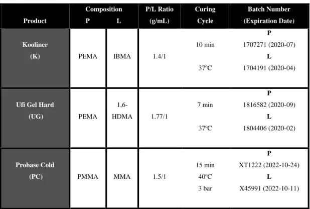
The name, composition, power/liquid ratio, polymerization condition, batch number and expiration date of which one of them are presented in Table 3.1.
Table 3.1 – Materials used in the study.
Product Composition P L P/L Ratio (g/mL) Curing Cycle Batch Number (Expiration Date) Kooliner (K) PEMA IBMA 1.4/1 10 min 37ºC P 1707271 (2020-07) L 1704191 (2020-04)
Ufi Gel Hard
(UG) PEMA 1,6-HDMA 1.77/1 7 min 37ºC P 1816582 (2020-09) L 1804406 (2020-02) Probase Cold (PC) PMMA MMA 1.5/1 15 min 40ºC 3 bar P XT1222 (2022-10-24) L X45991 (2022-10-11)
P = Powder, L = Liquid; PEMA = Poly(ethyl methacrylate), IBMA = Isobutyl methacrylate HDMA = Hexanedioldimethacrylate, PMMA = Poly(methyl methacrylate), MMA = Methyl methacrylate
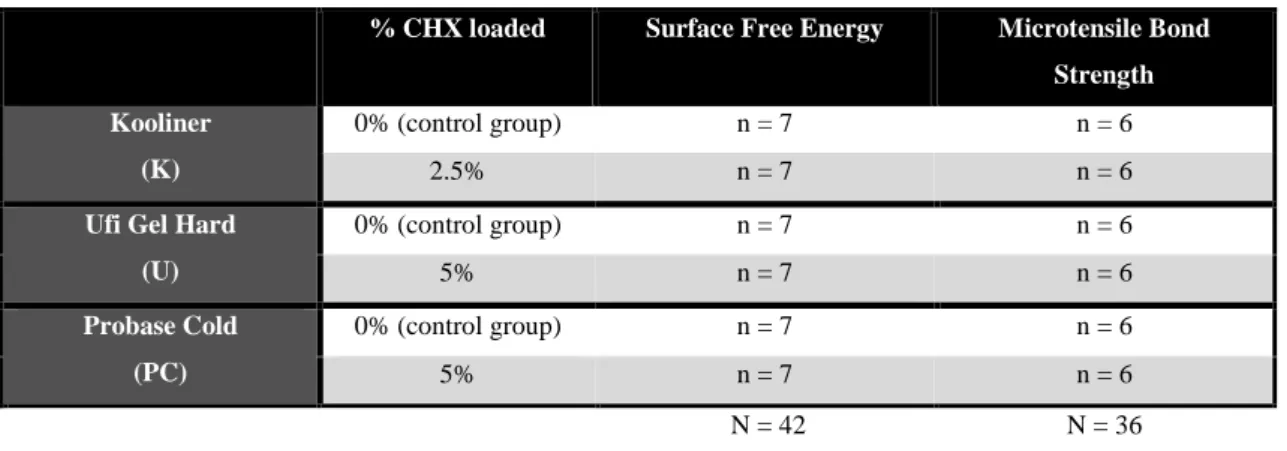
7 Chlorhexidine Diacetate Monohydrate (Panreac AppliChem, Darmstadt, Germany) (Appendix 2, Figure 4) was incorporated in the previous listed acrylic reline resins with a specific concentration presented in Table 3.2.
Table 3.2 – CHX Concentration and number of specimens for each material.
% CHX loaded Surface Free Energy Microtensile Bond Strength Kooliner
(K)
0% (control group) n = 7 n = 6 2.5% n = 7 n = 6
Ufi Gel Hard (U) 0% (control group) n = 7 n = 6 5% n = 7 n = 6 Probase Cold (PC) 0% (control group) n = 7 n = 6 5% n = 7 n = 6 N = 42 N = 36
The powder of acrylic reline resin and CHX (Figure 3.1.1-a) was weighted using a precision balance with internal calibration (A&D FZ-200i) (Appendix 2, Figure 5) and the liquid was measured using a graduated pipette. The mixture of the two materials was done according to the acrylic reline resin weight (w/w) and mixed with a mortar and a pestle until homogenization was achieved (Figure 3.1.1-b). Then the mixture was blend with the correspondent amount of liquid and the polymerization was taken by the recommendations of the manufacturer. Concerning the direct reline resins, specimens were maintained under compression in an incubator at 372ºC (Appendix 2, Figure 6), in order to simulate the intraoral polymerization conditions of the materials (Ehret, Mahlberg, Germany). Otherwise, for the indirect acrylic Probase Cold the relined specimens were placed inside an Ivomat pressure device (IvoclarVivadent, Liechtenstein) (Appendix 2, Figure 7) during 15 min at a temperature of 40ºC and 3 bar.
Figure 3.1.1 – Chlorhexidine diacetate monohydrate: a) Package;
b) Incorporation and homogenization.
8
3.2. Surface Free Energy
Preparation of the specimens
For each material two groups of seven specimens (n=7) were produced (one control group without CHX and one experimental group with the CHX percentages mentioned before), resulting in fourteen specimens per material and a total of forty-two specimens (Table 3.2).
Specimens were obtained by placing the mixed material into metallic rectangular shapes (125×25×1 mm) and then clamped together in order to spread the excess of the material (Figure 3.2.1-a). After polymerization with specific conditions according to the manufacturer’s instructions and cited before (Table 3.1), all the samples were removed from the molds material (Figure 3.2.1-b) and cut with a turbine cylindrical drill to the dimensions of approximately 25 mm width, 16 mm height and 1 mm thickness. The edges of each sample were polished manually with 600-grit silicon carbide paper (Carbimet Paper Discs, Buehler Ltd., Lake Bluff, IL) (Appendix 2, Figure 8) in order to remove irregularities.
At this point, the specimens were submitted to chemical ageing procedure.
Chemical ageing procedure
The procedure of chemical ageing consists of immerging each specimen in a 50 mL graduated falcon tube filled with artificial saliva, respecting a 1g/5mL ratio (Figure 3.2.2-a). To respect this proportion, specimens were weighted (A&D Company, Limited, Tokyo, Japan) and the calculation was obtained.
The solution used in the present study was artificial saliva at pH=7 and pH=3, prepared according to a Faculty of Pharmacy University of Lisbon formula, courtesy of Professor Joana Marto:
a b
Figure 3.2.1 – Preparation of the specimens: a) Compression of the resin through metal mold compression;
9 1) Determination of deionized water volume and PBS quantity (9,6g/1000mL) needed. Mixture both and boiling (F12-ED Refrigerated/Heating Circulator) half of the volume prepared at 60ºC (solution 1). Placing the magnet agitator inside the solution, turning the motor on at 700 rpm.
2) Sprinkling the quantity calculated of Xanthan gum (0,05g/100mL) into boiling buffer and stirring until total of xanthan gum was dissolved.
3) Dissolving of Calcium chloride dihydrate (0,04g/100mL) (EW-N/EG-N balance), Sodium chloride (0,08g/100mL) and Potassium chloride (0,08g/100mL) in solution 1 and stirring until total of materials were dissolved.
5) Dissolving the quantity calculated of Propylene glycol (15,0g/100mL) in solution 2 and stirring until total of Propylene glycol was dissolved.
7) Pouring the solution 3 into a graduated beaker and complete the solution with phosphate buffer pH=7.0 to the volume initially calculated. Removing the magnet agitator.
8) Adjusting the pH (Crison micro pH 2001) (Appendix 2, Figure 9) of artificial saliva to 3 with HCl 1N, since for pH=7 there is no need for adjustment.
9) Keep the solutions out of light, at room temperature.
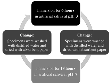
Protocol of chemical ageing consisted in simulating oral conditions by placing the falcons into an incubator at 37ºC (Memmert, Schwabach, Germany) with constant gentle shaking (300 rpm) (Figure 3.2.2-b), following the sequence in Figure 3.2.3, until an ageing period of 28 days or 672 hours was achieved.
Figure 3.2.2 – Incubation of the specimens: a) in graduated falcon tubes with artificial saliva;
b) in incubator at 37ºC.
b
10 .
Surface free energy assessment
After the chemical ageing procedure was complete, the dimensions of each specimen (height, width and thickness) (Appendix 1, Tables 1.1, 1.2 and 1.3) were measured with digital micrometer (Mitutoyo Digimatic, MFG.Co., Ltd Tokyo, Japan) (Appendix 2 , Figure 10) with precision ± 0.01mm and introduced in the software of a computer connected with a Tensiometer K12 (Kruss, Hamburg, Germany) (Appendix 2, Figure 11).
Firstly, the specimen was suspended on the balance (sensitivity = 10-4 g) of the equipment, following the immersion of 4 mm in the liquid (water and 1,2-propanediol) at a speed of 20μms-1 (Appendix 2, Figure 12). In all the procedure, careful was taken not to handle the surfaces of the specimens to reduce the chance of contamination. The measurement of contact angles of distilled water and 1,2-propanediol of the specimens, at room temperature, were obtained applying the Wilhelmy plate technique.(55)Advancing contact angles were used to estimate total surface free energy (γ) of all specimens, as well as its dispersive (γd) and polar components (γp), based on the harmonic mean method proposed by Wu.(56)
The 1,2-propanediol used in this study had a total surface free energy (γ) of 38 mN/m, with a dispersive component (γd) of 28.6 mN/m and a polar component (γp) of 9.4 mN/m. The density was 1.04 kg/m3 and the respective molar mass was 76.09 g/mol (1-2 Propanediol R.822324-1L; Merck, Germany) (Appendix 2, Figure 13). The water used was of Milli-RX quality (Merck Millipore, Germany).
Immersion for 6 hours in artificial saliva at pH=3
Change:
Specimens were washed with distilled water and dried with absorbent paper
Immersion for 18 hours in artificial saliva at pH=7
Change:
Specimens were washed with distilled water and dried with absorbent paper
11
3.3. Microtensile Bond Strenght
Preparation of denture base specimens
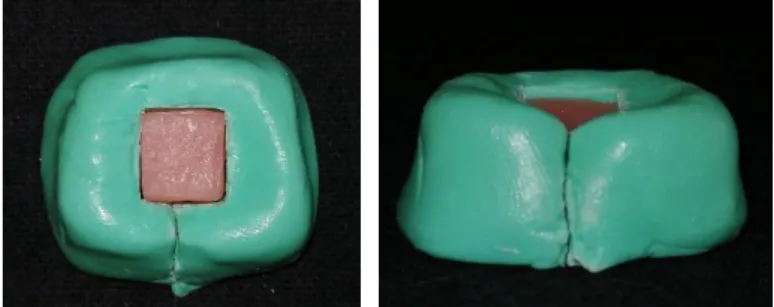
A total of thirty-six specimens of heat-polymerizing denture base acrylic resin Probase Hot (Ivoclar Vivadent AG, Liechtenstein) (Appendix 2, Figure 14) were produced. A conventional flasking technique was used, in which all the wax specimens obtained with a putty elastomer mold with a quadrangular shape (10 × 10 × 10 mm) were flasked and placed above a stratum of gypsum type II. Afterwards, a coat of vaseline above the primary stratum of gypsum was applied, placing another compound of gypsum type II and III mixture on the superior half, covering the specimens. Then the top of the flask was positioned, allowing the excess of gypsum to flow throw the holes. After the complete set of the gypsum was achieved, the flask was placed under boiling water between 4 to 6 minutes and, once removed from the boiling water, it was opened to clear the wax. A separating fluid was applied on the impressed gypsum (Ivoclar Vivadent AG, Liechtenstein), and then a heat-polymerizing resin (Probase Hot, Ivoclar Vivadent AG, Liechtenstein) was prepared and packed into the flask with a powder/liquid ratio of 22.5/10 g/mL. The set was subjected to polymerization through a hydraulic system which guaranteed the conditions indicated by the manufacturer (heat up to 100ºC and boil for 45 min). After being removed from the water, the set was let to cool at room temperature before removing the specimens.
In order to simulate a three month ageing process inside the oral cavity, all specimens were submitted to 2500 thermocycling cycles composed of alternating submersions of 20 seconds at 5ºC and 55ºC, with an interval of 5 seconds between each bath, on a thermocycling machine (Refri 200-E, Aralab, Cascais, Portugal) (Figure 3.3.1).
12 Relining procedure
The measures of the denture base specimens were confirmed using a digital micrometer (Mitutoyo Digimatic, MFG.Co, Ltd. Tokyo, Japan) with a precision of ± 0.01mm and adjusted in a rotational polishing machine (DAP- U, Struers, Denmark) with a 600-grit silicon carbide paper (Carbimet Paper Discs, Buehler Ltd., Lake Bluff, IL).
A denture base specimen was placed in a putty elastomer mold (Figure 3.3.2) and, prior to relining with Kooliner or Probase Cold, correspondent monomer of these reline resins was soaked on the bonding area. In Ufi Gel Hard relining, a specific conditioner was applied and then dried in the air for about 30 seconds, as recommend by the manufacturer.
Two groups (control and experimental group with CHX) of six specimens (n=6) were prepared for each material, as presented in Table 3.2.
The relining procedure was carried out placing the mixed material above the denture base cube and with specific conditions, according to the manufacturer’s instructions (Table 3.1). After polymerization, all the samples were removed from the molds and were polished manually with 600-grit silicon carbide paper (Carbimet Paper Discs, Buehler Ltd., Lake Bluff, IL) in order to remove irregularities. The face corresponding to the denture base was identified with nail varnish, applying a different color for each experimental group.
Preparation of specimens for microtensile bond strength assessment
The relined cubes were assembled perpendicularly to the large axis of an acrylic resin cylinder, with the varnished base up, and fixed with sticky wax. Then the relined cubes were positioned on an Isomet cutting machine 1000 Precision Saw (Serial No. 666-IPS-03518; Buehler, Lake Bluff, IL, USA) (Figure 3.3.3-a) parallel to the diamond cutting blade (Lapcraft, OH, EUA; 4” x .012” x ½”) and sectioned with 550 rpm and cooling, first on the X axis and
c
13 then on the Y axis to obtain sticks (parallelepiped specimens) (Figure 3.3.3-b,c) with a sectional area of 1mm2.
Measurements of each stick were taken with a digital micrometer (Mitutoyo Digimatic, MFG.Co, Ltd. Tokyo, Japan) with a precision of ± 0.01 and the five most uniform were selected.
Chemical ageing procedure
At this point, the five selected sticks of each relined cube (n=6) were allocated in a eppendorf falcon tubes of 1.5mL filled with artificial saliva (Figure 3.3.4), respecting a 1g/5mL ratio and submitted to the same chemical ageing procedure explained above in section 3.2 – Chemical Ageing Procedure and exemplified in Figure 3.2.3.
Microtensile bond strength assessment
After the ageing process each stick was placed on a stainless-steel device, Geraldeli’s
Jig, in which the extremities were fixed with cyanoacrylate glue (PERMABOND, Permabond
Adhesive, S. Paulo, Brazil) (Appendix 2, Figure 15). The placement of the sticks was performed
Figure 3.3.3 – Preparation of specimens: a) Position on Isomet cutting machine; b) and c) After section in X
and Y axis to obtain sticks.
a b
Figure 3.3.4 – specimens in graduated falcon tubes with artificial saliva.
14 with the help of a stereomicroscope (EMZ-8TR, Meiji Techno Co, Saitama, Japan) (Appendix 2, Figure 16), in order to ensure that the interface was placed at the center of the device (Figure 3.3.5). Also, the side of the device correspondent to the denture base resin was identified with a permanent marker.
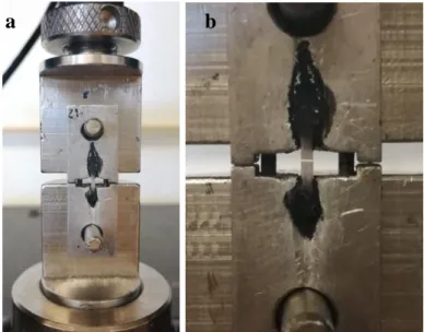
The device was installed in a universal testing machine model 4502 (Instron Ltd., Bucks, HP 12 3SY, England) (Figure 3.3.6) and the test was runned with 1kN load cell and crosshead speed of 1mm/min until fracture.
After fracture occurred, the measures of the bonding area were registered using a digital micrometer (Mitutoyo Digimatic, MFG.Co, Ltd. Tokyo, Japan) with a precision of ± 0.01mm (Figure 3.3.7). The microtensile bond strength (μTBS) value, expressed in MPa, was obtained by the Series IX program (Series IX, Automated materials test system, version 8.34.00, serial
Figure 3.3.6 – Sticks fixed to Geraldeli’s Jig with cyanoacrylate glue and placed at Instron universal testing
machine. a) Before fracture; b) After fracture.
a b
Figure 3.3.5 – Positioning the stick in the Geraldeli’s Jig with the interface centered, using the
15 number 21744H, Instron Corporation, Grove City, PA, EUA), through the relation between the load at the time of fracture and the stick interfacial area.
Failure mode assessment
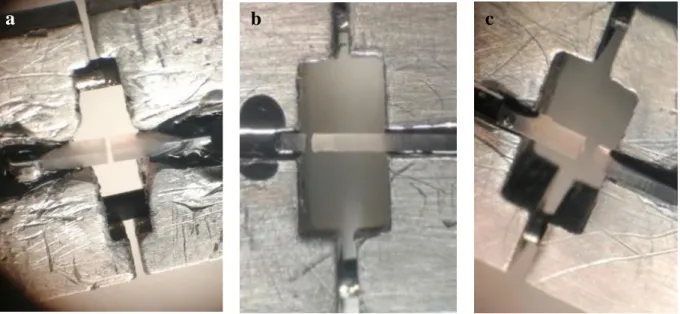
The failure mode on the separated surfaces was assessed by two observers with a stereomicroscope and classified as adhesive, cohesive or mixed (Figure 3.3.8). The failures were considered adhesive if occurred between the reline resin and the denture base resin and cohesive if the fracture occurred exclusively within one of the resins. If the fracture occurred in the interface of the two resins but included vestiges of reline resin, it was considered mixed.
Figure 3.3.8 – Stereomicroscope’s images of the three types of failures: a) Adhesive; b) Cohesive; c) Mixed. Figure 3.3.7 – Measurement of stick’s bonding area with a digital micrometer.
16
3.4. Statistical Analysis
For microtensile bond strength test, each cube was considered as an experimental unit, assuming the mean of the values obtained from all the sticks of the same cube as an independent observation for the purpose of statistical analysis. In the case of failure mode assessment, each stick was considered as an experimental unit for the purpose of statistical analysis.
Descriptive statistics of surface free energy and microtensile bond strength values were carried out being determined the mean, median, standard deviation and interquartile range per group.
Data were statistically analyzed using SPSS Statistics 20 (SPSS Inc., Chicago, IL, USA) and did not follow a normal distribution for the studied variables in the Shapiro-Wilk normality test. Therefore, the results were submitted to the nonparametric tests according to the Kruskal-Wallis method, followed by multiple corrections using Mann-Whitney tests. To determine the association between the failure mode and the incorporation of CHX, chi-square test and the Fisher’s exact test were applied.
17
4. Results
4.1. Surface Free Energy
Descriptive analysis of the data was carried out for each material, including mean, median, standard deviation and minimum and maximum values for contact angle (Appendix 1, Table 1.4) and surface free energy (Appendix 1, Tables 1.5, 1.6 and 1.7).
The values of the total surface free energy (γ) and their components, the dispersive (γd) and polar (γp), are summarized in Table 4.1. Likewise, the mean, median, standard deviations and interquartile range of the groups by reline resin were registered.
Table 4.1 – Total surface free energy data, as well as the dispersive and polar components, by reline resin.
Material % CHX
loaded
Surface Free Energy (γ) (mN/m)
γ
γ
dγ
p Kooliner0%
M±SD 32.1 ± 3.19a 15.8 ± 6.64a 16.3 ± 9.23a m (IR) 31.8 (4.20) 16.7 (4.30) 14.0 (7.60)2.5%
M±SD 34.1 ± 2.26b 16.2 ± 3.04b 17.9 ± 4.79b m (IR) 34.4 (2.30) 15.2 (5.00) 19.2 (4.20) Ufi Gel Hard0%
M±SD 39.89 ± 3.48a 19.04 ± 2.39a 20.81 ± 5.48a m (IR) 41.5 (4.40) 18.7 (2.40) 21.1 (6.70)5%
M±SD 41.9 ± 1.09b 18.1 ± 2.69b 23.8 ± 2.92b m (IR) 42.0 (1.80) 19.0 (4.70) 24.2 (3.80) Probase Cold0%
M±SD 36.7 ± 4.60a 12.3 ± 5.50a 24.4 ± 6.78a m (IR) 37.2 (4.40) 15.4 (9.50) 23.3 (10.4)5%
M±SD 37.2 ± 1.75b 19.1 ± 3.74a 18.1 ± 4.59b m (IR) 36.6 (2.80) 18.1 (2.90) 19.3 (3.50)γ=Total surface free energy; γd=Dispersive surface free energy; γp=Polar surface free energy;
18 Vertically identical superscripted letters denote significant differences among each group of the same material (p<0.05).
Considering Kooliner specimens (Table 4.1), significant differences have not occurred either in the total surface free energy or in its correspondent components, dispersive (γd) and polar (γp) (p>0.05).
For Ufi Gel Hard (Table 4.1), as well as in the previous acrylic reline resin, there were no statistical differences in total surface free energy and in the dispersive (γd) and polar (γp) components (p>0.05).
Regarding Probase Cold specimens (Table 4.1), a statistically significant difference (p=0.025) in the dispersive component (γd) was found, with specimens loaded with 5% of CHX
exhibiting significant higher values than the control group. In the total surface free energy and in the polar component (γp), no significant differences were found (p>0.05) between groups.
19
4.2. Microtensile Bond Strenght
The mean (M) values of microtensile bond strength for each group are summarized in Table 4.2, as well as the standard deviation (SD), median (m) and interquartile range (IR).
Table 4.2 – Microtensile bond strength data by reline resin (n=6).
Material % CHX
loaded
Microtensile Bond Strenght (MPa)
M ± (SD) m IR Kooliner 0% 13,0 ± 3,7 12,7 5,9 2.5% 13,5 ± 3,6 13,4 6,0 Ufi Gel Hard 0% 22,6 ± 7,4 22,2 14,0 5% 18,3 ± 5,6 16,8 9,6 Probase Cold 0% 45,0 ± 3,3 44,0 5,8 5% 33,7 ± 1,9 33,1 3,3
Regarding Kooliner specimens (Figure 4.2.1), no statistically significant differences were found on microtensile bond strength between the control group and the 5% CHX loaded group (p>0.05).
Figure 4.2.1 – Box plot of microtensile bond strength (MPa) of Kooliner. No statistically significant differences were found between groups (p>0.05).
0 5 10 15 20 25 30 35 40 45 50 Mi cr ot ensi le B ond Str engt h ( MP a)
Kooliner
20 Also, for Ufi Gel Hard (Figure 4.2.2) significant differences have not occurred on microtensile bond strength values between 5% CHX loaded group and control group (p>0.05).
Figure 4.2.2 – Box plot of microtensile bond strength (MPa) of Ufi Gel Hard. No statistically significant
differences were found between groups (p>0.05).
Considering Probase Cold (Figure 4.2.3), 5% CHX group had lower microtensile bond strength values compared to the control group (p=0.004). Horizontal line below the boxes denote significant differences among groups (p<0.05).
Figure 4.2.3 – Box plot of microtensile bond strength (MPa) of Probase Cold. Statistically significant
differences were found between control group and 5% CHX group (p=0.004).
0 5 10 15 20 25 30 35 40 45 50 Mi cr ot ensi le B ond Str engt h (MP a)
Probase Cold
0 5 10 15 20 25 30 35 40 45 50 Mi cr ot ensi le B ond Str engt h ( MP a)Ufi Gel Hard
21 All specimens were observed in a stereomicroscope to assess the type of bonding failure, which percentages within each group are specified in Figure 4.2.4.
Figure 4.2.4 – Percentage of failure according to the acrylic reline resin and proportion of CHX loaded.
The predominant type of failure in the study was adhesive, with 79.4% of the sticks tested (N=180) showing this type of failure.
Considering Kooliner, in both experimental and control group 96.7% of the failures were adhesive and no cohesive failures occurred, having no statiscally significant differences between them (p>0.05).
In the case of Ufi Gel Hard all types of failures occurred in both groups, however, the predominant type of failure in the control group was adhesive (63.3%), while in the experimental group was cohesive (43.3%). It was observed a decrease in the adhesive failures and an increase of cohesive failures in the experimental group. However, these differences were not statistically significant when compared to the control group (p>0.05).
For both Probase Cold groups 90% of the failures where adhesive. Mixed failures were higher on the experimental group (10%) but this difference was not statistically significant (p>0.05). P C 5 % P C 0 % U G 5 % U G 0 % K 2 , 5 % K 0 % 90 90 40 63.3 96.7 96.7 3.3 43.3 20 10 6.7 16.7 16.7 3.3 3.3
FAILURE MODE (%)
22
5. Discussion
The present study evaluated the effect of CHX loading on surface properties of different acrylic reline resins, specifically the surface free energy, microtensile bond strength and the type of failure seen.
Incorporation of CHX acrylic resins is a therapeutic approach for denture stomatitis in which a slow and sustained-releasing device is created. It has been widely evidenced in microbiological and release studies due to its broad-spectrum antimicrobial activity, including against C. albicans.(19,21,22,33,43) The antifungal effect of CHX was found to be more effective than other drugs, such as fluconazole, both on releasing and microbiological tests.(22,27,31) However, the incorporation of antimicrobial agents such as CHX into polymeric materials may affect their mechanical properties, making their evaluation imperative.(21,23,57-60) CHX concentrations used in this study were selected based on the results of previous studies that evaluated the 10%, 7.5%, 5%, 2.5 % and 1% concentrations, excluding the ones that influenced negatively the mechanical and physical properties of each material. A recent preliminary microbiological study by Costa established a concentration of 2.5% for Kooliner and 5% for both Ufi Gel Hard and Probase Cold as the minimal concentration effective against Candida albicans.(36) Others authors found that these concentrations have no negative influence on the acrylic resins mechanical properties.(51,61,62) Thus, this concentrations were selected for this study.
The three reline resins studied were chosen for their differences in chemical composition and structural arrangement. Direct reline resins Kooliner and Ufi Gel Hard are both poly(methyl methacrylate) based materials and are known for an anomalous water uptake behaviour (21,63,64), with higher drug release characteristics.(64) Kooliner forms a simple non-crosslinking net when polymerization is complete, while Ufi Gel Hard forms a more complex crosslinking net. Indirect reline resin Probase Cold is a poly(methyl methacrylate) based material forming a net with a reduced percentage of uncured monomer methyl methacrylate.(65-68) Since these resins have different physical structure and chemical composition, CHX molecules when incorporated in the net can create different links to the polymeric chains and change their properties in distinct magnitudes. Also, CHX incorporation can increase the distance between polymer molecules, resulting in an expected weaker polymer net.(61)
23 These biomaterials are submitted to biodegradation processes that can change their physical and biomechanical properties due to the oral environment conditions (16,67,69), being important to simulate oral cavity conditions in vitro.(43) Some authors studied the effect of thermal ageing (51,61,62), but it is also importante to mimic the conditions of the oral cavity through a chemical ageing process, which was an objective of this study. Other studies have concluded that the release of CHX from acrylic resins showed a high initial rate of elution from the material followed by a slower and steadier diffusion throughout at least 28 days.(19,21,22,27,31,35,70) Also, another study concluded that maximum cumulative release of CHX was higher at pH=3 and pH=7 for the this three materials.(71) Taking these results into account, in this study a cyclic procedure of 6 hours at pH 3 interchanging with 18 hours at pH 7 was applied for 28 days, because it has also been suggested that an individual with a cariogenic diet is subject, daily, to approximately 6 hours of acid environment.(20,37,68)
The first objective of this investigation was to assess the influence of loading CHX on the surface free energy of reline resins, after a chemical ageing procedure.
The total surface free energy of a solid consists in the sum of components arising from dispersive (apolar) and polar contributions. The technique to determine the surface free energy in this study is an indirect method, in which the contact angles formed on the acrylic resins were measured by immersing each specimen 4mm into two distinct liquids (water and 1,2-propanediol). Then, the contact angles were used to calculate the surface free energy by the Wu method.(55,56) The method enable the calculation of the unknown solid surface energy components (polar and dispersive) from contact angle measurements with the two mentioned liquids.(72-75)Changes in the surface free energy of the acrylic resin will have an impact in its surface wettability and, consequently, in the denture retention to support mucosa and adherence of microorganisms to removable dentures.(30,39,74,76,77)
Considering Kooliner and Ufi Gel Hard, the results showed that there were no statistical differences in total surface free energy, dispersive and polar components between the control and the experimental groups of both materials, similarly to Costa results.(51) However, the total surface free energy values obtained by Costa relatively to the CHX concentrations applied in this study were lower, demonstrating a different effect of a chemical ageing as opposed to thermal ageing process.(51)This could be explained by the results of Alexandre, in which higher release of CHX was seen at a pH 3.(71)
Similarly to a study by Arima, in this study CHX incorporation in Kooliner seems to reveal lower total surface free energy values than the other two materials.(65) The fact that both
24 Ufi Gel Hard and Kooliner groups weren’t affected by the CHX loading might be explained by their similar chemical constitution, being both composed of pre-polymerized poly(ethyl methacrylate) particles.(65)At this time, it may be concluded that the first and second null hypothesis cannot be rejected, meaning that surface free energy seems to not be affected by CHX loading in both Kooliner and Ufi Gel Hard groups.
On Probase Cold, inspite of the CHX group showed significant higher values of the dispersive component compared to the control group (meaning that it could become more apolar with CHX incorporation), there were no significant differences in the total surface free energy.(25) With this knowledge, it may be concluded that the third null hypothesis can not be rejected, since the loading of CHX does not seem to affect the total surface free energy of the acrylic reline resin Probase Cold.
The other objective of this study was to evaluate whether the loading of CHX would interfere or not with the microtensile bond strength between the reline resins and the denture base resin, after a chemical ageing procedure.
Adequate bonding between denture base resin and reline material is essential, since a failure can harbor bacteria, promote staining, decrease the strength of the denture and cause fractures.(23,40,41,43,78,79) In past studies, reline resin adhesion to denture base resin has been determined by test methods such as tensile and shear bond strength. However, according to the current literature, there isn’t a consensus on the most reliable test for evaluating the bond strength between denture base and reline resins, because they are not truly testing the bonded interface and tend to induce cohesive fractures.(41,80-82)
Microtensile has been suggested as the first-choice method to determine the bond strength of interfaces between other dental materials because of the reduced testing area and more uniform distribution interfacial stresses, often leading to more adesive failures(45) No studies were found in the existing literature that evaluated the effect of CHX incorporation on microtensile bond strength of acrylic reline resins to denture base resins, although some authors presented it as a viable method.(80,82)Therefore, this study is innovating by applying this test method.
In the present study a crosshead speed of 1mm/min until the separation of the denture base resin and the reline resin was used, since it is considered to be the speed that distributes a more uniform force in the adhesive interface.(83)Is it know that with higher crosshead speed the microtensile bond strength values tend to increase.(8,78)However, there are no previous studies that indicates the most suitable velocity for testing μTBS between acrylic resins. In this
25 work each cube was treated as an experimental unit for μTBS, in which an average of the values obtained from all sticks of the same cube was used for statistical analysis.(48)Meanwhile for failure mode assessment, the stick was considered as an experimental unit for statistical analysis.(48)Though, the use of sticks as an experimental unit is controversial, as it is associated with pseudoreplication of the results and compromises the independence condition of the specimens.(84)
Regarding the Kooliner and Ufi Gel Hard groups no statistically significant differences were found between experimental and control groups mean values of μTBS. These results are similar than early studies that tested shear bond test strength after thermal ageing from other authors.(23,50,51)Also the mean values obtained in Kooliner for both control (13,0 ± 3,7 MPa) and 2.5% loaded CHX (13,5 ± 3,6 MPa) groups seems lower than the other resins, such as Costa obtained with shear bond test.(51) This may be due to the composition of its monomer isobutylmethacrylate, with a high molecular weight monomer that makes the dissolution of PMMA denture base resin surface difficult and leads to a less effective penetration of the reline resin into the denture base.(66,83) These findings sustained the theory that bond strength is dependent on the chemical composition of both materials (7,66,78,85-88), since bonding of autopolymerizing resins to denture base resin seems to be achieved by penetration and diffusion of monomer into the last one.
With these findings the fourth and fifth null hypothesis can not be rejected, since there were no differences between microtensile bond strength of experimental groups compared to the control in Kolliner and Ufi Gel Hard reline resins.
On the other hand, 5% CHX loaded Probase Cold group presented significantly lower mean microtensile bond strength values compared to the control group. This can be explained by the incorporation of CHX within the polymer matrix of the material, introducing more spaces, less homogeneity in the polymerized materials and weakening the bond strength.(23,31) Also the control group showed the highest mean microtensile bond values (45.0 ± 3.3 MPa) followed by the loaded CHX group with highest results (33.7 ± 1.9 MPa), once again accordingly to shear bond strength results of Costa.(51)This support Ahmad and colleagues hypotheses, since Probase Cold composition is identical to Probase Hot and a much easier diffusion and penetration of PMMA reline monomers of smaller molecular weight into denture base resin is achieved, forming an inter-penetrating polymer network.(44,66,78,85,86)At this point, the sixth null hypothesis can be rejected, since loading Probase Cold with 5% of CHX seem to affect the microtensile bond strength with the acrylic base resin.
26 In this study the predominant type of failure was adhesive, seen in 79.4% of the sticks when considering the entire sample (N=180). This is in accordance with the purpose of microtensile bond strength tests, where forces are directed towards the adhesive interface with a more uniform distribution, therefore validates the method chosen and applied in this study. An adhesive failure mode may indicate that the bond strength between the reline resin and the denture base is weaker than the reline material strength, which is an advantage if the objective is a temporary lining in practice.(7,31)
Contrary to what was done in this study, some authors defend to not include cohesive failure sticks in the statistical analysis, defending the non-overestimation of the results whose sticks did not fracture at the adhesive interface. However, according to Pashley and colleagues, the fact that the fractures did not occur at the adhesion interface does not mean that the adhesion is stronger than the intrinsic resistance of the substrate but rather that the test may not have been uniformly done and concentrated in a highly localized region.(89)
Regarding Kooliner, both 0% and 2.5% CHX groups, in 96.7% of the sticks an adhesive failure was observed and no cohesive failure. Also, a correlation between the type of failure and the microtensile bond strength values was detected, since a greater tendency for the occurrence of adhesive failures in sticks with lower μTBS values was observed both in 0% and 2.5% CHX Kooliner groups. This result is in agreement with the results of other authors.(48,90,91)
Concerning Ufi Gel Hard 5% CHX loaded specimens, the failure mode most obtained was cohesive (43.3%), higher than the 20% founded in the control group, followed by adhesive failures (40%). It can be stated that as the concentration of CHX incorporation increased a weakening of the internal structure of the UG occurred comparatively to the bond strength in the interface, leading to an increase of cohesive failures. Nevertheless, these differences were not statistically significant, meaning that more studies with a higher number of specimens are needed to confirm the conclusions of the present study, especially in the groups of Ufi Gel Hard were a high standard error was seen.
On the other hand, for the groups with higher μTBS values, such as Probase Cold, it would be expected to obtain more cohesive failures.(48,90,91) However, both experimental and control groups of this material showed a predominance of adhesive failures (90%). Also, a decrease in cohesive failures and an increase in mixed failures with the incorporation of 5% CHX was seen, possibly justified with the significant reduction of μTBS values occurred. A limitation of failure mode assessment in this material is the difficulty of observing the type of failures in the stereomicroscope, since the denture base resin and the reline resin have an