Liliana Raquel Leite Martins
Antimicrobial resistance analysis of fecal Escherichia coli and
Enterococcus spp. isolates from dogs and cats: prevalence,
assessment of potential risk factors and ability of
multidrug-resistant strains to spread within household
Tese de Candidatura ao grau de Doutor em Ciências Veterinárias submetida ao Instituto de Ciências Biomédicas de Abel Salazar da Universidade do Porto.
Orientador - Professor Doutor Paulo Manuel Rodrigues Martins da Costa
Categoria - Professor Auxiliar
Afiliação - Instituto de Ciências Biomédicas de Abel Salazar (ICBAS), Universidade do Porto (UP)
Co-orientador - Professor Doutor Augusto José Ferreira de Matos
Categoria - Professor Auxiliar
Afiliação - Instituto de Ciências Biomédicas de Abel Salazar (ICBAS), Universidade do Porto (UP)
iii
v
“O médico que só sabe Medicina, nem Medicina sabe” Abel de Lima Salazar
vii Publicações
Em obediência ao disposto no n.º 1 do Artigo 34º do Decreto-Lei n.º 74/2006, publicado em Diário da República, 1ª série, nº 60 de 24 de Março de 2006, e republicado pelo Decreto-Lei nº 115/2013, publicado em Diário da República, 1ª série, nº 151 de 07 de Agosto de 2013, que procede à terceira alteração do Decreto-Lei nº 74/2006, de 24 de Março de 2006, a autora declara que participou na conceção e na execução do trabalho experimental, bem como na interpretação dos resultados e na redação dos trabalhos, publicados e em publicação, que fazem parte integrante desta tese e/ou que dela advêm.
Artigos que fazem parte integrante da Tese
I. Martins, L.R., Pina, S.M., Simões, R.L., de Matos, A.J., Rodrigues, P., da Costa, P.M. (2013). Common phenotypic and genotypic antimicrobial resistance patterns found in a case study of multiresistant E. coli from cohabitant pets, humans, and household surfaces. Journal of Environmental Health. 75(6):74-81.
II. Leite-Martins, L., Meireles, D., Bessa, L.J., Mendes, A., de Matos, A.J., Martins da Costa, P. (2014). Spread of Multidrug-Resistant Enterococcus faecalis Within the Household Setting. Microbial Drug Resistance. March 11. (Epub ahead of print).
III. Leite-Martins, L., Meireles, D., Beça, N., Bessa, L.J., de Matos, A.J.F., da Costa, P.M. (2014). Spread of multidrug-resistant Escherichia coli within Domestic Aggregates (humans, pets and household environment). Microbes and Environments (Submitted).
IV. Leite-Martins, L., Mahú, M.I., Costa, A.L., Mendes, A., Lopes, E., Mendonça, D.M.V., Niza-Ribeiro, J., de Matos, A.J.F., da Costa, P.M. (2014). Prevalence of antimicrobial resistance in enteric Escherichia coli from domestic pets and assessment of associated risk markers using a Generalized Linear Mixed Model. Preventive Veterinary Medicine (Submitted).
V. Leite-Martins, L., Mahú, M.I., Costa, A.L., Vaz-Pires, P., Niza-Ribeiro, J., de Matos, A.J.F., da Costa, P.M. (2014). Prevalence of antimicrobial resistance in enteric Enterococcus spp. from domestic pets and assessment of associated risk factors using a Generalized Linear Mixed Model. Journal of Medical Microbiology (Submitted).
viii
Outros trabalhos científicos motivados pela presente Tese
Artigo em revista de circulação internacional com arbitragem científica
I. Beça, N.M., Bessa, L., Mendes, A., Santos, J., Leite-Martins, L., Matos, A., Martins da Costa, P. (2014). Coagulase-positive Staphylococcus - prevalence and antimicrobial resistance in companion animals, veterinary professionals and clinical environment. Journal of the American Animal Hospital Association. (In press).
Artigo em revista de circulação nacional com arbitragem científica
II. Martins da Costa, P., Leite-Martins, L., Antunes, F., Simões, R. (2010). Transferência de bactérias resistentes aos antimicrobianos entre nichos ecológicos interligados: homem, animais e ambiente. Revista da Faculdade de Medicina de Lisboa, série III 15 (5/6): 319-326.
Publicações em atas de encontros científicos
Comunicações Orais
III. Leite-Martins, L. (2013). Prevalência da resistência aos antimicrobianos em Escherichia coli e enterococcus spp. isolados em cães e gatos e estudo dos respetivos fatores de risco. VIII Congresso OMV. Lisboa, Portugal. 30 de Novembro a 01 de Dezembro de 2013.
IV. Martins da Costa, P., Simões, R., Martins, L., Matos, A.J. (2011). O ciclo ambiental das resistências antimicrobianas (Environmental dissemination of drug-resistant bacteria between intermingled ecological niches). V Congresso de Ciências Veterinárias 2011. Sociedade Portuguesa de Ciências Veterinárias. Santarém, Portugal. 14 de Outubro de 2011, (Pp.57).
ix Comunicações Posters
V. Meireles, D.M., Martins, L.R., Bessa, L.J., Mendes, Â.J., Cunha, S.A., Matos, A., da Costa, P.M. (2014). Estudo da partilha de clones bacterianos entre animais de companhia, coabitantes humanos e superfícies domésticas. VI Congresso da Sociedade Portuguesa de Ciências Veterinárias: Praxis e futuro, Oeiras, Portugal, 3-5 de Abril. (Pp.127).
VI. Leite-Martins, L., Beça, N., Lopes, E., Frias, C., Matos, A., Martins da Costa, P. (2012). In-home and through-home transmission of antimicrobial resistance between human and pets. II International Conference on Antimicrobial Research – ICAR 2012, Lisbon, Portugal, 21-23 November. (Pp:410).
VII. Beça, N.M., Simões, R.L., Santos, J.C., Lopes, E., Leite-Martins, L., Matos, A., Martins da Costa, P. (2012). Culture media isolation of Staphylococcus pseudointermedius and Staphylococcus spp. coagulase positive prevalence in domestic animals, Veterinary practitioners, Veterinary auxiliary workers and environment of a Veterinary hospital. II International Conference on Antimicrobial Research – ICAR 2012, Lisbon, Portugal, 21-23 November. (Pp:387).
xi AGRADECIMENTOS
Esta etapa é o culminar de uma opção de vida tomada uns bons anos atrás, com o apoio de pais e marido, alicerces e pilares da minha vida. Foram anos intensos, repletos de momentos difíceis, mas também abençoados por dois acontecimentos maravilhosos: o Gonçalo e o Francisco. É a este núcleo familiar fascinante que eu devo este momento…
Um doutoramento é um trabalho longo, pejado de pesquisa, de ideias, de esforço, de paciência e de pessoas. Um doutoramento nunca é um trabalho individual e, por isso, viverei mais intensamente esta fase, se conseguir difundir a minha gratidão a todos os que, de uma forma ou de outra, para ele contribuíram. Como tal, quero expressar o meu mais genuíno agradecimento:
Ao meu orientador, o Professor Doutor Paulo Martins da Costa, por me ter recebido e acolhido no seu laboratório, assim como pela oportunidade de integrar o seu grupo de investigação. Agradeço a confiança que em mim depositou, a responsabilidade que me incutiu e a sua dedicada orientação. Acima de tudo, agradeço todo o apoio e compreensão, nos bons e nos maus momentos. Muito obrigado pelo profissionalismo, pela sincera amizade e pela grande disponibilidade que sempre revelou.
Ao Professor Doutor Augusto Ferreira de Matos, meu co-orientador e diretor clínico da UPVet, agradeço ter aceitado colaborar na orientação do presente estudo e estou grata pelo sempre precioso e atempado apoio. Agradeço também ter permitido que a amostragem fosse proveniente da casuística da UPVet.
À enorme Família que constitui os Colaboradores do Laboratório de Microbiologia e Tecnologia Alimentar do ICBAS, com um apreço especial para a D. Elisabete, uma das pessoas mais proactivas, dinâmicas, competentes e responsáveis que eu conheço, ao Romeu, ao Beça, à Sónia, à Ana Lia, à Inês Mahú, à Diana Meireles, e aos incansáveis Ângelo e Lucinda, quero agradecer o caloroso acolhimento, a ajuda, a partilha de conhecimentos, os bons momentos e o companheirismo de todos estes anos; este trabalho é também vosso!
Ao Professor Doutor Paulo Vaz Pires, pelo acolhimento e hospitalidade incondicionais, à Susana Pina, pelos preciosos primeiros passos na genética e aos Professores Doutores Niza Ribeiro e Denisa Mendonça pelo apoio no tratamento estatístico dos dados.
xii
A toda a Equipa da UPVet agradeço a colaboração e a tolerância pelo prolongamento de inúmeras consultas na realização dos meus inquéritos e colheitas. Agradeço também a paciência nas infinitas vezes em que o cansaço me havia já arrebatado toda a energia. Ao Frias deixo uma palavra especial, pelo que é e pelo apoio que deu em períodos imprescindíveis.
A todos os companheiros de 4 patas que deram corpo a este trabalho, alegre e incondicionalmente, assim como aos seus proprietários, principalmente aos que se voluntariaram para participar nos estudos que envolveram todo o agregado doméstico.
Ao Instituto de Ciências Biomédicas Abel Salazar da Universidade do Porto, “casa mãe” onde me formei, cresci para a ciência e tenho a honra de trabalhar; à UPVet, Clínica Veterinária de Animais de Companhia do ICBAS e ao Laboratório de Microbiologia e Tecnologia Alimentar do ICBAS, pela oportunidade e pelos meios físicos, técnicos e humanos disponibilizados.
Aos meus pais, a quem tudo devo: modelo de justiça, honestidade, bondade, amizade e capacidade de trabalho. Foi sobre este alicerce, e com o seu incondicional apoio, que avançaram todas as realizações da minha vida.
Ao Carlos, pelo apoio incondicional ao longo destes anos, nos bons e nos maus momentos, companheiro de todas a horas! Pelas risadas e brincadeiras, pela paciência e tolerância, pelos abraços e desabafos, pelos silêncios escutados… Bom marido e bom pai… Amo-te Muito!
Aos meus filhos, Gonçalo e Francisco, o que de mais precioso eu tenho na vida, não quero agradecer, quero pedir desculpa… Privados da minha atenção e do meu tempo em anos tão importantes do vosso crescimento, tantas vezes tiveram de reprimir a vossa carência de mimo e afeto para suportar-me apressada, cansada e sem paciência… Adoro-vos acima de qualquer coisa e prometo que não voltará a acontecer…
xiii ABBREVATIONS
AMC Amoxicillin-clavulanic acid
AMK Amikacin
AMP Ampicillin
AMR Antimicrobial resistance
ATM Aztreonam
AZM Azithromycin
CAMV Centro de atendimento Médico-Veterinário
CAZ Ceftazidime
CDC Centers for Disease Control and Prevention
CEF Cephalothin
CHL Chloramphenicol
CIP Ciprofloxacin
CTX Cefotaxime
DANMAP Danish Integrated Antimicrobial Resistance Monitoring and Research Program
DGAV Direção-Geral de Alimentação e Veterinária DNA Deoxyribonucleic acid
E. coli Escherichia coli
E. faecalis Enterococcus faecalis E. faecium Enterococcus faecium
e.g.
exempli gratia
EAAD European Antibiotic Awareness Day
ERI Erythromycin
ESBL Extended-Spectrum Beta-Lactamases
et al. et alii
ExPEC Extra-intestinal Pathogenic E. coli FAO Food and Agriculture Organization
Fig. Figure
FOX Cephoxitin
GEN Gentamicin
GSP Good Stewardship Practice HGT Horizontal Gene Transfer
IPM Imipenem
xiv
NAL Nalidixic acid
NARMS National Antimicrobial Resistance Monitoring System
NIT Nitrofurantoin
OMV Ordem dos Médicos Veterinários
QD Quinupristin/dalfopristin
RIF Rifampicin
SCOPE Surveillance and Control of Pathogens of Epidemiologic Importance SENTRY Antimicrobial surveillance Program
STR Streptomycin
SVARM Swedish Veterinary Antimicrobial Resistance Monitoring Program SWEDRES Antibiotic Consumption and Resistance in Sweden
SXT Trimethoprim-sulfamethoxazol
TEC Teicoplanin
TET Tetracycline
TOB Tobramycin
UK United Kingdom
UPVet Clínica Veterinária de Animais de Companhia do ICBAS / UP USA United States of America
UTI Urinary Tract Infection
VAN Vancomycin
xv TABLE OF CONTENTS Resumo………...…. 1 Abstract……….... 3 Chapter 1………. 5 1.1. General introduction………... 7
1.1.1. The phenomenon of antimicrobial resistance…..….... 7
1.1.2. The importance of Escherichia coli and enterococci 11 1.1.3. The role of companion animals……….….… 13
1.2. Rationale and aims………..….… 15
Chapter 2………... 17
2.1. Antimicrobial resistance prevalence and risk factors
……….….…
192.1.1. Paper I ……….….. Prevalence of antimicrobial resistance in enteric Escherichia coli from domestic pets and assessment of associated risk markers using a Generalized Linear Mixed Model 19 2.1.2. Paper II ………...……..…..….. Prevalence of antimicrobial resistance in enterococcus spp. from feces of domestic pets and assessment of associated risk factors using a Generalized Linear Mixed Model 47 2.2. Household antimicrobial resistance share and spread ……….. 69
2.1.3. Paper III ……….……….………....….. Common phenotypic and genotypic antimicrobial resistance patterns found in a case study of multiresistant E. coli from cohabitant pets, humans, and household surfaces.
69
2.1.3. Paper IV ……….……….... Spread of multidrug-resistant Escherichia coli through domestic aggregates (humans, pets and household environment).
xvi
2.1.3. Paper V ……….………..…....….. Spread of multidrug-resistant Enterococcus faecalis within the household setting.
101
Chapter 3………. 111
3.1. General discussion ………..…..……. 113
3.2. Final remarks and future perspectives ………..……...……… 123
Chapter 4………. 125
4.1. References ………...…...……. 127
Chapter 5………. 137
5.1. Annexes ………...………..…... 139 Outros trabalhos científicos motivados pela presente Tese
1 RESUMO
A resistência aos antimicrobianos (AMR) é atualmente um dos principais problemas de saúde pública a nível mundial. Sem que se vislumbrem medidas corretivas imediatas, a conjugação da emergência de bactérias multirresistentes com o enfraquecimento do interesse da indústria farmacêutica na descoberta de novos compostos antimicrobianos invoca o espetro de estarmos a progredir em direção a uma era pós-antimicrobiana, que nos deixará indefesos mesmo perante as infeções bacterianas mais vulgares. A emergência e a disseminação massiva dos determinantes de resistência é resultado de décadas de uso de antibióticos, no homem e nos animais, sem um conhecimento cabal do impacto ecológico destes compostos na flora bacteriana. A evolução da medicina veterinária e a sensibilização da população para a saúde e bem-estar animais conduziram a um incremento quer da longevidade dos animais de companhia, quer da frequência de patologias crónicas e imunodebilitantes, amplamente associadas a maior probabilidade de carecerem de tratamentos antimicrobianos que, por sua vez, promoveram a emergência de AMR nestes animais. Para defesa da saúde humana e animal, é importante recolher informação epidemiológica, relativa a cães e gatos, que auxilie a antibioterapia empírica e que, ao mesmo tempo, apoie o desenvolvimento de estratégias conservativas para o controlo dos riscos de transmissão de estirpes multirresistentes entre animais de companhia e os seus coabitantes humanos.
Considerando as referidas preocupações, dois objetivos foram propostos para o presente estudo: i) a monitorização dos perfis de AMR de Escherichia coli e Enterococcus spp. isolados em fezes de cães e gatos atendidos na Clínica Veterinária da Universidade do Porto (UPVet), Portugal, e estudo dos respetivos fatores de risco; e ii) a avaliação da disseminação e partilha de bactérias ou de determinantes genéticos de resistência antimicrobiana através do ambiente doméstico, considerando coabitantes humanos, animais de companhia e superfícies e objetos frequentemente tocados por ambos.
Para o trabalho de monitorização recolheram-se zaragatoas rectais em 81 cães e 30 gatos que não haviam sido submetidos a qualquer tratamento antibioterapêutico nos quatro meses que antecederam a colheita. A seleção dos animais foi efetuada por um método sistemático aleatório, entre Setembro de 2009 e Maio de 2012. Os proprietários assinaram um termo de consentimento, preencheram um questionário e permitiram a amostragem dos animais, através de zaragatoa rectal, para posterior isolamento de E. coli e enterococos. A Comissão de Ética do Instituto de Ciências Biomédicas Abel Salazar da Universidade do Porto deu a sua aprovação prévia à realização do estudo.
2
Obtiveram-se 396 isolados de E. coli e 315 isolados de Enterococcus spp. Uma proporção considerável de isolados de E. coli revelou resistência à ampicilina (51,3%), à cefalotina (46,7%), à tetraciclina (45,2%) e à estreptomicina (43,4%). Os enterococos mostraram-se mais resistentes à tetraciclina (67,0%), à rifampicina (60,3%), ao aztreonam (58,4%), à quinupristina/dalfopristina (54,0%) e à eritromicina (53,0%). Não se encontraram resistências à nitrofurantoína nem ao imipenem. O “tratamento prévio com quinolonas” foi considerado o principal fator de risco para a presença de AMR em 12 (ampicilina, cefalotina, ceftazidima, cefotaxima, ácido nalidíxico, ciprofloxacina, gentamicina, tetraciclina, estreptomicina, cloramfenicol, trimetoprim-sulfametoxazol e aztreonam) dos 15 antimicrobianos testados para E. coli e em 3 (cloranfenicol, ciprofloxacina e azitromicina) dos 9 antimicrobianos testados para enterococos. Os “hábitos de coprofagia” foram também positivamente associados a um maior risco de AMR para E. coli (ampicilina, amoxicilina-ácido clavulânico, cefamicina, ciprofloxacina, estreptomicina e trimetoprim-sulfametoxazol) e para os enterococos, relativamente à tetraciclina, rifampicina, gentamicina, cloranfenicol, ciprofloxacina, eritromicina e azitromicina.
Em função dos perfis de resistência antimicrobiana encontrados e/ou historial antibioterapêutico dos animais, alguns proprietários foram abordados no sentido de colaborarem na segunda fase do estudo, para se proceder à recolha de amostras nos coabitantes humanos e animais, assim como em algumas superfícies e objetos de uso frequente no quotidiano doméstico. Realizaram-se três estudos para avaliação da potencial disseminação de enterococos em agregados domésticos, originários de dois cães e um gato amostrados para o estudo de prevalência; para os trabalhos com E. coli participaram três agregados selecionados a partir do universo de 81 cães amostrados. Os resultados obtidos evidenciaram a disseminação de E. coli e Enterococcus faecalis multirresistentes entre animais de companhia (cães e gatos) e respetivos proprietários. As mesmas estirpes foram também encontradas disseminadas em diversos objetos e superfícies do ambiente doméstico.
Os resultados do presente estudo deveriam alertar a classe médico-veterinária para o problema da emergência da AMR nos animais de companhia, para os fatores de risco que a regulam, assim como para a possibilidade de disseminação intra- e inter-espécies.
3 ABSTRACT
Antimicrobial resistance (AMR) is currently a major threat to public health around the world. In the absence of urgent corrective and protective actions, the worrying conjuncture of bacteria developing resistance against all known classes of antibiotics at a time that pharmaceutical industry was weakening investment in discovering new ones, mankind is heading towards a post-antibiotic era, in which many common bacterial infections will no longer have a cure. The increasing emergence and spread of AMR is the result of decades of usage of antibiotics in humans and animals with a misperception of the ecological impact of this usage on the bacterial flora. Advances in veterinary medicine and heightened sensibility of population towards the health and welfare of pets conducted to a rise in pets’ longevity with a substantial augment in chronic debilitating and immunocompromising conditions and higher probability for needing antimicrobial treatments, guiding to the emergence of AMR amongst these animals. Due to both animal and human health concerns, investigation efforts involving dogs and cats are needed to provide epidemiological information that could guide antimicrobial empiric therapy and help the development of conservative risk management strategies to mitigate the transmission of multidrug-resistant strains between them and their human cohabitants.
Bearing in mind the above concerns, two main purposes were addressed for the present work: i) a survey study of AMR profiles of fecal Escherichia coli and Enterococcus spp. from dogs and cats attending the Small Animal Veterinary Clinic of Porto University (UPVet) in Portugal, with an estimation of the respective risk factors; and ii) the assessment of within household spread and share of antimicrobial resistant determinants or bacteria, taking into consideration cohabitant humans and pets and common touched objects and surfaces.
For the surveillance work, fecal samples were obtained from 81 dogs and 30 cats that were not submitted to any antimicrobial therapy within the preceding four months. A random systematic approach was adopted to select the animals for the survey study at the UPVet, from September 2009 to May 2012. The owners were asked to sign in a term of acceptance, to fill a questionnaire and to allow the collection of fecal samples from their pets using rectal swabs in order to perform E. coli and enterococci isolation. A previous approval was obtained from the Ethics Committee of the Abel Salazar Institute for the Biomedical Sciences, University of Porto.
4
Three hundred and ninety six E. coli and 315 enterococci isolates were obtained. A considerable proportion of E. coli isolates displayed resistance to ampicillin (51.3%), cephalothin (46.7%), tetracycline (45.2%) and streptomycin (43.4%). Enterococci were more resistant to tetracycline (67.0%), rifampicin (60.3%), aztreonam (58.4%), quinupristin/dalfopristin (54.0%) and erythromycin (53.0%). No resistances were found to nitrofurantoin and imipenem. It was found that “Previous quinolone treatment” was the main risk factor for the presence of AMR in 12 (ampicillin, cephalothin, ceftazidime, cefotaxime, nalidixic acid, ciprofloxacin, gentamicin, tetracycline, streptomycin, chloramphenicol, trimethoprim-sulfamethoxazol and aztreonam) out of the 15 antimicrobials assessed for E. coli and in 3 (chloramphenicol, ciprofloxacin and azithromycin) out of the 9 of the antimicrobials assessed for enterococci. “Coprophagic habits” were also positively associated with an increased risk of AMR in E. coli (for ampicillin, amoxicillin-clavulanic acid, cephamycin, ciprofloxacin, streptomycin, and trimethoprim-sulfamethoxazol) and in enterococci (for tetracycline, rifampicin, gentamicin, chloramphenicol, ciprofloxacin, erythromycin and azithromycin).
Considering the resistance profiles found into some enteric bacteria and/or the previous clinical records of the animals, some of the owners were asked to enter the second branch of the study, expanding the investigation to the humans and pets cohabitants as well as to some frequently touched household objects and surfaces. Domestic aggregates from two dogs and one cat agreed to collaborate in the enterococci spread investigation whereas three dog owners’ endorsed the E. coli dissemination study. Results showed that multidrug-resistant E. coli and Enterococcus faecalis can happen between pets (dogs and cats) and owners. Those strains were also disseminated throughout home and household objects and surfaces.
Results from the present study should alert veterinarians for the AMR emergence problem in small animals, the risk factors that regulate it as well as of its ways of intra- and inter-species spread.
5
Chapter 1
__________________________________________
GENERAL INTRODUCTION
7 1.1. GENERAL INTRODUCTION
1.1.1. The phenomenon of antimicrobial resistance
Antibiotics are one of the most important therapeutic discoveries in medical history. When antibiotics were first introduced in the 1940s, they were called “wonder drugs”, the “miracle” of modern medicine (WHO, 2011). Major diseases that killed millions of people could then be treated. Its widespread use for over 70 years, however, “educated” bacteria to become resistant and, apparently, the global resistance phenomenon has caught everyone unprepared (Prescott, 2014). According to the World Health Organization (WHO, 2012a), the world is heading towards a post-antibiotic era, in which many common infections will no longer be cured with antibiotics because bacteria are becoming largely resistant to them (Andersson and Hughes, 2010; EAAD, 2013). The increasing global resistance rates in many bacterial species, responsible for both community- and hospital-related infections (Enterobacteriaceae, staphylococci and enterococci,), as well as the emergence and rapid dissemination of new mechanisms of resistance (e.g. extended-spectrum beta-lactamases (ESBL) and carbapenemases), are two staggering phenomena (Carlet et al., 2012). Infections by resistant bacteria are currently quite common, and some pathogens are resistant to multiple types or classes of antibiotics (CDC, 2013). Portugal is not immune to this problem, with alarming detection rates of ESBL producing and fluoroquinolone-resistant E. coli isolates in both nosocomial and community infections (Machado et al., 2006; Mendonça et al., 2007; Guimarães et al., 2009). Resistance dramatically reduces first-line and second-line antibiotic treatment options, forcing healthcare providers to use antibiotics that may be more toxic to the patient, more expensive and frequently less effective, thus increasing the risk of complications, delayed recuperation, long-term disability and even fatal outcomes (Andersson and Hughes, 2010; Carlet et al., 2012; CDC, 2013). Additionally, the increasing resistance to last-line antibiotics, such as carbapenems used to treat healthcare-associated infections, means that presently carbapenem-resistant infections are being treated with old and toxic drugs, which may be considered a drawback in antimicrobial therapy (EAAD, 2013).
The implications of AMR emergence go beyond the resurgence of deadly infections; it will also threat many life-saving and life-prolonging interventions attending to the emergence of highly-resistant pathogens in hospital settings (Bassetti and Righi, 2013; EAAD, 2013).
8
To address these issues, it is imperative that novel classes of antibiotics demonstrating activity against bacterial strains resistant to the existing ones are introduced into the clinical practice (Georgopapadakou, 2013). Nonetheless, only a small number are currently in development and most belong to the existing classes: lipoglycopeptides, cephalosporins, amino-glycosides, ketolides, oxazolidinones and antifolates (Projan and Bradford, 2007). Worryingly, antibiotics under development target almost exclusively Gram-positive bacteria (O’Neill, 2008). There is thus an urgent need for compounds active against Gram-negative bacteria, particularly Enterobacteriaceae displaying resistance against currently available drugs (Bassetti and Righi, 2013).
The threatening hospital-emerging “superbugs” are just the extreme expression of a much broader and disturbing phenomenon. The development of resistance is a natural biological process that will occur, sooner or later, for every drug. It is based on the genetic plasticity of bacteria and has emerged as the consequence of a “selective pressure” exerted by the antimicrobial usage in human and veterinary medicine, animal and fish production, agriculture and food technology (van de Sande-Bruinsma et al., 2008; da Costa et al., 2013). There is considerable evidence that antimicrobial use selects for resistance in commensals and zoonotic pathogens of both humans (Enne, 2010; da Costa et al., 2013; EAAD, 2013) and animals (McEwen and Fedorka-Cray, 2002; Berge et al., 2006).
The development of antibiotic resistance is usually associated with genetic changes, either mutations in elements relevant for the activity of the antibiotic, or the acquisition of resistance genes. The later may occur by transduction (mediated by bacteriophages), conjugation (which involves direct cell-to-cell contact and transfer of plasmids or transposons) or transformation, involving the uptake of free DNA that results from bacterial lysis (da Costa et al., 2013). Horizontal gene transfer (HGT) among bacteria is crucial for resistance spreading, particularly within mixed bacterial populations such as intestinal microbiota (McDermott et al., 2003; Smillie et al., 2011). Co-selection of resistance to more than one antibiotic, owing to genetic linkage of the resistance genes (that can be present in the same plasmid or transposon), is a common feature of resistance acquired by HGT. For that reason, the frequency of resistance to an antibiotic may augment, even if that antibiotic is no longer used (O’Brien, 2002; Summers, 2002; Andersson and Hughes, 2010). In some situations, resistance can be achieved without genetic alterations. These non-inherited resistances are associated to specific phenotypic processes such as growth in biofilms, a stationary growth phase or persistence, swarming motility, and surfactant or flagella synthesis (Kearns, 2010).
9
In summary, AMR emergence is a natural process that has been vastly accelerated and amplified by several human practices, behaviors and policy failures. Unreasonable and inappropriate use of antimicrobials is by far the major driver of drug resistance (Turnidge and Christiansen, 2005; Enne, 2010; da Costa et al., 2013; EAAD, 2013). Thus, it is extremely important to simultaneously adopt numerous interventions or actions in order to restrain or stabilize resistance and gain time while new antibiotics can be developed (Prescott, 2014). Such interventions are based on public health strategies like immunization, infection control, protection of food supplies, antibiotic stewardship, and reduction of person-to-person spread through screening, treatment and education (CDC, 2013). Among those, the ethics of Good Stewardship Practice (GSP) is being highlighted as an active and dynamic process of continuous improvement in antibiotic use that must be approached by all antibiotic users (Weese et al., 2013; Prescott, 2014).
The presence of AMR in the commensal microbiota of animals can have a serious impact in human health because these bacteria are most likely to be transferred to humans through i) direct; or ii) indirect contact; iii) the food chain and iv) transference of genetic resistance determinants to zoonotic pathogens (McEwen and Fedorka-Cray, 2002; Guardabassi et al., 2004; da Costa et al., 2013).
According to Prescott (2014), the complex epidemiology of resistance is such that potentially ‘‘resistance anywhere is resistance everywhere’’. This concept is reflected in Figure 1.
Figure 1. Schematic representation of the global dissemination of antimicrobial resistance (bacteria and resistance genes). Adapted from Prescott (2014).
10
The food chain is believed to be the most effective way for antimicrobial-resistant bacteria transmission from animals to humans. Relevant data were achieved for E. coli (Hammerum et al., 2010) as well as for Enterococcus spp. (Heuer et al., 2006). Resistant pathogenic or non-pathogenic bacteria are selected in the intestinal flora of animals, contaminate foods of animal origin and colonize or transfer resistance to other bacteria in the human gut (van den Bogaard et al., 2000). However, resistant bacteria or their genetic determinants, originated from direct or indirect contact with other sources (e.g. contaminated hands, foods, drinks or water) can also achieve and colonize human intestine through the alimentary pathway (Prescott, 2014).
Direct contact is probably the most frequent form for antimicrobial resistant bacteria to pass from animals to humans. Farm workers have frequent contact with skin, feces and urine as well as secretions from oral, nasal or genital cavities of animals. Some reports support the possibility for E. coli (or its resistance genes) to be transmitted through direct contact between humans and farm animals such as cattle (Madec et al., 2012), pigs (van den Bogaard et al., 2000; Zhao et al., 2010), chicken and poultry (Zhao et al., 2010; Girlich et al., 2007; Huang et al., 2009); the same was reported for Enterococcus spp. (van den Bogaard et al., 2000). A similar situation happens with companion animals: direct interaction between pets and owners enables the contact with the animals’ skin, residues of urine and feces, and oral, auricular and nasal secretions. Several reports have documented the presence of fecal multidrug-resistant E. coli and Enterococcus spp. in dogs and cats (Nam et al., 2010; Wieler et al., 2011; Leonard et al., 2012; Hamilton et al., 2013) and some of them reported that such multidrug-resistant E. coli strains were shared between humans and pets (Stenske et al., 2009; Harada et al., 2012) or between cohabitant pets (Leonard et al., 2012) whereas others have postulated that pets could be reservoirs of Enterococcus spp. associated with human infections (Damborg et al., 2009; Kwon et al., 2012; Tremblay et al., 2013).
More recently, the environmental pathways were recognized as important routes for AMR spread between different biomes (van Elsas et al., 2011). In addition, antibiotics used in human and veterinary medicine may contaminate the environment via wastewater treatment plant effluents, hospital and processing plant effluents, application of agricultural wastes and biosolids to fields, and leakage from waste-storage containers and landfills (Williams et al., 2005; da Costa et al., 2008; Kümmerer, 2009; Chagas et al., 2011; Wang et al., 2012). Thus, the emergence of resistant pathogens could occur distantly from the original place where such drugs were used and a long time after the original selection pressure. The pool of antibiotic resistant microorganisms in the environment is thought to
11
be enormous and capable of sharing genes and resistance mechanisms (Martínez, 2012; Perry and Wright, 2013) between them. Every use of an antimicrobial drug has provided the selective pressure necessary to capture, accommodate and turn these complex structures functional, not affecting the bacterial fitness in diverse environments (van Elsas et al., 2011). Presently, this ecological framework is receiving much more attention, with research focused on the assessment of all pathways of indirect transmission, which may be very broad (e.g. water cycle) or narrow (e. g. hand contact surfaces in hospitals (Kramer et al., (2006)). Various studies have reported dissemination of multidrug-resistant microorganisms through veterinary clinical settings (Murphy et al., 2010; Hamilton et al., 2012; Kukanich et al., 2012).
1.1.2. The importance of Escherichia coli and enterococci
Few microorganisms are as versatile as E. coli. Most strains are harmless and an important part of the normal intestinal microflora of humans and other mammals, being able to compete with the abundant facultative anaerobe intestinal microflora (Kaper et al., 2004). However, there are several highly adapted E. coli clones that have acquired specific virulence attributes, which confers an increased ability to adapt to new niches and allow them to cause a broad spectrum of disease. These virulence traits, frequently encoded on genetic elements that can be shifted into different strains to create novel combinations of virulence factors, or on genetic elements that might once have been mobile, but have now evolved to become ‘locked’ into the genome. Only the most successful combinations of virulence factors have persisted to become specific pathotypes of E. coli that are capable of causing disease in healthy individuals (Kaper et al., 2004). Extra-intestinal pathogenic E. coli (ExPEC), despite being part of the intestinal microflora of a fraction of the healthy population, they can reach and colonize niches outside of the gut, causing disease such as urinary tract infection (UTI), septicemia or meningitis in newborns, as well as UTI or systemic disease in many animals (Köhler and Dobrindt, 2011). Although the host fecal flora is usually the immediate source of ExPEC strains, external reservoirs from which hosts can acquire such strains, as well as the relevant mechanisms of transmission, are still poorly understood (Johnson et al., 2008).
Similar to the acquisition of virulence attributes, the evolution of resistance reflects the genomic plasticity of E. coli, which results from the frequent acquisition and loss of genomic information as well as the high recombination rates within the flexible genome
12
(Brzuszkiewicz et al., 2009; Tenaillon et al., 2010). Such features make this bacterium an important “indicator” that could be used to track the evolution and dissemination of antibiotic resistance in different ecosystems (van den Bogaard and Stobberingh, 2000; Sáenz et al., 2004; Costa et al., 2008a; Murphy et al., 2009).
Enterococci are also commensals of the intestinal microbiota of people and animals; however, they have emerged as one of the most prevalent nosocomial pathogens worldwide, mostly due to their metabolic versatility and intrinsic resistance to inhospitable conditions, which allow them to extensively colonize different environments. Although unable to form spores, enterococci are highly tolerant to desiccation and can persist for months on dried surfaces. Enterococci also tolerate extremes of pH, ionizing radiation, osmotic and oxidative stresses, high heavy metal concentrations, and antibiotics (Ramsey et al., 2014). Moreover, enterococci can also survive or grow over a wide range of temperatures for mesophilic bacteria, from 10 to 45°C. Finally, some strains of enterococci have emerged worldwide as multidrug-resistant and hospital-acquired pathogens (Damborg et al., 2009; Ghosh et al., 2011; Kwon et al., 2012; Tremblay et al., 2013; Werner et al., 2013). The species of the highest clinical importance are Enterococcus faecalis and Enterococcus faecium. Generally, the resistance characteristics of these two species can be categorized as intrinsic resistance, acquired resistance, and tolerance (Kristich et al., 2014).
Although the prevalence of human hospital-acquired enterococci infections is being assessed, little is known about the prevalence of enterococci infections acquired in veterinary hospitals and clinics. Gosh et al., (2011) found that dogs discharged from intensive care units on antimicrobial treatment, harboured a large community of multidrug-resistant enterococci. These were probably originated from the endogenous flora of animals with compromised immunity or from the environmental bacteria (KuKanich et al., 2012).
In addition, E. coli and Enterococcus spp. are able to enter into various transmission cycles, such as: i) the in- and through-household intra- and inter-species transmission; ii) have the ability to exchange resistance genetic determinants with a broad diversity of microbial flora, and iii) survive in the environment (objects, surfaces, food) for enough time to have the opportunity to colonize a new host. In Portugal, multidrug-resistant E. coli were recently identified in feces of seagulls (Poeta et al., 2008; Simões et al., 2010), wild boars (Poeta et al., 2009) and other wild animals (Costa et al., 2008b). Moreover, multidrug-resistant isolates of Enterococcus spp. and E. coli were recovered from 30 fecal samples of the wild Iberian lynx from South Spain (Gonçalves et al., 2013).
13
There are currently irrefutable evidences of distant AMR dissemination, such as the findings of antimicrobial-resistant strains in animals from inhospitable places worldwide, as in the Arctic birds (Sjölund et al., 2008) or in the Iguanas from Galapagos Islands (Thaller et al., 2010).
Taking into account the previous considerations, E. coli and Enterococcus spp. are invaluable bacteria to assess the burden of antibiotic resistance within a certain population. Regular monitoring of the level of AMR in pathogens and normal flora has been recommended by the World Health Organization since 2001 at WHO Global Strategy for Containment of AMR (WHO 2012b). Some national and international surveillance programs on AMR have been established for people as well as for food-producing animals (SENTRY, SCOPE, SWEDRES, SVARM, FAO, DANMAP and NARMS) although pet animals have been ordinarily excluded from such programs (Gosh et al., 2011).
1.1.3. The role of companion animals
A crucial importance has been attributed to the transmission of multidrug-resistant bacteria (or genetic material) between food-producing animals and humans, while little attention has been given to the contribution of companion animals to the scenario of AMR. It is expectable that dogs and cats, by sharing the same household, being exposed to the same substances and contacting with the same objects and surfaces as their owners, influence the AMR status of the domestic aggregate. Therefore, such pets play a role in the supply of bacteria to the household pool of microorganisms, hence contributing to the spread and even share of household antimicrobial resistant bacteria or genetic determinants.
During the last decades, a change in the social role of companion animals has taken place, resulting in closer contacts between owners and their pets. The evolution of veterinary practice, the improved living conditions of the community and the increased sense of social responsibility for the welfare and health of pets have conducted to an extended pets’ longevity with a substantial augment of oncologic and geriatric patients, more prone to chronic, debilitating and immunocompromising diseases and in need for antimicrobial treatments (da Costa et al., 2013).
14
Portugal accompanied the global veterinary medicine evolution and has, at the present time, 4798 active veterinary practitioners and 1240 approved small animal veterinary attendance centers (CAMVs) (OMV, 2014). In the recent years, more and new veterinary approved antimicrobial formulations became available to the Portuguese professionals. Nowadays, Portugal has 168 approved antimicrobial medicament presentations for veterinary use (DGAV, 2014) with quinolones comprising almost half of them (47.0%), whereas amoxicillin and clavulanic acid constitute 24.4% and cephalosporins 11.3% (all first generation with one exception: cefovecin). According to DGAV data (2010), during the last years massive quantities of veterinary antimicrobials were consumed in Portugal, reaching a maximum in 2010 (179,874 kilograms) with a gradual decline since then (158,906 kilograms in 2012). Unfortunately, detailed data are unavailable, hampering the in depth analysis of the specific use of antimicrobials in the Portuguese veterinary field. Examples are the lack of information about which antimicrobials are the most administered to a particular species or to what extent are antibiotics used in human medicine also administered to companion animals.
The general focus on agriculture and food-producing animals as a source of resistant bacteria and resistance genes for human pathogens may underestimate the role of companion animals as one of the contributors to resistance in human pathogens. However, the close contact between companion animals and humans builds up a unique and critical aspect related to antimicrobial resistance that creates opportunities for inter-species transmission of multidrug-resistant bacteria (EMA, 2013). Furthermore, similar to human medicine, the high prevalence of pets’ infections by resistant microorganisms is limiting the veterinary therapeutic options. In fact, resistant strains to last-line antibiotics of exclusive human use, such as carbapenems, were already recovered from companion animals (Shaheen et al., 2013). Therefore, veterinarians play an important role in the global approach for combating AMR. The assessment of the real need for antimicrobial treatment; rational and appropriate choice of the drugs; knowledge of the resistance transmission pathways; sharing of surveillance data; and animal owners information on preventive measures during and after antimicrobial treatment are important stress points in the clinical activity of small animal practitioners that are essential in such context.
In summary, antimicrobial resistance is a kind of snow ball that is rolling down the hill and embodying everyone that is nearby.
15 1.2. RATIONALE AND AIMS
One of the current challenges in AMR is to assess the public health burden that companion animals’ resistant bacteria or resistance genes represent. This assessment is made difficult by the lack of data, as well as by the fact that transmission of antimicrobial resistance is a complex and largely unpredictable phenomenon involving different routes and mechanisms.
Given the importance of antibiotics for human and animal health, this difficulty cannot be considered an insurmountable obstacle. Rather, it should be understood as an object of study for which every single contribution is important.
Two purposes were planned for the present work:
I – A survey study of the antimicrobial resistance profiles of fecal E. coli and Enterococcus spp. from domestic dogs and cats in Portugal and the estimation of risk factors for antimicrobial resistance development;
II – The assessment of the spread and share of antimicrobial resistant determinants or bacteria within household, comprising human and pet cohabitants and common touched objects and surfaces.
17 Chapter 2
__________________________________________
ANTIMICROBIAL RESISTANCE PREVALENCE AND RISK FACTORS
19
2.1. ANTIMICROBIAL RESISTANCE PREVALENCE AND RISK FACTORS
2.1.1. Paper I
PREVALENCE OF ANTIMICROBIAL RESISTANCE IN ENTERIC ESCHERICHIA COLI FROM DOMESTIC PETS AND ASSESSMENT OF ASSOCIATED RISK MARKERS
USING A GENERALIZED LINEAR MIXED MODEL
Leite-Martins, L., Mahú, M.I., Costa, A.L., Mendes, A., Lopes, E., Mendonça, D.M.V., Niza-Ribeiro, J., de Matos, A.J.F., da Costa, P.M
21
Prevalence of antimicrobial resistance in enteric Escherichia coli from domestic pets and assessment of associated risk markers using a generalized linear mixed
model
Liliana Leite-Martins a,*, Maria I. Mahú b, Ana L. Costa b, Ângelo Mendes b, Elisabete Lopes b, Denisa M. V. Mendonça c, d, João Niza-Ribeiro c, d, Augusto J. F. de Matos a,
Paulo Martins da Costa b
a Veterinary Clinics Department, Abel Salazar Institute for the Biomedical Sciences (ICBAS), Porto University (UP), Portugal b Microbiology and Food Technology Department, Abel Salazar Institute for the Biomedical Sciences (ICBAS), Porto
University (UP), Portugal
c Population Studies Department, Abel Salazar Institute for the Biomedical Sciences (ICBAS), Porto University (UP),
Portugal
22 Abstract
Antimicrobial resistance (AMR) is a growing global public health problem for which the use of antimicrobials both in human and animal medical practice have an important contribution. The objectives of the present cross-sectional study were: 1) to determine the prevalence of resistance in Escherichia coli isolated from feces of pets from Porto region, in Portugal, against 19 antimicrobial agents and 2) to assess individual, clinical and environmental characteristics associated with each pet as risk markers for the AMR found in E. coli isolates.
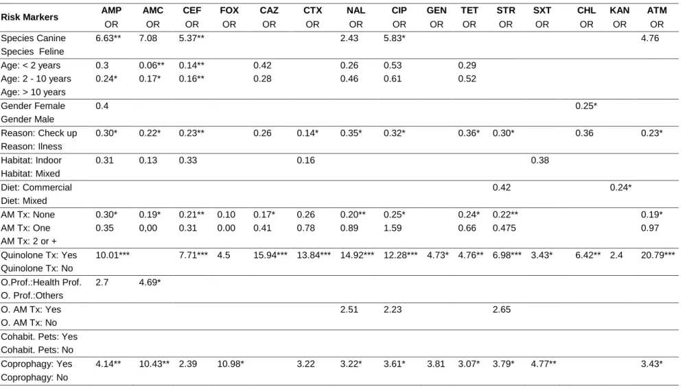
From September 2009 to May 2012, rectal swabs were collected from pets selected using a systematic random procedure from the ordinary population of animals attending the Veterinary Hospital of Porto University. A total of 78 dogs and 22 cats were sampled with the aim of isolating E. coli. Animal owners, who allowed the collection of fecal samples from their pets, answered a questionnaire to collect information about the markers that could influence the AMR of enteric E. coli. Chromocult tryptone bile X-glucuronide agar was employed for E. coli isolation and disc diffusion method was used to determine antimicrobial susceptibility. The data were analyzed using a multilevel, univariable and multivariable generalized linear mixed model (GLMM). Several (49.7%) out of the 396 isolates obtained in this study were multidrug-resistant. Antimicrobial agents for which many E. coli isolates exhibited resistance were ampicillin (51.3%), cephalothin (46.7%), tetracycline (45.2%) and streptomycin (43.4%). Previous quinolone treatment was the main risk marker for the presence of AMR in 12 (ampicillin, cephalothin, ceftazidime, cefotaxime, nalidixic acid, ciprofloxacin, gentamicin, tetracycline, streptomycin, chloramphenicol, trimethoprim-sulfamethoxazol and aztreonam) out of the 15 antimicrobials assessed. Coprophagic habits were also positively associated with an increased risk of AMR for 6 drugs: ampicillin, amoxicillin-clavulanic acid, cephamycin, ciprofloxacin, streptomycin, and trimethoprim-sulfamethoxazol.
In summary, pets with record of one or more previous treatments with quinolones and exhibiting coprophagic habits were at increased risk of harboring multidrug-resistant E. coli strains in their feces when compared with pets having not such characteristics. AMR is a serious global problem and assessing the risk markers for the presence of drug-resistant bacteria in pets, a very close source of resistance determinants to humans, is essential for the implementation of safe handling procedures for companion animals and prudent selection of antimicrobial substances in veterinary practice.
23 1. Introduction
Antimicrobial resistance (AMR) will probably be one of the main global public health problems of the next decade(Carlet et al., 2012). The phenomenon of AMR, which is based on the genetic plasticity of bacteria, has the selective pressure exerted by the antimicrobial usage in human and veterinary medicine, animal production, fish production, agriculture and food technology, the main driver force for its emergence (Kearns, 2010; EAAD, 2013; Martins da Costa et al., 2013). Resistant bacteria may be transmitted between interdependent hosts and spread into the environment, contributing to the worldwide increase of AMR (CDC, 2013). The progress in veterinary medicine and the number of domestic pets treated by specialized practitioners generated an increased usage of antimicrobial treatments (Martins da Costa et al., 2013). Additionally, pets live longer and are in closer contact with their owners, favoring the mutual transfer of microbial flora, directly by skin or bacteria-containing material contact (e.g. saliva and feces) and indirectly, via the household environment (Martins et al., 2013). When reaching the new host, resistant bacteria can colonize, infect, or remain in that particular environment for very short periods of time. In all cases, resistant bacteria can either spread their resistance genes to host-resident bacteria (commensals or pathogens) or accept resistance genes from such microorganisms (Jernberg et al., 2010). As a consequence, AMR in companion animals is simultaneously an important veterinary medical issue and a public health concern (Lloyd, 2007).
The regular monitoring of AMR in pathogenic and normal flora has been recommended by the World Health Organization and the European Centre for Disease Prevention and Control. For this purpose, the European Antimicrobial Resistance Surveillance Network (EARS-Net), involving 53 countries, was created (EFSA and ECDPC, 2013). Similar programs have been proposed for veterinary medicine, leading to the development of field studies on food animals (Aarestrup, 2004; Taylor et al., 2008) and pets (Moyaert et al., 2006; Lloyd, 2007; Costa et al., 2008; Murphy et al., 2009; Leonard et al., 2012). However, to our knowledge, no studies have included clinical histories of both pets and their cohabitants neither household features in order to assess potential AMR risk markers.
Escherichia coli is an important member of the normal intestinal microflora of humans and other mammals, but it can be also a highly versatile pathogen, causing diverse intestinal and extra intestinal diseases by means of virulence factors that affect a wide range of cellular processes (Kaper et al., 2004). Carriage of AMR has been
24
associated with several treatment failures in both human and veterinary patients (Toutain et al., 2010; Vigil et al., 2009).
The present study intended to determine the proportion of antimicrobial resistance of E. coli isolated from feces of pets from Porto region, in Portugal, as well as to assess individual, clinical and environmental characteristics of pets as risk markers for the AMR found in the isolated strains. It is hypothesized that animals with relevant clinical background will harbor more resistance E. coli isolates.
2. Materials and methods
2.1. Enrollment and sampling
A random systematic approach was used to select animals to the present cross-sectional study, performed at the Veterinary Hospital of Porto University (UPVET). From September 2009 to May 2012, on Monday or Tuesday, one among the first five pets to arrive at the UPVET attending room was randomly selected to be included in the study. If the owners refused to collaborate in the study the next pet, by order of arrival, was included, without following any criteria. To be eligible to be enrolled in the study, the animal should not have received any antimicrobial therapy within the preceding 4 months. The owners were asked to sign a consent form, to fill a questionnaire and to allow the collection of fecal samples from their pets using rectal swabs. Approval to conduct the study was previously obtained from the Ethics Committee of the Abel Salazar Institute for the Biomedical Sciences, University of Porto.
2.2. Questionnaire
By a brief questionnaire, owners were asked to provide information about possible risk markers for multidrug-resistant (Magiorakos et al., 2012) E. coli colonization. The questionnaire was constructed following similar studies in animals (Akwar et al., 2007; Ahmed et al., 2012; Boothe, 2012)and humans (McDonald et al., 2001; Sotto et al., 2001; Lietzau et al., 2007; Kalter et al., 2010; Lastours et al., 2010). To evaluate the potential risk markers, questionnaires included individual and clinical characteristics, such as 1) species, 2) gender, 3) age, 4) daily access to the outside environment (indoor habitat refers to those animals which live predominantly at home or with very restricted access
25
outdoor), 5) diet (“commercial” refers to the animals that were fed with strictly commercial dry or wet food), 6) coprophagic habits (ingestion of feces, both their own or from other animals), 7) previous systemic antimicrobial treatments with particular emphasis on 8) previous systemic quinolone treatments (assessed through the clinical file of the pet), 9) existence of cohabitant pets in the household, 10) previous antimicrobial treatments received by owners, 11) owners’ professional connection with healthcare units such as human or veterinary hospitals, clinics or health centers (such elements were classified as “Health Professionals”, 12) “reason for veterinary visit” (recorded by the veterinary surgeon following a complete physical examination).
2.3. Escherichia coli isolation
Fecal samples were obtained using saline wet swabs that were introduced with circular movements into the rectum of each animal. Swabs were immediately immersed on Buffered Peptone Water (BPW) (Oxoid, Basingstoke, Hampshire, England), transported to the laboratory and stored at room temperature for 1 h. Then, for E. coli isolation, an aliquot of 5 µl was streaked on Chromocult tryptone bile X-glucuronide (TBX) agar (Biokar Diagnostics, Allonne, Beauvais, France) and incubated at 37 °C for 24 h. Two to five confirmed pure colonies with typical appearance of E. coli were selected on the basis of colony size and morphology. The described procedure and the biochemical confirmation of isolates were adapted from standard protocols used in similar studies, aiming to achieve the most reliable and accurate E. coli detection (Costa et al., 2008; Simões et al., 2010; Martins et al., 2013).
2.4. Antimicrobial susceptibility characterization
Disk diffusion assay, following standard guidelines (CLSI, 2012), was performed to assess the antimicrobial susceptibility of each isolate. Antimicrobial drugs were selected in order to include those regularly used in both human and veterinary medicine and to provide diversity by representing different antimicrobial classes (Goossens et al., 2005; Elseviers et al., 2007; EFSA and ECDPC, 2013). A total of 19 antimicrobial agents (AM) (Oxoid, Basingstoke, Hampshire, England) were used: ampicillin (AMP, 10 µg), amoxicillin-clavulanic acid (AMC, 30 µg), cephalothin (CEF, 30 µg), cefoxitin (FOX, 30 µg), ceftazidime (CAZ, 30 µg), cefotaxime (CTX, 30 µg), nalidixic acid (NAL, 30 µg), ciprofloxacin (CIP, 5 µg), gentamicin (GEN, 10 µg), tetracycline (TET, 30 µg), streptomycin (STR, 10 µg), amikacin (AMK, 30 µg), trimethoprim-sulfamethoxazol (SXT,
26
25 µg), chloramphenicol (CHL, 30 µg), tobramycin (TOB, 10 µg), kanamycin (KAN, 30 µg), aztreonam (ATM, 30 µg), imipenem (IPM, 10 µg), and nitrofurantoin (NIT, 300 µg). The interpretation of the inhibition zone length followed the Clinical and Laboratory Standards Institute (CLSI) recommendations and breakpoints for Enterobacteriaceae (CLSI, 2012).
2.5. Data analysis
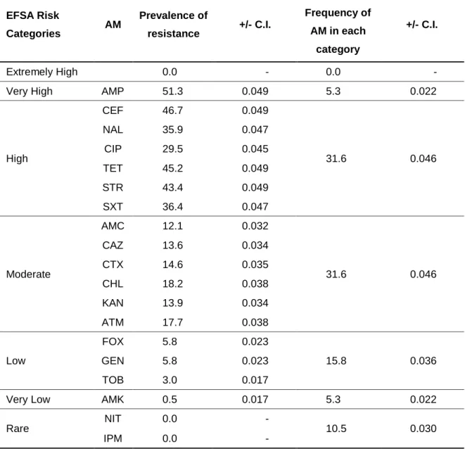
The prevalence of AMR regarding each AM was calculated by dividing the number of resistant E. coli isolates by the total number of E. coli tested. The potential risk markers obtained from the questionnaire were analyzed as categorical variables as follows: dichotomous variables, such as species (canine, feline), gender (male, female), reason for veterinary visit (routine check-up, illness signs), habitat type (indoor, mixed), diet (commercial, mixed), previous quinolone treatments (yes, no), health professionals owners (yes, no), owners submitted to previous antimicrobial treatments (yes, no), cohabitant pets (yes, no), coprophagy habits (yes, no); the exposure of the animal to any previous antimicrobial treatment was transformed into a categorical variable with three levels: “none”, “just one”, and “two or more”. Age was also categorized with three levels: “young” (before 2 years of age), “adult” (between 2 and 10 years old), and “old” (with more than 10 years old). The outcome in the analysis was the result of AMR which was dichotomized in either resistant or sensitive; intermediate results were categorized as sensitive. Using the European Food Safety Authority criteria, each antimicrobial was further classified into one of the following categories of prevalence of AMR: extremely high: >70%; very high: 50-70%; high: 20-50%; moderate: 10-20%; low: 1-10%; very low: 0.1-1% and rare: <0.1% (EFSA and ECDPC, 2013).
A descriptive analysis of AMR prevalence and frequency of risk markers was conducted. To analyze these markers and to assess the strength of their association with the AMR, a Multilevel Generalized Linear Mixed Model (GLMM) was used.
The logit link function was used to model the probability of occurrence of resistance to an antibiotic. To take into account the multilevel structure of the data in which more than one E. coli strain (i) was isolated from each animal (j), a two level structure in the data was assumed in which E. coli strains (first level) were nested within the animal from which they were isolated (second level).
27 The data were modeled in the following way:
𝑌 = { 0 (𝑛𝑜 𝐴𝑀𝑅) 1 (𝐴𝑀𝑅) Where Y is the response variable.
Pr(Y) = pij, i = 1, …, 396 and j = 1, ..., 100.
The generic model used the following equation: 𝑙𝑜𝑔𝑖𝑡(𝑝𝑖𝑗) = 𝑎 + 𝑐𝑗+ 𝛽 𝑎𝑛𝑖𝑚𝑎𝑙 𝑣𝑎𝑟𝑖𝑎𝑏𝑙𝑒𝑠𝑗
The model, the animal (the pet) was allowed to be random. The second level random effect is given by cj ∼ N(0,σ2 ) where σ2 is the variance of the random effects at the
animal level.
The basic multivariable multilevel model was as follows:
𝑙𝑜𝑔𝑖𝑡(𝑝𝑖𝑗) = 𝑎 + 𝑐𝑗+ 𝛽1𝑆𝑝𝑒𝑐𝑖𝑒𝑠𝑗+ 𝛽2𝐴𝑔𝑒𝑗+ 𝛽3𝐺𝑒𝑛𝑑𝑒𝑟𝑗+ 𝛽4𝑅𝑒𝑎𝑠𝑜𝑛 𝑜𝑓 𝑣𝑖𝑠𝑖𝑡𝑗+ 𝛽5𝐻𝑎𝑏𝑖𝑡𝑎𝑡𝑗
+ 𝛽6𝐷𝑖𝑒𝑡𝑗+ 𝛽7𝑁𝑢𝑚𝑏𝑒𝑟 𝐴𝑀 𝑡𝑟𝑒𝑎𝑡𝑚𝑒𝑛𝑡𝑠𝑗
+ 𝛽8𝑃𝑟𝑒𝑣𝑖𝑜𝑢𝑠 𝑄𝑢𝑖𝑛𝑜𝑙𝑜𝑛𝑒𝑠 𝑡𝑟𝑒𝑎𝑡𝑚𝑒𝑛𝑡𝑠𝑗+ 𝛽9𝑂𝑤𝑛𝑒𝑟′𝑠 𝑝𝑟𝑜𝑓𝑒𝑠𝑠𝑖𝑜𝑛𝑗
+ 𝛽10𝑂𝑤𝑛𝑒𝑟′𝑠 𝐴𝑀 𝑡𝑟𝑒𝑎𝑡𝑚𝑒𝑛𝑡𝑠𝑗+ 𝛽11𝐶𝑜ℎ𝑎𝑏𝑖𝑡𝑎𝑛𝑡𝑠 𝑃𝑒𝑡𝑠𝑗
+ 𝛽12𝐶𝑜𝑝𝑟𝑜𝑝ℎ𝑎𝑔𝑦 ℎ𝑎𝑏𝑖𝑡𝑠𝑗
Variables codes are presented in Tables 4 to 6.
A three step procedure was taken as follows: firstly, a univariable multilevel GLMM analysis was conducted to assess the individual relationship between each potential risk factor and the presence of AMR; a second step was performed to conduct a multivariable multilevel GLMM analysis with all the variables that had a p < 0.15 in the previous analyses followed by a manual backward and forward procedure to obtain a final model where each factor effect was adjusted for the remaining factors. Only factors with a p < 0.05 were retained in the final model. The data were analyzed using the procedure GEE in the SPSS Software V. 21.0 (IBM SPSS statistical 21 package, IBM Corporation, NY).
28 3. Results
A total of 78 dogs and 22 cats belonging to 100 distinct households were enrolled. Overall 396 E. coli isolates were obtained, 307 (77.5%) were isolated from dogs and 89 (22.5%) from cats, ranging from two to five per animal; on average, 3.96 isolates per animal were obtained.
3.1. Antimicrobial resistance profiles
Our results showed that 28.8% of the isolates were susceptible to all compounds; the median number of AMR among the isolates was three and the isolate in the 75th percentile harbored seven resistances. Extreme resistance towards 14 or 15 AM was present in five isolates (1,3 %). The histogram displaying the absolute number of resistances suggests the existence of two or perhaps three subpopulations of E. coli (Fig. 1): one group with less than four resistances, a second one with five to 10 resistances, and a conceivable third group with more than 10 resistance results.
29 3.2. Antimicrobial resistance prevalence
The prevalence of AMR varied from 0% found for nitrofurantoin and imipenem up to 51.3% (+/- 0.049) for ampicillin. After categorization according to the EFSA (EFSA and ECDPC, 2013) recommendations, 5.3% (+/- 0.022) of the tested AM were in the very high resistance category; 31.6% (+/- 0.046) in the high resistance group and a similar proportion were in the moderate resistance category, as displayed in Table 1.
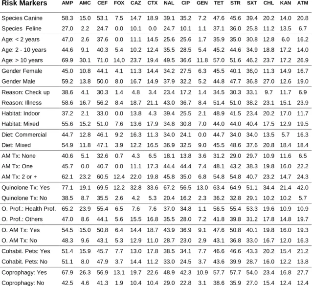
3.3. Distribution of potential risk markers associated with pets
The frequency of each tested potential risk marker is shown in Table 2. After comparing the factors species, age, sex and reason for the visit, it was clear that the population of pets enrolled in our study resembles the population of cats and dogs attending the hospital. Twenty-three dogs (29.5 %) and 15 cats (68.2%) were healthy animals admitted for regular check-up or prophylactic actions, while the remaining animals attended the hospital for clinical reasons.
3.4. Distribution of potential risk markers among E. coli isolates
The distribution of potential risk markers amongst E. coli isolates are displayed in Table 3. The largest numbers of isolates were obtained from pets owned by non-health professionals (n = 304; 76.8%) and animals with outdoor access (n = 302; 76.3%). Some characteristics associated with categories with small proportion of isolates were having just one antimicrobial treatment (n = 81; 20.5%), being older than 10 years (n = 93; 23.5%) and living indoor (n =9 4; 23.7%).
30
Table 1. Categorization of the tested antimicrobials (AM) in Escherichia coli isolates according to EFSA risk categories for prevalence of resistances.
EFSA Risk Categories AM Prevalence of resistance +/- C.I. Frequency of AM in each category +/- C.I. Extremely High 0.0 - 0.0 -
Very High AMP 51.3 0.049 5.3 0.022
High CEF 46.7 0.049 31.6 0.046 NAL 35.9 0.047 CIP 29.5 0.045 TET 45.2 0.049 STR 43.4 0.049 SXT 36.4 0.047 Moderate AMC 12.1 0.032 31.6 0.046 CAZ 13.6 0.034 CTX 14.6 0.035 CHL 18.2 0.038 KAN 13.9 0.034 ATM 17.7 0.038 Low FOX 5.8 0.023 15.8 0.036 GEN 5.8 0.023 TOB 3.0 0.017
Very Low AMK 0.5 0.017 5.3 0.022
Rare NIT 0.0 - 10.5 0.030
IPM 0.0 -
Legend: AM – antimicrobial agent; C.I. – Confidence interval; AMP – ampicillin; AMC – amoxicillin-clavulanic acid; CEF – cephalothin; FOX – cephoxitin; CAZ – ceftazidime; CTX – cefotaxime; NAL – nalidixic acid; CIP – ciprofloxacin; GEN – gentamicin; NIT – nitrofurantoin; TET – tetracycline; STR – streptomycin; AMK – amikacin; SXT – trimethoprim-sulfamethoxazol; CHL – chloramphenicol; TOB – tobramycin; KAN – kanamycin; IPM – imipenem; ATM – aztreonam. Values are expressed in percentages.