Analysis of the induction of the cytoprotective Nrf2 signalling pathway in reticuloendothelial cells from iron-treated mice and HFE Haemochromatosis patients
86
0
0
Texto
(2)
(3) FILIPE FERNANDES DE SOUSA. Analysis of the induction of the cytoprotective Nrf2 signalling pathway in reticuloendothelial cells from iron-treated mice and HFE Haemochromatosis patients. Dissertação de Candidatura ao grau de Mestre em Bioquímica da Universidade do Porto. Orientador – Doutor Tiago Pereira de Lacerda Costa Duarte Categoria – Investigador Auxiliar Afiliação – Instituto Biologia Molecular e Celular, Universidade do Porto Co-orientador – Maria da Graça Beça Porto Categoria – Professora Catedrática Convidada Afiliação – Instituto de Ciências Biomédicas Abel Salazar, Universidade do Porto. 2012 i.
(4) ii.
(5) “He who ceases to be better, ceases to be good.” Oliver Cromwell. “To be conscious that you are ignorant is a great step to knowledge.” Benjamin Disraeli. iii.
(6) iv.
(7) Acknowledgements. v.
(8) vi.
(9) Acknowledgements. I want to thank my parents who have always supported me in all my decisions and academic life. I am grateful for all the resources they provided me with, which have certainly influenced the quality of my work. A very special deep gratitude to my mother, Maria Manuela, for her love, tireless courage and exceptional wisdom; to my father, Vitor Sousa, a profound appreciation for his love, constant encouragement and comprehension; to my brother, Ricardo, for his everyday presence and joy; to Liliana Carvalho, for her love, dedication and for cheering me up through the stressful times. I would also wish to express my recognition to Susana Oliveira for her kindness and availability in the lab. I am deeply indebted to her, as she helped me develop several scientific and artistic skills that will definitely influence the quality of my future work. In addition, I want to thank her holy patience because having someone always messing around the desk must be pretty annoying. Furthermore, I would like to thank Inês Vieira and João Arezes for being excellent neighbours and establishing a nice working atmosphere. I would also wish to express my sincere gratitude to Graça Porto for her scientific assistance and clinical experience. Words cannot fully describe my admiration for her, and I hope one day I can be a clinician as good as her. Also, I want to thank nurse Graça Melo for her sympathy and special care with my patient samples. Naturally, I want to specially thank my supervisor Tiago Duarte for his continuous encouragement and assistance. His suggestions and insightful views were a most fundamental help throughout the year. Furthermore, he was certainly a most valuable input of experience, knowledge and opinion. Moreover, I would like to point out his regular good mood which motivated me even more to be in the lab. I was also his trainee in my BSc thesis and I believe it is time to cut the umbilical cord. I would like to show my greatest appreciation to Iron and Innate Immunity group members Pedro Rodrigues, João Neves, Sandro Gomes, Carolina Caldas, Sílvia Costa and Ana Gomes for encouragement and guidance throughout the year. I would, as well, like to register a collective recognition to all my colleagues and friends that have accompanied me along this path, for their most meaningful support.. vii.
(10) viii.
(11) Index. ix.
(12) x.
(13) Index. Acknowledgements .........................................................................................................v Index ................................................................................................................................ix. Summary ..........................................................................................................................1 Sumário ............................................................................................................................5 Introduction .....................................................................................................................9 1. Iron Ubiquity in Biological processes .................................................................11 2. Oxidative stress: iron misbehaving .....................................................................12 3. Iron Metabolism .................................................................................................12 3.1. Iron absorption ......................................................................................12 3.2. Iron transport and storage ......................................................................13 3.3. Iron regulation .......................................................................................14 4. Hereditary Haemochromatosis (HH)...................................................................16 4.1. 4.2. 4.3. 4.4. 4.5.. HFE – related HH ..................................................................................16 Other forms of iron overload: HJV, FPN & HAMP mutations .................. 16 Pathophysiology .....................................................................................17 Diagnostics and treatment .....................................................................18 Prediction of fibrosis and cirrhosis ..........................................................18. 5. The Nrf2 pathway ...............................................................................................19 5.1. Crosstalk with the Unfolded Protein Response ....................................... 20 6. Disease modifiers of HFE - HH ..........................................................................22 6.1. Environmental modifiers .........................................................................22 6.2. Genetic modifiers ...................................................................................23 Aims ................................................................................................................................27 Materials & Methods ......................................................................................................29 Results and Discussion ................................................................................................37 Part 1 – Animal Model ..........................................................................................39 1.1. Characterization of the animal model ......................................................41 1.2. Nrf2 activation in iron dextran treated - mice ..........................................45 Part 2 – Clinical Work ..........................................................................................51 2.1. Isolation of CD14+ blood cells from patients and control subjects ........... 53 2.2. Expression of markers of UPR and Nrf2 activation in blood CD14+ cells 56 Final Remarks ...............................................................................................................61 References ....................................................................................................................65. xi.
(14) xii.
(15) Summary. 1.
(16) 2.
(17) Summary. HFE−associated Hereditary Haemochromatosis (HH) is a common genetic disorder that leads to total body iron overload with secondary tissue damage, attributed to oxidative stress. Most HH patients are homozygous for the C282Y mutation in the HFE gene, resulting in continued iron absorption. A major challenge in the HH field is to understand why some patients are at increased risk for developing severe iron overload−associated clinical symptoms while most C282Y homozygotes are apparently healthy. We hypothesize that resistance to oxidative stress is a modifier of disease progression in iron overload−related pathologies. Transcription factor Nrf2 is an important modifier of chronic diseases involving oxidative stress, including inflammatory and neurodegenerative diseases, and cancer. In theory, Nrf2 could coordinate the transcriptional induction of cytoprotective/antioxidant genes in response to iron overload and/or as result of the previously described unfolded protein response (UPR) elicited by the HFE C282Y mutation in vitro. Whilst the liver is often the most affected organ in HFEHH patients, access to liver biopsies is difficult and poses ethical issues. The aim of the work was to investigate if Nrf2 is activated by iron overload in reticuloendothelial cells, which play an active role in iron metabolism and clearance, and could represent a surrogate tissue. Firstly, we investigated Nrf2 activation in response to iron delivery to tissue macrophages via injection of Fe-dextran in mice. Subsequently, we analyzed the expression of relevant genes in blood monocytes (CD14+) from HFE-HH patients. Injection of Fe-dextran in mice caused a significant increase in serum iron and a concomitant deposition of iron in splenic macrophages and liver Kupffer cells. Liver and spleen RNA was collected 1d post injection. The expression of 2 cytoprotective genes (Nqo1, Gclc) and of the iron exporter Fpn1 was significantly elevated in the liver and spleen of mice injected with Fe-dextran when comparing with animals injected with dextran (control). Notably, no differences were observed in Nrf2 KO mice, demonstrating that the activation of these genes was Nrf2-dependent. CD14+ cells were isolated from the blood of a group of 7 C282Y HFE homozygous subjects with elevated iron indices (serum iron, ferritin and transferrin saturation) followed at Centro Hospitalar do Porto, and 7 apparently healthy controls with normal iron indices and absence of C282Y HFE mutation. We found no significant changes in the steady-state mRNA expression levels of NRF2 or a panel of Nrf2-regulated cytoprotective genes (NQO1, GCLC, HMOX1, MnSOD, CAT, TXNRD1, FPN1, FTH1, FTL). No evidence of an UPR was observed either, as determined by analysis of XBP1 splicing and BIP mRNA. Whilst Nrf2 is activated by iron loading of tissue macrophages in mice treated with Fe-dextran, CD14+ cells from HFE-HH patients apparently do not respond to increased serum iron. 3.
(18) 4.
(19) Sumário. 5.
(20) 6.
(21) Sumário. A hemocromatose hereditária (HH) associada ao gene HFE é uma doença genética comum que origina sobrecarga de ferro e dano secundário nos tecidos derivado do stress oxidativo. A maioria dos pacientes com HH é homozigótica para a mutação C282Y do HFE, resultando numa absorção contínua de ferro. Um grande desafio da investigação na área da HH é perceber porque é que alguns pacientes apresentam um risco acrescido de desenvolver sintomas clínicos resultantes da sobrecarga severa de ferro, enquanto que a maioria dos indivíduos homozigóticos para a mutação C282Y é aparentemente saudável. A nossa hipótese é que a resistência ao stress oxidativo é um modificador da progressão de patologias associadas com sobrecarga de ferro. O fator de transcrição Nrf2 é um importante modificador em doenças crónicas relacionadas com stress oxidativo, incluindo doenças inflamatórias, neurodegenerativas e cancro. Em teoria,. o. Nrf2. poderia. coordenar. a. indução. transcripcional. de. genes. antioxidantes/citoprotetores em resposta à sobrecarga de ferro ou à Unfolded Protein Response (UPR), que foi previamente demonstrada como resultado da mutação C282Y in vitro. Embora o fígado seja o órgão mais afetado nos pacientes com HFE – HH, o acesso a biopsias é difícil e introduz problemas éticos. O objetivo deste trabalho foi investigar se o Nrf2 é ativado pela sobrecarga de ferro em células reticuloendoteliais, que intervêm ativamente no metabolismo do ferro e podem representar um modelo de estudo alternativo. Inicialmente investigámos a ativação do Nrf2 em resposta à sobrecarga de ferro nos macrófagos residentes nos tecidos através da injeção de ferro-dextrano em murganhos. Subsequentemente, analisámos a expressão de genes relevantes em monócitos do sangue (CD14+) de pacientes com HFE – HH. A injeção de ferro-dextrano nos murganhos causou um aumento significativo do ferro no soro e uma deposição concomitante nos macrófagos esplénicos e células de Kupffer. O RNA de fígado e baço foi recolhido 1d após a injeção. A expressão de 2 genes citoprotetores. (Nqo1. e. Gclc) e. do. exportador de ferro. Fpn1. encontrava-se. significativamente elevada no fígado e baço de murganhos injetados com ferro-dextrano comparando com animais injetados com dextrano (controlo). Notavelmente, nenhuma diferença foi observada em murganhos Nrf2 KO, demonstrando que a ativação desses genes foi dependente de Nrf2. As células CD14+ foram isoladas do sangue de um grupo de 7 sujeitos C282Y HFE homozigóticos com índices de ferro elevados (ferro no soro, ferritina e saturação de transferrina) seguidos no Centro Hospitalar do Porto, e 7 indivíduos aparentemente saudáveis, com índices de ferro normais e sem mutação C282Y no HFE (grupo controlo). Não foram detetadas diferenças significativas na expressão de mRNA do NRF2 ou de um painel de genes citoprotetores regulados pelo Nrf2 (NQO1, GCLC, HMOX1, MnSOD, 7.
(22) Sumário. CAT, TXNRD1, FPN1, FTH1, FTL). Não foi igualmente detetada nenhuma evidência de UPR, tanto pela análise de splicing da XBP1 como pelos níveis de mRNA da BIP. Pode-se concluir deste estudo que, embora o Nrf2 seja activado por sobrecarga de ferro nos macrófagos tecidulares em murganhos tratados com ferro-dextrano, as células CD14+ do sangue de pacientes HFE – HH aparentemente não respondem ao aumento de ferro no soro.. .. 8.
(23) Introduction. 9.
(24) 10.
(25) Introduction. 1. Iron ubiquity in biological processes Mammalian development and health rely on optimal nutrition. Iron is classified as a micronutrient and, as a result, it is required throughout life in low quantities. It has the ability to orchestrate a whole range of physiological processes such as oxygen transport, respiration, the tricarboxylic acid cycle, lipid metabolism, gene regulation and DNA synthesis (Cairo et al., 2006). Either iron overload or deficiency may cause genome instability. If present in excess, iron is capable of reacting profusely with H 2 O 2 to produce DNA strand breaks and to substantially increase 8-hydroxydeoxyguanosine (8-OHdG) lesions. On the other hand, iron is a critical co-factor for most of the enzymes involved in nucleic acid metabolism and its deficiency may impair this process (Pra et al., 2012). Iron metabolism also intersects the regulation of other trace elements such as copper. For yet unknown mechanisms, liver copper levels vary inversely with iron status. One possible explanation states that copper functions as a prosthetic group of critical enzymes of the iron metabolism (Collins et al., 2010). Iron also plays a critical role at the immune system level. Microorganisms require iron for growth and the host immune response evolved countermeasures to restrict its availability such as iron chelator proteins, trap siderophores and iron transport/modulator molecules (Ganz, 2009). Moreover, during the inflammation phase and its resolution, macrophage polarization phenotypes present opposite expression of several genes related to iron storage (ferritin), traffic (ferroportin, ceruplasmin) and regulation (IRP2, haem-oxygenase). As a consequence, large differences in both intra- and extracellular iron availability are achieved (Recalcati et al., 2010 and Cairo et al., 2011). Acquired immunity is also affected by iron levels, whose influence extends from the expansion of specific T lymphocyte subpopulations to the modulation of T cell surface markers expression (Walker & Walker, 2000). In fact, intravenous iron preparations were shown to inhibit the survival of the immune cell populations CD4+ and CD16+ (Gupta et al., 2010). Furthermore, low CD8+ T lymphocyte numbers have been related to the severity of iron overload in HFE-related hereditary haemochromatosis (Porto et al., 1994). These few examples illustrate how cell homeostasis crucially depends on iron tight regulation and why any defect in its partakers may cause serious systemic damage.. 11.
(26) Introduction. 2. Oxidative stress: iron misbehaving As stated above, iron is essential for life but only in trace amounts. Once present in excess, it can be toxic and produce free radicals through the Fenton reaction: Fe2+ + H 2 O 2 → Fe3+ + OH• + OH−. [1]. Iron homeostasis must therefore be finely tuned. The hydroxyl radical (OH•) is very reactive and highly damaging. Once produced it may directly interact with many biological macro and small molecules, including DNA, proteins and unsaturated lipids. Thus Fe (II) reacts with lipid hidroperoxides splitting the O-O bond. This gives rise to an alkoxyl radical which can abstract H• from polyunsaturated fatty acids and hydroperoxides. Subsequently, peroxyl radicals can continue propagation of lipid peroxidation. Oxidative stress leads also to considerable DNA damage and denaturation of proteins. The Fenton reaction described above states that in natural environment ferrous ions react spontaneously with oxygen to produced ferric ions. To face the problems of both solubility and toxicity, organisms developed siderophores which are iron chelator molecules such as transferrin or ferritin (Kell, 2009).. 3. Iron metabolism 3.1. Iron absorption Iron is a vital biocatalyst that coexists in either an oxidized insoluble form (Fe3+) or a reduced soluble form (Fe2+). In healthy individuals, approximately 1-2 mg of iron are absorbed from the diet per day to maintain iron balance. Absorption occurs in the duodenum and upper jejunum via the apical surface of the enterocytes in two possible forms: haem or non-haem iron. Haem iron is obtained mostly from meat sources and its putative transporter, the haem carrier protein (HCP), was recently described (Shayeghi et al., 2005). This remains controversial as there is evidence stating that it functions as a proton-coupled folate transporter (Qiu et al., 2006). Haem oxygenase activity would be responsible for liberating the haem iron content. Non-haem iron must be reduced from ferric to ferrous ion by a ferrireductase at the apical surface of the brush border. Duodenal cytochrome B (DcytB) has ferrireductase activity and reduces dietary ferric iron which has not been previously reduced by reductants such as ascorbate. Once reduced, ferrous iron is internalized through the divalent metal transporter 1 (DMT1). This transporter also has the ability to mediate the internalization of other divalent 12.
(27) Introduction. metals, such as copper and zinc (Gunshin et al., 1997). Ferrous iron is then transported across the basolateral membrane of the enterocyte by ferroportin. To be bound to plasma transferrin (Tf), ferrous iron is oxidized by hephaestin which is a multicopper oxidase located near ferroportin (Olynyk et al., 2008). In this manner, iron is nonreactive and transported to the tissues where it is either utilized for the synthesis of haem or non haem proteins, or stored as ferritin. Uptake of holotransferrin – transferrin fully loaded with 2 iron atoms per molecule – is mediated through the transferrin receptor 1 (TfR1). Tf-TfR1 complexes are internalized through clathrin dependent endocytosis, and endosomes are acidified by a proton pump which promotes conformational changes in both Tf and TfR1 releasing the iron content (Hentze et al., 2004). Ferric iron is then reduced by the six-transmembrane epithelial antigen of prostate protein 3 (STEAP3) and transported across the endossomal membrane by DMT1. Furthermore, apotransferrin and TfR1 are recycled back to the cell surface where they can restart further cycles of iron binding and uptake (Olynyk et al., 2008). A second transferrin receptor (TfR2) is capable of mediating the internalization and recycling of transferrin although with lower affinity. However, while TrfR1 is widely expressed, TfR2 is expressed predominantly in hepatocytes, hematopoietic cells and duodenal crypt cells. Iron export through ferroportin from non-intestinal cells requires ceruloplasmin to oxidize Fe2+ to Fe3+ before loading inside plasma transferrin (Harris et al., 1999). As stated above, the daily iron uptake is 1-2 mg which is clearly not sufficient to supply iron needs for erythropoiesis (20-25 mg) (Hentze et al., 2004). Inevitably, iron recycling is necessary. Senescent erythrocytes are fagocytosed by members of the reticuloendothelial system, mainly in the liver and spleen, which subsequently release or store iron from haem using the same molecular machinery present in the enterocyte (Evstatiev & Gasche, 2012).. 3.2. Iron transport and storage Transferrin is the major plasma protein that transports iron between the sites of absorption, storage and utilization. It has two lobes each comprising an iron binding site joined by a flexible hinge. Both lobes bind iron reversibly and with high affinity and can also bind manganese, cobalt, copper and cadmium with lower affinity. Transferrin is present in the plasma as a mixture of apotransferrin (iron-free), monoferric transferrin and diferric transferrin depending on plasma concentrations of iron. Certain diseases are characterized by iron overload and free binding sites in transferrin provide a primary safeguard. Normal transferrin saturation in circulation is approximately 30% and so the body has a high buffer capacity for iron (Chua et al., 2007). When the binding capacity of 13.
(28) Introduction. transferrin is saturated, iron can be bound to low molecular weight ligands being defined as non transferrin bound iron (NTBI). The hepatocyte, which has an intrinsic ability to accumulate high amounts of iron, internalizes the NTBI through DMT1 or ZIP14 (Graham et al., 2007). In addition, hepatocytes can also take up haem iron bound to haptoglobin (Kristiansen et al., 2001) or haemopexin (Hvidberg et al., 2005). Inside the cell, iron storage is performed by a specialized ubiquitous protein known as ferritin. Mammalian ferritin (Ft) is a heteropolymer composed of 24 subunits of two types (H and L), which assemble to make a hollow spherical shell in which iron accumulates in a readily available but non-toxic form (Harrison & Arosio, 1996). H and L chains are tissue specific and may generate several isoforms with different metabolism and function. Isoferritins rich in the L subunit predominate in iron storing tissues whereas H subunit rich ferritins are found in cells which rapidly take up and release iron (Cairo et al., 2006).. 3.3. Iron regulation Systemically, iron homeostasis involves meticulous control of intestinal absorption, erythropoiesis, recycling and storage. Hepcidin, which is now known to be the primary modulator of iron metabolism, is a cysteine-rich peptide cleaved from a larger precursor and is secreted by the liver and excreted through the kidneys (Krause et al., 2000 and Park et al., 2001). Hepcidin regulates cellular iron efflux by binding to ferroportin and promoting its internalization and degradation (Nemeth et al., 2004). This is particularly important in the intestine and in iron-recycling macrophages of the reticular endothelial system: inactivation of basolateral ferroportin leads to retention of iron in the intestinal epithelium and inactivation of ferroportin interrupts release of iron recovered from senescent red cells (Andrews, 2008). Overall, there is a reduction of serum iron. Disturbances in hepcidin signalling or regulation originate several disorders of iron overload (Viatte & Vaulont, 2009). Important regulatory roles are also attributed to the IRE/IRP regulator system. The two iron regulatory proteins IRP1 and IRP2 sense the iron labile pool and bind to the Iron Responsive Element (IRE) located on target mRNA controlling its expression (Rouault, 2006). IRE/IRP interactions regulate the translation of the mRNAs encoding proteins for iron acquisition (TfR1, DMT1), storage (FtH and FtL) and export (Fpn). IRE/IRP complexes formed within the 5’UTR of an mRNA (such as FtH, FtL and Fpn1) inhibit translation, whereas IRP binding to IREs in the 3’UTR of TFR1 mRNA prevents its degradation. In conclusion, the IRE-binding activities of IRP1 and 2 assure the appropriate expression of IRP target genes and cellular iron balance (Muckenthaler et al., 2008). An overview of iron homeostasis is depicted in Figure 1. 14.
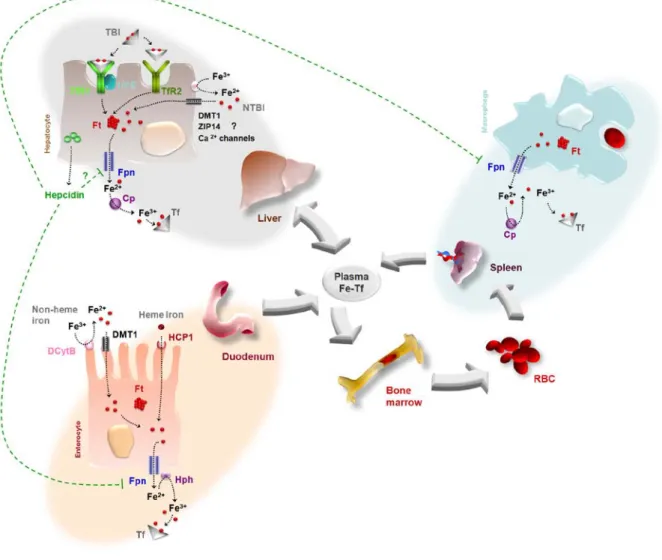
(29) Introduction. Figure 1. Overview of Iron Metabolism. Cp, Cerulopalsmin;. DCytB, duodenal cytochrome b;. DMT1, divalent metal transporter 1; FPN, ferroportin; Ft, ferritin; HCP1, haem carrier protein 1; Hph, hephaestin; NTBI, non transferrin bound iron; TBI, transferrin bound iron; Tf, transferrin; TfR1, transferrin receptor 1; TfR2, transferrin receptor 2.. 15.
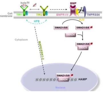
(30) Introduction. 4. Hereditary Haemochromatosis (HH) 4.1. HFE-related HH HFE-related HH is an autossomal recessive disorder resulting in iron overload and multiorgan dysfunction. The most common mutation of HFE–HH is the missense mutation where a cysteine is replaced for a tyrosine at amino acid 282 (C282Y). Another mutation, although with limited clinical effect, is the substitution of histidine for aspartate at amino acid 63 (H63D). Nevertheless, compound heterozygosity for C282Y and H63D seems to predispose individuals for disease expression (Pietrangelo, 2004). HFE is a major histocompatibility class I-like protein (Feder et al., 1996) and, as a result, requires β2microglobulin for proper assembly on the membrane. C282Y mutation disrupts a disulphide bond between the α3 domain of the HFE protein and β2-microglobulin preventing cell surface expression. H63D mutation does not prevent β2-microglobulin association or cell surface expression (Bennett et al., 2000). The role of HFE in iron metabolism is not completely understood, but it is believed to compete with diferric transferrin, lowering its affinity for binding to the TfR1. When serum iron levels are high, iron loaded transferrin can displace HFE from TfR1 which migrates to interact with TfR2 mediating hepcidin signalling (Goswami & Andrews, 2006). Mutations in either HFE or TfR2 impair the signalling cascade causing a failure to properly upregulate hepcidin, resulting in increased parenchymal iron deposition leading to clinical haemochromatosis (Gao et al., 2009 and Schmidt et al., 2008). In vitro studies demonstrated that the HFE C282Y mutated protein is retained in the ER leading to ER stress and consequent activation of the UPR (de Almeida et al., 2007).. 4.2. Other forms of iron overload: HVJ, FPN & HAMP mutations Juvenile haemochromatosis has its onset in the first few decades of life and is a particularly. severe. form. of. iron. overload.. Hemojuvelin. (HJV). is. a. glycosylphosphatidylinositol (GPI) - anchored protein and a Bone Morphogenetic Protein (BMP) co-receptor. BMPs are members of the TGFβ family which induce hepcidin gene transcription through the SMAD1,5,8/SMAD4 pathway. As a GPI-anchored protein, HVJ can be cleaved by a furin (TMPRSS6) and released in a soluble form, sHJV. In contrast to membrane-bound HJV, sHJV inhibits iron-induced hepcidin production and consequently promotes massive iron absorption (Figure 2).. This genetic defect turns a normal. physiologic response into a sustained pathological process (Camaschella, 2005).. 16.
(31) Introduction. Hepcidin (HAMP) mutations decrease the levels of hepcidin and its downstream response in iron homeostasis. It also leads to a severe iron overload condition. Nevertheless, it should be emphasized that the vast majority of HFE-HH patients do not have hepcidin mutations (Lee et al., 2002). Ferroportin mutations are inherited in a dominant fashion and are characterized by a mild anemia. In most cases, iron storage occurs predominantly in Kupffer cells and not in the hepatocytes, as happens in most forms of haemochromatosis. Iron is trapped inside macrophages as ferroportin is the only iron exporter (Beutler, 2006).. Figure 2. Hepcidin signalling through HFE and Hemojuvelin (HJV).. 4.3. Pathophysiology HFE-HH leads to the progressive deposition of iron in the liver and, if untreated, results in fibrosis and ultimately cirrhosis. Iron is accumulated within the hepatocyte and there is a hepatic iron concentration threshold associated with the development of cirrhosis. While the liver is the most damaged organ in HH, iron also accumulates in the parenchymal. cells. of. various. organs. resulting. in. hypogonadism,. diabetes,. cardiomyopathy, arthropathy and skin pigmentation. When intracellular iron exceeds ferritin capacity it leads to the production of highly ROS that damage intracellular structures and promotes multi-organ failure (Pietrangelo, 2010). Diagnosis of HFE-HH is performed in most cases on the basis of genotyping and biochemical evidence of iron loading, without liver biopsies. Populational studies show that genetic predisposition to HH is common, with one in 200 homozygous for the C282Y mutation in Caucasian 17.
(32) Introduction. populations of northern Europe. Penetrance and progression rates are variable, though. Some homozygotes do not develop iron loading and most remain asymptomatic (Griffiths, 2007).. 4.4. Diagnostics and treatment The clinical presentation of HFE-HH occurs in middle aged patients and ranges from simple biochemical abnormalities to severe organ damage and disease. The most common symptoms include fatigue, malaise, arthralgia and hepatomegaly. Transferrin saturation and ferritin levels are elevated in these patients. Serum ferritin is an indicator of body iron deposition (Andrews & Levy, 1998). Penetrance is usually higher among male patients than females, possibly because of menstruation.. Current treatment involves. phlebotomy for patients with an elevated serum ferritin (in males > 300 µg/L and in females > 200 µg/L). There is not a biochemical threshold above which phlebotomy is absolutely required: it is assumed that a C282Y homozygote with an elevated ferritin and transferrin levels is on a track of progressive iron overload (Adams, 2009). Indeed, evaluation of liver biopsies before and after phlebotomy therapy showed that hepatic fibrosis can be reversed, demonstrating its clinical benefits (Adams & Barton, 2010).. 4.5. Prediction of fibrosis and cirrhosis Noninvasive prediction of cirrhosis in C282Y linked haemochromatosis has been a major challenge. Guyader and colleagues showed that the absence of severe fibrosis could be accurately predicted based on a combination of clinical and biochemical variables (Guyader et al., 1998). The major drawbacks of this study were the subjective evaluations of fibrosis and hepatomegaly. Several other models have been developed such as taking into account ferritin levels, platelets counts and aspartate aminotransferase (AST) values. This combination could correctly classify most individuals that were a priori known to have liver cirrhosis (Beaton et al., 2002). Recently, an observational study stated that probands are at increased risk of death from iron overload by having serum levels of ferritin above 1000 µg/L at diagnosis (Barton et al., 2012). Clearly, statistical methodology must be chosen carefully to correctly summarize all clinical variables and result in valid outputs.. 18.
(33) Introduction. 5. The Nrf2 pathway Many chronic diseases are associated with a constant stress provoked by electrophiles and oxidants. They are often characterized by high levels of oxidized proteins, phospholipids and DNA. Once again, cells have developed dynamic mechanisms to counteract these environmental stresses. These mechanisms can be divided into four categories: (a) oxidations and reductions, (b) nucleophilic trapping processes that add glutathione or other nucleophiles, (c) efflux transporters that export toxic metabolites and (d) thiol containing molecules, such as thioredoxin (Kensler et al., 2007). This protective response requires antioxidant response elements (AREs) and the nuclear factor erythroid 2-related factor 2 (Nrf2), which is the principal transcription factor that binds to the ARE along with the Maf family of transcription factors (Figure 3). The Kelch ECH associating protein 1 (Keap1) is a cytosolic repressor protein which, through its Kelch domain, retains Nrf2 in the cytoplasm and promotes its ubiquitylation and proteasomal degradation (Itoh et al., 1999). Moreover, Keap1 also associates with the actin-cytoskeleton to further stabilize the interaction with Nrf2, maintaining it in the cytosol (Kang et al., 2004). One key distinguishing feature of Keap1 is the high number of cysteine residues. Human Keap1 contains 27 cysteine residues, which constitute the primary sensors of oxidative stress. The number of cysteine residues absolutely required to trigger the Nrf2 response is still controversial, although it is known that more than one are involved (Holland & Fishbein, 2010). Upon stressors or inducers such as ROS, the association between Keap1 – Nrf2 is disrupted leading to an enhanced nuclear accumulation of Nrf2 and target gene expression. Nrf2 promotes the transcription of enzymes that directly inactivate oxidants (glutathione-S-transferases, GST; NAD(P)H dehydrogenase, quinone 1, NQO1), increases levels of glutathione and NADPH synthesis (glutamate - cysteine ligase catalytic subunit, GCLC), enhances toxin export via multidrug response transporters (multidrug resistance-associated proteins, Mrps) and enhances the recognition, repair and removal of damaged proteins (heat shock proteins, HSP) (Kensler et al., 2007). In addition, interactions with the NF-κB, p53 and Notch pathways have been described (Wakabayashi et al., 2010). In the liver, not only Nrf2 mediates longer lifespan and reduced hepatotoxicity, it is also a regulator of cellular lipid disposition (Kitteringham et al., 2010).. 19.
(34) Introduction. Figure 3. Cytoprotective/Antioxidant Nrf2 pathway. ARE, Antioxidant Response Element; Keap1, kelch-like ECH-associated protein 1; NO•, nitric oxide free radical; Nrf2, nuclear factor erythroid 2related factor 2; ROS, reactive oxygen species; Ub, ubiquitin.. 5.1.. Crosstalk with the Unfolded Protein Response. The endoplasmic reticulum (ER) serves many general functions, including the facilitation of protein folding and the transport of synthesized proteins. Upon disruption in either protein folding or modification within the ER a state of stress is induced. The failure to sense and respond to these perturbations ultimately results in cell death. As a result, all cells have evolved accurate mechanisms to ensure proper protein folding occurs and to dispose of irreversible misfolded proteins. Quality control is a surveillance mechanism that permits only properly folded proteins to exit the ER en route to other intracellular organelles and the cell surface (Malhotra & Kaufman, 2007). Efficient protein-folding reactions depend on particular environmental conditions within the ER. Its lumen must have an oxidizing environment to permit dissulfide bond formation, high levels of calcium ion (Ca2+), which boost chaperone functions, and ATP, which provides the necessary energy for these reactions to occur (Higa & Chevet, 2012). Accumulation of unfolded proteins initiates activation of an adaptive signalling cascade known as the unfolded protein response (UPR).. 20.
(35) Introduction. Figure 4. Major players of the UPR pathway. The endoplasmic reticulum (ER)-transmembrane proteins PERK, IRE1 and ATF6 act as proximal sensors of ER stress, initiating distinct downstream signalling cascades. Accumulated misfolded proteins titrate BiP away from the luminal domain of each sensor thereby enabling their activation. PERK, double stranded RNA-dependent protein kinase-like ER kinase; IRE1, inositol-requiring enzyme 1; ATF6, activating transcription factor 6; BiP, immunoglobulin heavy chain-binding protein; eIF2α, α subunit of eukaryotic initiation factor 2; ATF4, activating transcription factor 4; CHOP, CCAAT/enhancer-binding protein homologous protein; XBP1, X-box-binding protein 1; ERAD, ER associated degradation; S1P, site-1 protease; S2P, site-2 protease; AARE, amino acid response element; UPRE, unfolded protein response element; ERSE, ER stress response element.. The UPR comprises three principal branches which operate in parallel and use distinct mechanisms of signal transduction: activating transcription factor 6 (ATF6), inositol requiring 1 (Ire1) and PKR-like endoplasmic reticulum kinase (PERK) (Figure 4). The three branches share a common feature: the signalling molecules are anchored by BiP. As an ER resident chaperone, BiP assists in the proper folding of nascent proteins within the ER, and following the induction of the UPR, BiP production increases dramatically in order to increase protein folding capacity (Cullinan & Diehl, 2006). In response to stress, unfolded proteins accumulate and bind BiP thereby promoting the release of those 21.
(36) Introduction. sensors that were in an inactive state. ATF6 is a transcription factor which upon activation migrates to the Golgi apparatus where it is cleaved by two proteases. Then it migrates to the nucleus, where it activates the transcription of ER resident proteins involved in protein folding such as BiP and disulfide isomerase. PERK is a protein kinase which is known to phosphorylate the eukaryotic initiation factor 2α (eIF2α) and mediates the translational response of the UPR. eIF2α phosphorylation inhibits the guanine nucleotide exchange factor eIF2B that recycles eIF2 complex to its active GTP-bond form. The formation of the ternary translation initiation complex eIF2α-GTP-tRNAMet is required for AUG initiation codon recognition and joining to the 60S ribosome subunit that occurs during initiation phase of polypeptide chain synthesis. Lower levels of active ternary complex result in lower levels of translation initiation. However, some mRNA’s containing short upstream open reading frames uORFs in their 5’ unstranslated regions are preferentially translated when eIF2 is limited. ATF4 is an example of this regulation, and is very important as it controls genes encoding components involved in apoptosis (Walter & Ron, 2011). Ire1, once liberated from BiP, is free to dimerize, trans-autophosphorylate and thereby activate its RNase activity. It performs a nonconventional mRNA splicing of the X-Box binding protein (XBP1) mRNA, turning it into a more potent transcription factor (Yoshida et al., 2001). The spliced form of XBP1 is involved in the regulation of lipid biosynthetic enzymes and ER-associated degradation components (Travers et al., 2000). Overall, the role of the UPR is to protect cells against accumulation of misfolded proteins and maintain their viability. Phosphorylation of Nrf2 by a series of kinases also affects its fate and distribution. Nrf2 is a PERK substrate. Similar to oxidative stress-inducing agents, ER stress induces Nrf2 nuclear translocation in a PERK-dependent manner. This suggests that Nrf2 activation during ER stress conditions will be equivalent to activation during oxidative stress (Cullinan et al., 2003).. 6. Disease Modifiers of HFE-HH 6.1.. Environmental modifiers. Some environmental factors are known to modify iron loading and hence expression of the disease, such as excess alcohol consumption, the amount of bioavailable iron in the diet and iron losses, for example, due to blood donation or gastrointestinal pathology (Fletcher et al., 2002). Age can also have an important impact on the course of iron overload: it is possible that secretion of hepcidin decreases with age 22.
(37) Introduction. and there is an impairment of the balance between generation and scavenging of ROS (Bonekamp et al., 2009). The two ferroxidades described in the iron metabolism section, ceruplasmin and hephaestin, require copper as a prosthetic group. Patients with low circulating levels of copper have higher hepatic and serum iron concentrations, demonstrating that copper can be a disease modifier independent of HFE genotype (Rochette et al., 2010).. 6.2.. Genetic modifiers. Genetic modifiers could also influence disease progression. For example, TMPRSS6 loss was shown to increase Bmp/Smad signalling in an HFE-independent manner in mice, raising the possibility that the human orthologue can alter clinical penetrance (Finberg et al., 2011). One hypothesis that the host laboratory currently investigates is that the individual tolerance to oxidative stress could be a disease modifier in. HFE-HH. and. other. iron. overload-related. diseases. (TL. Duarte,. personal. communication). Whilst the direct activation of Nrf2 by iron overload or via the induction of a putative HFE C282Y-induced UPR (so far only described in vitro) has not been experimentally demonstrated, it is conceivable that the activation of Nrf2-regulated genes could represent a defense against iron-mediated oxidative damage. Single nucleotide polymorphisms in the antioxidant response element could compromise the ability of Nrf2 to promote the expression of antioxidant and phase II detoxification enzymes/proteins (Wang et al., 2007), thus resulting in a poor disease prognosis.. 23.
(38) 24.
(39) Aims. 25.
(40) 26.
(41) Aims. The aim of this project is to investigate if Nrf2 is activated by iron overload in vivo. Firstly, Nrf2 activation will be investigated in mice that receive an injection of Fe-dextran. This treatment results in the delivery of iron to reticuloendothelial cells (splenic macrophages and hepatic Kupffer cells) (Fishbane et al., 1996), which play an active role in iron metabolism and clearance. Subsequently, the expression of relevant genes will be analyzed in blood monocytes (CD14+) from HFE-HH patients. Whilst the liver is the major organ where iron accumulates in HFE-HH patients, access to liver biopsies is difficult and would pose ethical issues. Blood monocytes, which are exposed to increased levels of serum iron in these patients and which are capable of evoking an UPR and/or Nrf2 activation (Carroll et al., 2010), will thus be tested as a possible surrogate tissue for detecting markers of both UPR and Nrf2 activation.. 27.
(42) 28.
(43) Materials & Methods. 29.
(44) 30.
(45) Materials & Methods. Animal housing and treatments C57BL/6 (B6) and Nrf2-/- mice (male, 6-8 week old, B6 background) were injected intraperitoneally with 4mg Fe-dextran or an equivalent amount of dextran (control). Animals were sacrificed by competent laboratory staff for tissue collection at 1, 7 or 30 days post injection. Animals were housed at the IBMC animal facility and experiments were carried out in compliance with the animal ethics guidelines of the institute, and the national and European regulations for the care and handling of laboratory animals. Biochemical parameters of iron metabolism in animal serum were determined at Centro Hospitalar do Porto (Department of Clinical Chemistry) and liver non-heme iron levels were measured by the batho phenanthroline method.. Patient recruitment A group of 7 C282Y HFE homozygous subjects with elevated iron indices (serum iron, ferritin and transferrin saturation) followed at Centro Hospitalar do Porto, and 7 apparently healthy controls with normal iron indices and absence of C282Y HFE mutation were recruited. Whenever possible, patients and blood donors were sex- and agematched. Informed consent was obtained from all recruited subjects, according to the 1975 Declaration of Helsinki and with the approval of the CHP-HSA Ethical Committee. Biochemical parameters of iron metabolism in patients’ serum were determined at Centro Hospitalar do Porto.. Isolation of human CD14+ cells from blood Blood (400 ml) was collected in the unit of Hematology of Hospital Geral Santo António by specialized hospital staff. Buffy coats were prepared at the Institute for Molecular and Cell Biology as follows. Blood was diluted 1:1 with PBS and centrifuged at 300 ×g for 45 minutes with an accelerator/break of 5/1. Plasma supernatant was partially discarded and the buffy coat was aspirated into another tube. To concentrate the white blood cells, the buffy coat was diluted with PBS once again and centrifuged with the same settings. The buffy coat was aspirated into a new tube with a Pasteur pipette and diluted with Isolation Buffer (PBS with 0.1 % BSA and 2 mM EDTA). Cells were centrifuged at 350 ×g for 35 minutes at 4˚C, with an accelerator/break of 5/1. The protocol for CD14+ cells isolation was also tested starting from Peripheral Blood Mononuclear Cells (PBMC’s). Blood was diluted 1:1 with PBS 1x and carefully drawn in a tube containing 31.
(46) Materials & Methods. Lymphoprep (Axis-Shield). It was centrifuged at 800 ×g for 30 minutes with an accelerator/break of 3/2. The buffy coat was aspirated into a new tube with a Pasteur pipette, diluted with Isolation Buffer and further centrifuged at 1800 rpm for 10 minutes at 4˚C. From this step onwards the two protocols converged. CD14+ cells were isolated with the Dynabeads FlowComp Human CD14 kit (Invitrogen) following the manufacturer’s instructions. Briefly, supernatants were discarded (leaving 1 centimeter above the surface of the buffy coat), 100 µl Flowcomp CD14 Antibody were added, samples were vortexed for 5 seconds and incubated for 15 minutes at ˚C. 4 The tube was refilled with Isolation Buffer and centrifuged with the settings above. The supernatant was discarded and 600 µl Flowcomp Dynabeads were added. The sample was vortexed for 5 seconds and incubated with rolling and tilting at 4 ˚C. The tube was refilled with Isolation Buffer and then placed in the DynaMag (Invitrogen) for 3 minutes. While still in the magnet, the supernatant containing the CD14 negative cells was carefully pipetted and discarded taking care not to disturb the bead pellet on the wall tube. The washing steps were repeated with decreasing volumes of Isolation Buffer (50mL, 32 mL and 16 mL). After discarding the last wash of Isolation Buffer, the bead pellet trapped on the tube wall was carefully resuspended in 4 mL of Release Buffer, transferred into a new tube to avoid red blood cell contamination and incubated for 10 minutes with rolling and tilting at 4˚C. The solution was pipetted up and down 10 times, taking care to avoid air bubbles, to efficiently release the cells and placed in the magnet for 3 minutes. The supernatant containing the bead-free CD14+ cells was carefully transferred into a new tube and placed again in the magnet for 3 minutes to remove residual beads. The supernatant was collected and cells were counted in a Neubauer chamber.. Assessment of CD14+ purity Five. hundred. thousand cells. were. harvested,. transferred. into. a. 1.5mL. microcentrifuge tube and stored overnight at 4°C in the dark. On the day after, cells were centrifuged at 400 ×g for 3 minutes. The supernatant was rejected and cells were resuspended in the vortex. 2µl of anti-human CD14 Phytoerythrin-conjugated clone MEM15 antibody (Immuno Tools) diluted in 50µl of PBS/0.2% BSA/0.1% NaN 3 were added and incubated for 20 minutes in the dark at 4°C. Cells were washed 3 times by adding 100µl of PBS/0.2% BSA/0.1% NaN 3, centrifuging, rejecting the supernatant and resuspending the pellet in the vortex. Subsequently, the supernatant was discarded and the cell pellet was resuspended in 400µl of PBS. The sample was then analyzed by flow cytometry in a FACS Calibur (Becton Dickinson). Cell populations were identified based on a Forward 32.
(47) Materials & Methods. Scatter (FSC) versus Side Scatter (SSC) plot using the Cell Quest software. CD14+ cells present in the gated populations were identified on a SSC versus FL2-H dot plot. The peak emission for Phytoerythrin is 575 nm and was detected in the FL2-H channel.. Total RNA extraction CD14+ cells were centrifuged at 400 ×g for 4 minutes and then lysed in 0.5 ml of Tri Reagent (Sigma-Aldrich) by repeated pipetting. RNA extraction was performed following the manufacturer’s instructions. Briefly, chloroform was added, the mixture was shaken vigorously for 15 seconds and allowed to stand for 10 minutes at room temperature. The resulting mixture was centrifuged at 12,000 ×g for 15 minutes at 4°C. The aqueous phase containing the RNA was transferred to a fresh tube and 0.25 ml of isopropanol were added. The sample was allowed to stand for 10 minutes at room temperature and then centrifuged at 12,000 ×g for 10 minutes at 4°C. The supernatant was removed and the RNA pellet was washed by adding 1 ml of 75% ethanol. Subsequently, the sample was vortexed and centrifuged at 7,500 ×g for 5 minutes at 4°C. The RNA pellet was air-dried for 5-10 minutes and then dissolved in RNase-free water by repeated pipetting with a micropipette at 60°C for 2-3 minutes. RNA was quantified by measuring the UV absorbance in the NanoDrop ND-1000 spectrophotometer (Thermo Scientific). RNA integrity was assessed in the Experion (Bio-Rad).. DNase digestion and first-strand cDNA synthesis Turbo DNA-free kit (Ambion) was used to digest contaminant genomic DNA from the RNA preparations. The first strand cDNA was generated with the ThermoScript RTPCR System kit (Invitrogen) following the manufacturer’s instructions. cDNA was synthesized from 0.63 µg of CD14+ cells RNA and 1.44 µg from mice liver or spleen RNA.. RT - PCR Amplification Real-time RT-PCR was performed on the iCycler iQ5 spectrofluorometric thermal cycler (Bio-Rad) using iQ SYBR Green Supermix (Bio-Rad) according to the manufacturer. A volume of 1µl of cDNA solution was used for the amplification of the specific cDNA targets and the PCR was carried out as follows: initial denaturation at 95°C for 3.5 minutes, followed by 40 cycles of 94°C for 30 seconds, 59°C for 45 seconds and 72°C for 30 seconds. The annealing temperature depended on the primer used as 33.
(48) Materials & Methods. indicated in Table 1. Subsequently to the amplification, a dissociation curve was generated by gradually increasing the temperature 1°C at a time (from 55 to 95°C) and by measuring the fluorescence at the end of each temperature increase. The amplification of a single product was verified by the existence of a single peak in the dissociation curve. All reactions were done in duplicate and a non-template control (NTC) was always included. Threshold cycle (C t ) values were determined by using an automatically set fluorescence threshold. Standard amplification curves were generated for each target by amplifying serial dilutions of a standard cDNA containing the transcribed gene of interest (dilution series was 1:1, 1:10 and 1:100) to verify that there was a linear relationship between the Ct and the log (RNA input). The quantity of each target gene was estimated from the respective standard curve and normalized against the quantity of the endogenous control gene: HPRT1 (for human samples) or β-actin (murine samples). For analysis of XBP-1 splicing, 0.03 µg of RNA was reverse transcribed using ThermoScript RT-PCR system (Invitrogen Life Technologies), following manufacturer’s instructions. PCR amplification was performed with the TaqDNA polymerase (SigmaAldrich) hXBP1 forward primer: 5'-CCTTGTAGTTGAGAACCAGG-3' and hXBP1 reverse primer: 5'-GGGGCTTGGTATATATGTGG-3' primer, which are located outside the spliced fragment and differentiate between the sXBP1 and unspliced form of XBP1. Amplified products were resolved on 3% agarose gels. Positive control was cDNA of HepG2 cells treated with 2mM DTT for 5 hours, kindly provided by Susana Oliveira.. Histological detection of iron deposition Liver and spleen sections were deparaffinated with xylol, hydrated through descendent ethanol series (100%, 96% and 70% alcohol) and finally brought to distilled water. Staining was performed with Perls Prussian Blue solution (2% Potassium ferrocyanide and 2% Hydrochloric acid) prepared fresh and filtered before use, for 30 minutes. Sections were then washed 3 times in distilled water for 5 minutes each. Counterstaining was done with Nuclear Fast Red for 20 minutes. Sections were washed once again with distilled water, rapidly dehydrated through a series of increasing concentrations of ethanol and finally xylol. Microscope slides were mounted with Entellan and images captured with an Olimpus SFZ10 Stereomicroscope.. 34.
(49) Materials & Methods. Statistical analysis Statistical evaluation was performed using Graphpad Prism 5 software. Significance of the differences in mean values among two independent groups was estimated by two-tailed t-test. Statistical significance was assumed at P < 0.05.. 35.
(50) Materials & Methods. Gene Symbol. Gene Name. Forward primer (5’ – 3’). Reverse primer (5’ – 3’). Annealing Temperature (ºC). Homo sapiens. CAT GCLC HPRT1 NQO1 NFE2L2 (NRF2) TXNRD1 HMOX1 SOD2 (MnSOD) SLC40A1 (FPN1) HSPA5 (BIP) FTL FTH1. Catalase Glutamate-cysteine ligase, catalytic subunit Hypoxanthine phosphoribosyltransferase 1 NAD(P)H dehydrogenase, quinone 1 Nuclear factor erythroid-2 related factor 2 Thioredoxin reductase 1 Haem oxygenase 1 Superoxide dismutase 2, mitochondrial Solute carrier family 40, member 1 Heat shock 70kDa protein 5 Ferritin, light polypeptide Ferritin, heavy polypeptide 1. AGCCTTCGACCATGCC GCAGTGGTGGATGGTTGTGGCAAG GCAGACTTTGCTTTCCTTGGTCAG AGATGCTGACTGGCACTGGTGGTT AAGCTCTCCATATCCCATTCCCTGT GGCTCTATGCAGGTTCCACTGTCAA GCAGTCAGGCAGAGGGTGATAGAAG AAATTGCTGCTTGTCCAAATCAGGA ATGAATGCCACAATACGAAGGAT CCTGGGTGGCGGAACCTTCGATGTG CCTGAAGATGCAAAACCAGC CTGGAGCTCTACGCCTCCTA. AGGCGATGGCCAGGAT CCTTCCTTCCCATTGATGATGGTGT GTCTGGCTTATATCCAACACTTCGTG AATTGCAGTGAAGATGAAGGCAACA TGCTCTTTGGACATCATTTCGTTGA GCCACAAGCACCATATTCCAAAGG TGGTCCTTGGTGTCATGGGTCAG AGTAAGCGTGCTCCCACACATCAA GAAATAAAGCCACAGCCGATGAC CTGGACGGGCTTCATAGTAGACCGG GGTTCAGCTTTTTCTCCAGG TCTCAGCATGTTCCCTCTCC. 59 59 57 57 57 59 57 59 59 59 60 60. Mus musculus. Table 1. Oligonucleotide sequences of primers used in real-time RT-PCR.. Actb Gclc Nqo1 Slc40a1 (Fpn1). Actin, beta Glutamate-cysteine ligase, catalytic subunit NAD(P)H dehydrogenase, quinone 1 Solute carrier family 40, member 1. GGCGGACTGTTACTGAGCTGCGTTT AGGTTGACGAGAACATGAAAGTGGC GTGCAGAAGCGAGCTGGAAATACTC TTGGTGACTGGGTGGATAAGAATGC. CAAAGCCATGCCAATGTTGTCTCTT CCGCCTTTGCAGATGTCTTTCCTGA CGAATCTTGATGGAGGACTGGATGC CGCAGAGGATGACGGACACATTC. 59 59 59 59. 36.
(51) Results & Discussion. 37.
(52) 38.
(53) Part 1 Animal Model. 39.
(54) 40.
(55) Results & Discussion. 1.1. Characterization of the animal model The aim of this project was to investigate if Nrf2 is activated by iron overload in vivo. As a proof of principle, we first established a mouse model of iron overload by injecting Fe-dextran in C57BL/6 mice. Clinically, administration of parenteral Fe-dextran is a method of treatment for anemia due to iron deficiency. Also, it is known that after the injection serum iron reaches high levels and the complex is cleared from the plasma over three to four weeks and ultimately utilized for haemoglobin synthesis (Johnson, 1979). B6 and Nrf2-/- male, 6-8 week old mice were injected intraperitoneally with 4mg Fe-dextran or an equivalent amount of dextran as a control. Animals were sacrificed for spleen and liver collection at 1, 7 or 30 days post injection (Figure 5).. 4 mg Fe. Fe - Dx B6. Dx Fe - Dx. Nrf2 -/-. Dx. 1d. 7d. 30d. Figure 5. Overview of the experimental design. Liver and spleen were collected at the indicated time points.. Iron parameters (serum iron, transferrin saturation and liver non-haem iron) were assessed following these three time points. Serum iron, transferrin saturation and liver non-haem iron were all significantly elevated in mice treated with iron dextran at 1 day after injection (Figure 6).. 41.
(56) Results & Discussion. Figure 6. Iron parameters in mice at 1d, 7d and 30d post iron dextran injection. *P<0.05. At 7 days post injection, serum iron remained elevated only in Nrf2-/- mice treated with iron dextran, whereas transferrin saturation and liver non-haem iron were elevated in both strains. At 30 days post injection the serum iron and transferrin saturation values of iron dextran-treated mice were no longer different from those of the controls. On the other hand, liver non-haem iron remained elevated.. 42.
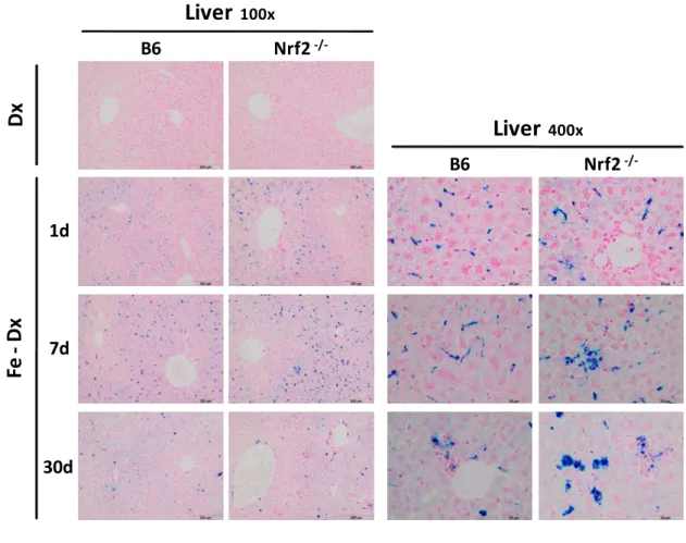
(57) Results & Discussion. Liver. Nrf2 -/-. Dx. B6. 100x. Liver. 400x. B6. Nrf2 -/-. Fe - Dx. 1d. 7d. 30d. Figure 7. Perls Prussian blue staining of mouse liver sections at 100x and 400x magnification.. Iron deposition was further detected with Perls Prussian blue stain of liver and spleen sections (Figure 7). Iron accumulation was evident in liver sections of iron dextraninjected animals, with a decreasing gradient in iron concentration from the periportal zones towards the centrolobular areas. A higher magnification clearly showed that the majority of iron is accumulated in reticuloendothelial cells of the liver, also known as Kupffer cells, which are located in the liver sinusoids. The pattern of iron deposition was similar throughout the whole course of the experiment (1d, 7d and 30d after injection), albeit after 7d and 30d a higher amount of iron was apparently deposited in parenchymal cells. In the spleen, iron was deposited mainly in the red pulp, but the difference between iron-treated mice and the controls was less prominent (Figure 8).. 43.
(58) Results & Discussion. Spleen Nrf2 -/-. Dx. B6. Fe - Dx. 1d. 7d. 30d. Figure 8. Perls Prussian blue staining of mouse spleen sections at 100x magnification.. The spleen plays important roles in regard to red blood cells, principally recycling their iron content through the metabolism of haemoglobin. In a healthy organism the balance between erythrocyte recycling and synthesis is maintained – the system is at equilibrium in terms of iron circulation, absorption and loss. Upon anemia or increased ingestion of iron the organism responds by restoring iron balance in erythropoiesis: in the former situation by mobilizing the input of injected iron sources and in the latter situation by not absorbing the surplus after ingestion. The mouse model used in this work is at equilibrium and as a result the iron injected is not primarily used for erythropoiesis, so the balance between erythrocyte recycling and synthesis is maintained. One can thus expect the spleen iron stores to remain constant, which histology of the spleen would not present. 44.
(59) Results & Discussion. a clear difference between the treatments. The main difference is to be found at the main body iron storage sites, which are replenished upon iron dextran injection. Iron dextran is thus removed from plasma by reticuloendothelial cells and it seems to be accumulated as non-haem iron in the liver.. 1.2. Nrf2 activation in iron dextran-treated mice. The activation of the Nrf2 pathway by iron dextran was investigated by measuring the mRNA levels of known transcriptional target genes in B6 (Nfr2 wt) and Nrf2-/- mice. This analysis was performed by real-time reverse transcriptase polymerase chain reaction, a technique based on the PCR that is used to amplify and simultaneously quantify a specific sequence in a complementary DNA (cDNA) sample. The reverse transcription step is necessary to convert the mRNA targets into cDNA, which can serve as a template for amplification. In a PCR reaction, the exponential amplification can be described according to equation [1], where C is the number of cycles, N C is the number of molecules at cycle C, N 0 is the initial number of molecules and E stands for the amplification efficiency: N C = N 0 x (E+1)C. [1]. Considering that all PCR reactions are compared at the same Fluorescence Threshold (FT), i.e. when the same number of molecules is reached, then N C becomes a constant value, as indicated in equation [2], where Ct is the threshold cycle and Nt is the number of molecules at FT. N 0 = Nt/(E+1)Ct. [2]. Quantification relies on standard curves constructed by amplification of known amounts of target DNA and plotting Ct values against the log template DNA [Log(N 0 )] (e.g Figure 9D). Log(N 0 ) = Log(Nt) – Log[(E+1)Ct]. [3]. Ct = - 1/Log(E+1)×Log(N 0 )+Log(Nt)/Log(E+1). [4]. E and Nt are assumed to be constants, so the equation above is the equation of the line (y = mx + b) obtained when plotting Log(N 0 ) versus Ct. In this equation, y is the Ct value, m is the slope of the line, x stands for Log(N 0 ) and b is the intercept. The relative quantity of each gene of interest was normalized against β-actin and later divided by the control 45.
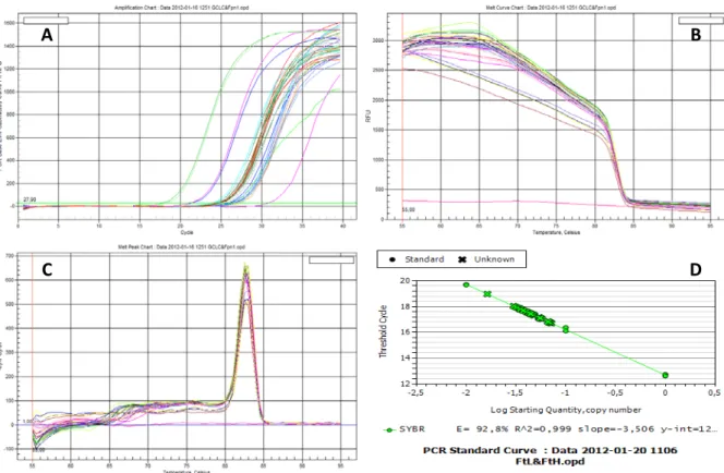
(60) Results & Discussion. sample to generate an expression fold change. The efficiency of the reaction (E) was calculated from the slope of the curve according to equation [5]: E = 10(-1/slope) – 1. [5]. N = 10(Ct – intercept)/slope. [6]. The amplification products are detected via SYBR Green fluorescence. SYBR Green is an asymmetrical Cyanine dye used as a nucleic acid stain in molecular biology and preferentially binds to double-stranded DNA. In a PCR reaction this occurs at the elongation and extension steps. Hence, fluorescence can be measured at each cycle of the polymerization reaction to monitor the amount of amplified DNA (Figure 9A). One major disadvantage of using SYBR Green is its non-specificity. It shows equal affinity to any double stranded DNA molecule formed during the amplification reaction such as primer dimers and non-specific products. Although primer dimers were avoided in the primer design stage by excluding primers with negative Gibbs free energy, the formation of non-specific products must be considered. Melting curve analysis of the PCR products was performed by gradually increasing the melting temperature (Figure 9B). The presence of non-specific products or primer dimers would generate extra peaks. The existence of a single dissociation peak confirmed the specificity of all reactions (Figure 9C). Relative quantification of the target genes was performed using the standard curve method. A relative standard curve was constructed by preparing serial dilutions of a cDNA sample containing the genes of interest (Figure 9D).. 46.
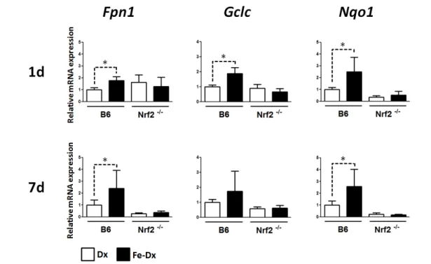
(61) Results & Discussion. A. B. C. D. Figure 9. Quantitative real-time RT-PCR analysis. A, Real-time detection of PCR amplification products by measurement of the fluorescence emitted by SYBR Green at the end of the elongation step of each amplification cycle. The horizontal green line is the fluorescence threshold (FT). These are amplification curves of multiple PCR products obtained by plotting cycle number versus fluorescence, in which the linear region represents the exponential phase of amplification. The FT is the point at which Ct values are determined for all individual samples. B, Dissociation curves of multiple PCR products. C, Derivative of the dissociation curves. The existence of a single melting point indicates the presence of a single amplification product. D, Standard curve generated by plotting the log of input material (template DNA concentration) versus threshold cycle (Ct).. The genes analyzed were quinone oxidoreductase 1 (Nqo1) and glutamatecysteine ligase catalytic subunit (Gclc), two prototypical Nrf2 targets, as well as ferroportin 1 (Fpn1), which has recently been described to be transcriptionally regulated by Nrf2 in cultured macrophages (Marro et al., 2010). A significant increase in the expression of Fpn1 and Nqo1 was found in the liver of iron-treated B6 mice at 1 and 7 days post injection, whereas Gclc was only elevated at 1 day after injection (Figure 10).. 47.
(62) Results & Discussion. Figure 10. Expression of Nrf2 target genes in the liver of mice treated with iron dextran. *p< 0.05. In the spleen, Gclc and Nqo1 transcripts were elevated at 1 day post injection. At 7 days post injection, mRNA levels were not different from the basal levels. On the other hand, Fpn1 mRNA levels were not regulated by iron dextran in the spleen. Importantly, none of the changes reported above for B6 liver (Nqo1, Gclc and Fpn1) and spleen (Nqo1 and Gclc) were seen in Nrf2-/- mice, where gene expression levels of iron dextran-treated animals were not significantly different from the control (dextran-treated) animals (Figure 11).. 48.
(63) Results & Discussion. Figure 11. Expression of Nrf2 target genes in the spleen of mice treated with iron dextran. *p<0.05. We can thus conclude that the expression of Fpn1, Gclc and Nqo1 is activated by iron dextran in mouse liver and spleen in an Nrf2-dependent manner. The data presented above represent the first evidence of Nrf2 activation by iron in cells of the reticuloendothelial system in vivo.. 49.
(64) 50.
(65) Part 2 Clinical Work. 51.
(66) 52.
(67) Results & Discussion. 2.1. Isolation of CD14+ blood cells from patients and control subjects A second aim of this project was to analyze the expression of Nrf2 target genes, UPR markers and iron-related genes in blood monocytes (CD14+) from HFE-HH patients. Whilst the liver is the major organ where iron accumulates in HFE-HH patients, access to liver biopsies is difficult and would pose ethical issues. Blood monocytes, which are exposed to increased levels of serum iron in these patients and which are capable of evoking an UPR and/or Nrf2 activation (Carroll et al., 2010), were thus tested as a possible surrogate tissue for detecting markers of both UPR and Nrf2 activation. A group of 7 C282Y HFE homozygous subjects with elevated iron indices (serum iron, ferritin and transferrin saturation) followed at Centro Hospitalar do Porto, and 7 apparently healthy controls with normal iron indices and absence of C282Y HFE mutation were recruited. Approximately 400 mL of blood were drawn and immediately processed for the isolation of CD14+ cells. This amount of blood is what is routinely drawn from HH patients undergoing iron-reduction phlebotomy therapy, where roughly 200 mg of iron are taken in each session. Whenever possible, patients and blood donors were sex- and agematched. As expected, the two groups were clearly distinguished in what regards the iron indices (Table 2 and Figure 12).. Table 2. Age and genetic profile of patient and control groups.. Profile. N. Age. Patient (C282Y/C282Y). 7. 64 ± 9. Control (Without C282Y). 7. 49 ± 5. 53.
(68) Results & Discussion. Figure 12. Serum iron parameters of patients and controls. *p<0.05. The isolation of CD14+ cells was performed either directly from whole blood or from peripheral blood mononuclear cells (PBMC’s) previously isolated with Lymphoprep. This comparison was meant to determine the most efficient method in terms of cell yield and specificity. The blood from one patient was split in two, one half followed the direct isolation protocol (Dynabeads FlowComp) and the other was submitted to a lymphoprep isolation step prior to the Dynabeads FlowComp protocol. The number of cells recovered was 6.3 ×10⁶ and 15.44 ×10⁶, respectively. These cells were subsequently analyzed for CD14 expression using a specific anti-CD14 antibody. CD14+ purity of cells isolated directly from whole blood and from PBMCs using Lymphoprep was assessed by flow cytometry, as depicted in Figure 13.. 54.
(69) Results & Discussion. Figure 13. Analysis for CD14 expression using a specific anti-CD14 antibody of cells isolated directly from whole blood or using a Lymphoprep intermediate step. The whole cell population was gated on an FSC vs. SSC dot plot and then analyzed for CD14 expression.. After gating the whole cell population on an FSC vs. SSC dot plot, it was apparent that 94.8% of the cells isolated directly from the whole blood and 89.9% of the cells isolated by the Lymphoprep method were positive for CD14. It is worth noting that not all CD14+ cells are monocytes, as two populations can be distinguished according to size/complexity. One of these populations shows some auto-fluorescence when nonlabeled (i.e. no anti-CD14 antibody) samples are acquired in the FACS and likely comprise CD14+ granulocytes. The percentage of these cells in the whole CD14+ population is higher when cells are isolated via the Lymphoprep method. Hence, we concluded that the Dynabeads FlowComp protocol in which CD14+ cells are isolated directly from whole blood was advantageous as it yielded a higher percentage of CD14+ cells and a lower degree of contamination of the CD14+ population with granulocytes. This isolation protocol was thus chosen for all subsequent work.. 55.
(70) Results & Discussion. 2.2. Expression of markers of UPR and Nrf2 activation in blood CD14+ cells The activation of an UPR in cells expressing C282Y HFE has been demonstrated in vitro (de Almeida et al., 2007) and ER stress may activate Nrf2 via PERK-mediated phosphorylation (Cullinan et al., 2003). As the induction of an UPR by C282Y HFE has not been demonstrated in vivo, BIP mRNA levels and XBP1 mRNA splicing were analyzed in CD14+ cells from C282Y HFE homozygous patients versus controls. As already reviewed in the introduction section, BiP is an ER resident chaperone which assists in the proper folding of nascent proteins within the ER, and following the induction of the UPR, BiP production increases dramatically in order to increase protein folding capacity (Cullinan & Diehl, 2006). BIP mRNA levels were assessed by real time RT-PCR and XBP1 splicing by a conventional PCR. Regarding BIP mRNA quantification, no significant differences were found between patients and controls (Figure 14).. Figure 14. BIP mRNA relative expression in CD14+ blood cells from patients versus controls.. The activation of an UPR releases a ribonuclease known as Ire1 which splices XBP1 mRNA. Our analysis of XBP1 splicing showed no evidence for the existence of the spliced form of XBP1 mRNA, neither in C282Y HFE homozygotes nor in the controls (Figure 15).. 56.
(71) Results & Discussion. Figure 15. XBP1 splicing in CD14+ blood cells from patients versus controls. The positive control consists of HepG2 cells treated with DTT. uXBP1 – unspliced form; sXBP1 – spliced form.. Overall, our data do not support the onset of an UPR in patient CD14+ blood cells. It is possible that the ER stress reported in cells overexpressing C282Y HFE has no physiological relevance in vivo, at least not in blood CD14+ cells. Alternatively, one can speculate that the endoplasmic reticulum associated degradation (ERAD) machinery is responsible for degrading the mutated protein soon after translation, not allowing ER chaperones such as BiP to activate the UPR. Finally, we can also assume that if Nrf2 was to be activated in patient blood monocytes it was not due to an UPR, but more likely to the elevated serum iron. Expression of iron-related genes, NRF2 and antioxidant/cytoprotective Nrf2 target genes was assessed by real-time RT-PCR. Iron-related genes were. the two ferritin. subunits, Ferritin L (FTL) and Ferritin H (FTH1), Ferroportin 1 (FPN1) and haemoxygenase 1 (HMOX1). As depicted in Figure 16 no significant differences were found between C282Y/C282Y patients and controls.. 57.
(72) Results & Discussion. Figure 16. Iron related genes expression in CD14+ blood cells from patients versus controls.. Antioxidant/cytoprotective Nrf2 target genes were Catalase (CAT), mitochondrial superoxide dismutase (MnSOD), thioredoxin reductase 1 (TRXNRD1), Glutamate cysteine ligase catalytic subunit (GCLC) and NAD(P)H dehydrogenase, quinone 1 (NQO1). As for the iron-related genes, we found no differences in the steady-state levels of NRF2 or its target genes when comparing C282Y/C282Y patients and controls (Figure 17).. 58.
(73) Results & Discussion. Figure 17. NRF2 and antioxidant/cytoprotective Nrf2 target genes expression in CD14+ blood cells from patients versus controls.. As liver biopsies are very difficult to obtain and pose ethical issues, we tried to use blood monocytes as an alternative model to measure Nrf2 activation in HH patients. This would allow us to assess whether Nrf2 activation correlated with the expression of HH symptoms in these patients and eventually establish whether Nrf2 may constitute a disease modifier for HFE-related HH. As demonstrated above, NRF2 is not activated by iron overload in CD14+ cells of HFE-HH patients. It is worth noting that the expression of iron-related genes is not elevated in these cells either, which suggests that blood monocytes do not respond to the significantly elevated serum iron levels. This result is in accordance with results from Graça Porto’s group. Transferrin saturation above 65% is associated with the formation of low molecular weight iron complexes not bound to transferrin (NTBI). NTBI retention in circulating immune system cells was negatively correlated with transferrin saturation of HH patients. Although highly variable, it is hypothesized that patients with higher retention of NTBI by immune cells are more protected from a more severe disease (Graça Porto, personal communication). As most HFE-HH patients in our work are clinically classified as symptomatic, their CD14+ cells may retain less iron, which could be one reason why Nrf2 is not activated by the iron59.
(74) Results & Discussion. induced oxidative stress. Whilst we cannot exclude the hypothesis that Nrf2 is a relevant disease modifier, it is likely that its activation needs to be analyzed in parenchymal cells such as hepatocytes, which accumulate large amounts of iron.. 60.
(75) Final Remarks. 61.
(76) 62.
Imagem


+7


Documentos relacionados
Mesmo para os indicadores mais simples (de quantidades e custos), devem os gestores acautelar-se de exprimirem relações causais: por exemplo, um maior número de serviços em dois
Com quem falam os adolescentes sobre sexualidade? Ilações com base nos resultados de um questionário
questões apresenta também diferenças significa- tivas. Os rapazes referem-se maioritariamente a postura do pai como receptiva, enquanto que as raparigas a declaram antes de
Eram chamados de Display a estes novos tipos de grandes dimensões usados em títulos.31 Vejamos os seguintes “Padrões de Fontes Vernaculares” que surgiram nesta época de acordo
The iron status of the animals treated with iron dextran confirmed our expecta- tions regarding the effect of serum and tissue iron overload on the baroreflex response (Table 1),
[r]
The hereditary hemochromatosis protein, HFE, lowers intracellular iron levels independently of transferrin receptor 1 in TRVb cells. Carpenter CE,
Although we could only perform subgroup meta-analysis of serum ferritin and hair iron levels but not peripheral serum iron or serum transferrin levels because of an insufficient
