ARTIGO ORIGINAL
RESUMO
Introdução: Os tumores das glândulas salivares incluem um vasto espetro de subtipos histológicos e comportamento clínico que pretendemos avaliar.
Material e Métodos: Realizou-se um estudo retrospetivo de todos os tumores das glândulas salivares diagnosticados e tratados no Centro Hospitalar São João, Porto, entre 2005 e 2015. Realizou-se reavaliação histológica de todos os casos. Os processos clínicos foram consultados e foram colhidos dados clínicos e de follow-up. A sobrevida livre de doença e a sobrevida global foram avaliadas usando as curvas de sobrevida de Kaplan-Meier e comparadas com o teste de log-rank Mantel-Cox. O limiar de significância foi es-tabelecido em 0,05.
Resultados: Selecionaram-se 295 casos, 150 no género masculino, com idade média de 50,4 anos. Nos tumores epiteliais benignos [n = 228 (77,3%)] predominaram os adenomas pleomórficos [n = 148 (64,9%)] e os tumores de Warthin [n = 61 (26,8%)]. Os tumores epiteliais malignos primários [n = 43 (14,8%)] incluíram carcinomas mucoepidermoides [n = 16 (37,2%)], adenoide-císticos [n = 6 (14,0%)] e de células acinares [n = 5 (11,6%)]; 32 (74,4%) localizaram-se na parótida, dois (4,6%) na submandibular e nove (21%) nas glândulas salivares minor. Os tumores epiteliais primários foram mais frequentemente malignos nas glândulas salivares minor (33,3%) do que nas major (13,9%). Observou-se recidiva local em 30,3% e metástases em 25,6%. O intervalo médio livre de doença foi 26 meses. As metástases mais frequentes ocorreram no pulmão e no sistema nervoso central. A taxa de sobrevida livre de doença aos cinco e 10 anos foi 63,4% e 50,1%, respetivamente; a sobrevida aos cinco e 10 anos foi 76,9% e 57,9%, respetivamente. Nos tumo-res primários identificaram-se também linfomas [n = 8 (2,7%)] e tumotumo-res mesenquimatosos [n = 5 (1,7%)]; nos tumotumo-res secundários identificaram-se metástases de carcinoma [n = 7 (2,4%)] e envolvimento por linfoma [n = 1 (0,3%)].
Discussão: Estes resultados assemelham-se aos da literatura europeia, designadamente quanto à idade, género, subtipo histológico e comportamento clínico. As divergências com séries portuguesas podem relacionar-se com critérios de inclusão, enviesamentos de referenciação clínica e período dos estudos.
Conclusão: Os tumores das glândulas salivares, embora raros, ocorrem numa faixa etária ampla, incluindo subtipos histológicos com prognósticos diferentes.
Palavras-chave: Glândulas Salivares/patologia; Neoplasias das Glândulas Salivares/patologia
Salivary Gland Tumors
Subtipos Histológicos e Avaliação do Comportamento
Clínico de Tumores das Glândulas Salivares
1. Department of Pathology. Centro Hospitalar São João. Porto. Portugal.
2. Unit of Pathology and Oncology. Department of Pathology. Faculdade de Medicina. Universidade do Porto. Porto. Portugal. Autor correspondente: Jorge Pinheiro. jorge.nature@gmail.com
Recebido: 30 de março de 2017 - Aceite: 16 de agosto de 2018 | Copyright © Ordem dos Médicos 2018
Jorge PINHEIRO1, Margarida SÁ FERNANDES1, Ana Rodrigues PEREIRA1, José Manuel LOPES1,2
Acta Med Port 2018 Nov;31(11):641-647 ▪ https://doi.org/10.20344/amp.9023
ABSTRACT
Introduction: Salivary gland tumors include a wide spectrum of histological subtypes and clinical behavior, which we aim to evaluate. Material and Methods: We performed a retrospective study of all salivary gland tumors diagnosed and treated at the Centro Hospitalar São João, Porto, between 2005 and 2015. Histological re-evaluation was performed in all cases and patient files were reviewed and both clinical and follow-up data were collected. Disease-free survival and overall survival were evaluated using Kaplan-Meier survival curves and compared using the Mantel-Cox log-rank test. The significance threshold was set at 0.05.
Results: We selected 295 cases, 150 males with a mean age at diagnosis of 50.4 (± 16.4) years. Primary benign epithelial tumors [n = 228 (77.3%)] were mostly pleomorphic adenomas [n = 148 (64.9%)] and Warthin tumors [n = 61 (26.8%)]. Primary malignant epithelial tumors [n = 43 (14.8%)] included mucoepidermoid [n = 16 (37.2%)], adenoid cystic [n = 6 (14.0%)] and acinic cell [n = 5 (11.6%)] carcinomas; 32 (74.4%) in parotid, 2 (4.6%) in submandibular and 9 (21%) in minor salivary glands. Primary epithelial tumors were more frequently malignant in minor (33.3%) than in major (13.9%) salivary glands. Local recurrence occurred in 30.2% and distant metastases in 25.6% tumors. The mean disease-free interval was 26 (± 37.5) months; most metastases were in lung and central nervous system. The 5 and 10 year disease-free survival rates were 63.4% and 50.1%, respectively; the 5 and 10 year disease-specific survival rates were 76.9% and 57.9%, respectively. Primary salivary tumors included also lymphomas [n = 8 (2.7%)] and soft tissue tumors [n = 5 (1.7%)]. Secondary tumors included metastases of carcinomas [n = 7 (2.4%)] and involvement by lymphoma [n = 1 (0.3%)].
Discussion: Our results concerning age, gender, histological subtype, frequency and clinical behavior of salivary tumors concur with European studies. Divergence with Portuguese studies might be related with the inclusion criteria, clinical referral and time lag variations.
Conclusion: Although uncommon, salivary gland tumors occur in a wide age range and include histological subtypes with diverse prognosis.
ARTIGO ORIGINAL INTRODUCTION Salivary gland tumors (SGT) are uncommon,
comprising about 3% - 5% of tumors in the head and neck region.1,2 However, these tumors encompass a wide range of histological subtypes and clinical behavior, challenging pathologists and clinicians in this complex and demanding area.
Reports and studies from various parts of the world reveal variations in the incidence and frequency of the different histological subtypes.2–7 In Portugal, there are a few previous studies addressing the features of malignant oral cancers,8 epithelial salivary gland tumors of children and adolescents,9 tumors of the parotid gland10 and prognostic factors of malignant SGT.11
Therefore, we performed a retrospective study to assess the relative frequency, location, patient demographics and clinical behavior of SGT diagnosed and treated at Centro Hospitalar São João (CHSJ), Porto, Portugal, including cases during a selected period of 11 years (2005 - 2015).
MATERIAL AND METHODS
All SGT diagnosed and treated at the Centro Hospitalar São João, Porto (a tertiary center in Northern Portugal for the diagnosis and treatment of non-oncologic and oncologic patients), between 2005 and 2015, were retrieved from the pathology department files. Only patients with major or minor salivary gland tumors that were subjected to surgical treatment were included. Patients who underwent surgical treatment due to primary skin or oral cancers with salivary gland direct invasion were excluded. Histological re-evaluation of the whole stored surgical specimen’s material was performed in all cases and additional histochemical and immunohistochemical studies were carried out whenever appropriate. Histopathological classification was accomplished according to the current World Health Organization (WHO) blue book,12 and pathological staging of malignant tumors according to the American Joint Committee on Cancer (AJCC) criteria (7th edition).13 The presence of extra-glandular extension or perineural and vascular invasion was systematically recorded. Risk stratification of malignant epithelial tumors was completed according to the criteria described by Seethala.14,15
Patient files were reviewed and clinical data, age, sex, location, clinical size of the tumor, type of treatment and follow-up were annotated.
Disease-specific survival time was calculated considering the date of surgical treatment and of the last clinical consultation or of patient death. Free-disease survival was calculated considering the date of surgical treatment and of the date of relapse, metastasis or of the last consultation. These data were evaluated using Kaplan-Meier survival curves due to variability in the follow-up time. Mantel-Cox log rank was used to perform a univariate analysis of potential prognostic parameters, such as age and gender, grading, staging or surgical margins. The significance threshold was set at 0.05. The low number of events prevented the assumption of proportional hazard.
The limited number of primary malignant epithelial SGT and the multicollinearity between relevant co-variates did not allow a multivariate analysis.
All statistical analysis was performed using IBM SPSS statistics for Windows, version 24.0.
RESULTS
A total of 295 salivary gland tumors (SGT) were identified with similar prevalence in both genders (150 men: 145 women). The mean age at diagnosis was 50.4 (± 16.4) years (range 13 - 83), similar in both genders; 9 cases (3.1%) occurred in pediatric age. The majority of cases were located in major salivary glands: 243 (82.4%) in the parotid, 20 (6.8%) in the submandibular and 2 (0.7%) cases in the sublingual glands; 30 cases (10.2%) located in minor salivary glands, the palate being the most frequent site [n= 18 (60%)]. Tumor size varied between 0.4 and 8 cm, with a median value of 2 cm, in the 257 cases in which this parameter was registered.
Primary tumors comprised most of the cases [n = 287 (97.3%)] (Table 1), the remaining being metastasis of carcinoma [n = 7 (2.4%)] and involvement by lymphoma [n = 1 (0.3%)]. The pediatric cases [n = 9 (3.1%)] included pleomorphic adenoma [n = 7 (77.8%)], mucoepidermoid carcinoma [n = 1 (11.1%)] and neurofibroma [n = 1 (11.1%)] in the setting of neurofibromatosis type1.
Primary benign epithelial SGT comprised 77.3% of cases (n = 228). The most frequent SGT subtype was pleomorphic adenoma accounting for 50.2 % (n = 148) of all cases. The peak incidence was in the 4th decade of life and it was predominant in women (women: men ratio = 1.84: 1) (Fig. 1). According to location, 92% (n = 136) occurred in major salivary glands: 81.8% (n = 121) in parotid, 8.8% (n = 13) in submandibular, and 1.4% (n = 2) in sublingual; and 8% (n = 12) in minor salivary glands. The mean tumor size was 2 (± 0.8) cm (range 0.5 - 4.7). Local recurrence was observed in two cases (1.4%).
Warthin tumor was the second most frequent SGT subtype, accounting for 20.7% (n = 61) of all tumors. Most of the cases occurred in men (men: women ratio = 6.6: 1) and peak incidence in the 6th decade of life (Fig. 2). The great majority (96.7%) of cases were located in the parotid. The mean tumor size was 2.3 cm (± 1.1; range 0.6 - 5.4). Multicentricity was present in 9 (14.8%) cases, of which 2 (22.2%) were synchronous and 7 (77.8%) metachronous; in 8 of these cases (88.9%) there was bilateral involvement. Primary malignant epithelial tumors represented 14.6% (n = 43) of all SGT (Table 1), i.e. 0.14% of all patients diagnosed with cancer in our department at CHSJ during the same period (2005 - 2015). They were slightly more common in the male gender (men: women ratio = 1.15: 1) and there was a bimodal distribution, with a first peak in the 4th decade of life and a second in elderly patients (Fig. 3). Considering the location, 74.4% (n = 32) arose in the parotid, 4.7% (n = 2) in the submandibular gland and 20.9% (n = 9) in minor salivary glands. However, primary epithelial SGT were more
ARTIGO ORIGINAL
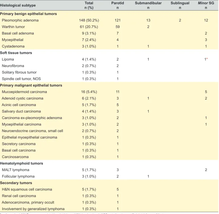
Table 1 – Histological subtypes of salivary gland tumors according to location. Values are presented by absolute number (n) and percentage (%).
Histological subtype Totaln (%) Parotidn Submandibular n Sublingualn Minor SGn
Primary benign epithelial tumors
Pleomorphic adenoma 148 (50.2%) 121 13 2 12
Warthin tumor 61 (20.7%) 59 2
Basal cell adenoma 9 (3.1%) 7 2
Myoepithelial 7 (2.4%) 4 3
Cystadenoma 3 (1.0%) 1 1 1
Soft tissue tumors
Lipoma 4 (1.4%) 2 1 1*
Neurofibroma 2 (0.7%) 2
Solitary fibrous tumor 1 (0.3%) 1
Spindle cell tumor, NOS 1 (0.3%) 1
Primary malignant epithelial tumors
Mucoepidermoid carcinoma 16 (5.4%) 11 5
Adenoid cystic carcinoma 6 (2.1%) 3 1 2
Acinic cell carcinoma 5 (1.7%) 5
Salivary duct carcinoma 4 (1.4%) 3 1
Carcinoma ex-pleomorphic adenoma 3 (1.0%) 2 1
Myoepithelial carcinoma 3 (1.0%) 2 1
Neuroendocrine carcinoma, small cell 2 (0.7%) 2
Epithelial myoepithelial carcinoma 1 (0.3%) 1
Secretory carcinoma 1 (0.3%) 1
Basal cell carcinoma 1 (0.3%) 1
Carcinosarcoma 1 (0.3%) 1
Hematolymphoid tumors
MALT lymphoma 5 (1.7%) 3 2
Follicular lymphoma 3 (1.0%) 2 1
Secondary tumors
H&N squamous cell carcinoma 5 (1.7%) 5
Renal cell carcinoma 1 (0.3%) 1
Adenocarcinoma, primary occult 1 (0.3%) 1
Involvement by generalized lymphoma 1 (0.3%) 1
SG: salivary gland; MALT: mucosa-associated lymphoid tissue; H&N: head and neck; NOS: not otherwise specified; * sialolipoma of the tongue
Figure 1 – Comparison of age distribution between men (n = 52) and women (n = 96) considering pleomorphic adenomas
≤ 19 0% 2% 4% 6% 8% 10% 12% 14% 16% ≥ 70 20 - 29 30 - 39 40 - 49 Men Women Age (years)50 - 59 60 - 69
Figure 2 – Comparison of age distribution between men (n = 53) and women (n = 8) considering Warthin tumors
≤ 19 0% 5% 10% 15% 20% 25% 30% 35% ≥ 70 20 - 29 30 - 39 40 - 49 Men Women Age (years)50 - 59 60 - 69
ARTIGO ORIGINAL
Figure 3 – Comparison of age distribution between men (n = 23) and women (n = 20) considering malignant epithelial salivary gland tumors ≤ 19 0% 2% 4% 6% 8% 10% 12% 18% 16% 14% ≥ 70 20 - 29 30 - 39 40 - 49 Men Women Age (years)50 - 59 60 - 69
frequently malignant in minor salivary glands (33.3%) than in major salivary glands (13.9%).
The most frequent subtypes of malignant epithelial SGT were mucoepidermoid [n = 16 (37.2%)], adenoid cystic [n = 6 (14.0%)] and acinic cell [n = 5 (11.6%)] carcinomas. According to the Seethala criteria for risk stratification of salivary gland cancers,8 53.4% (n = 23) of cases were low risk and 46.5% (n = 20) high risk tumors.
The AJCC pathological T value classification of 34 major SGT displayed 9 (26.4%) cases ≤ 2 cm in greatest dimension (pT1), 3 (8.8%) with 2 - 4 cm tumor size without extra parenchymal extension (pT2), 18 (52.9%) > 4 cm or extra parenchymal extension (pT3), and 4 (11.8%) invading adjacent structures (pT4). Histopathological evaluation of regional lymph nodes (N) was requested in 24 cases (55.8%), and metastasis were detected in 33.3% of cases (n = 8) (N1 = 5; N2a = 1; N2b = 2). Globally, perineural invasion was detected in 41.8% (n = 18) and vascular invasion in 30.2% (n = 13).
In the nine minor SGT, 5 (55.6%) were < 2 cm (pT1), two (22.2%) had 2 - 4 cm (pT2) and 2 (22.2%) were greater than
4 cm (pT3). Lymph node metastases were not detected in the three cases in which histopathological evaluation was requested. Globally, perineural invasion was detected in 33.3% (n = 3) and vascular invasion in 22.2% (n = 2). Distant metastases were not detected at the time of surgical treatment in any minor or major SGT.
Clinical staging at diagnosis displayed 32.6% (n = 14) cases stage I, 11.6% (n = 5) stage II, 41.9% (n = 18) stage III, and 13.9% (n = 6) stage IV.
Neoadjuvant therapy was not performed in any case. Adjuvant radiotherapy was performed in 27 (62.8%) cases, and in two of these cases it was combined with chemotherapy.
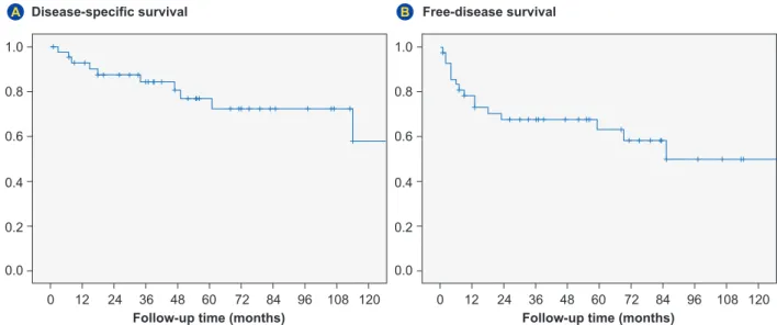
Globally, the 5 and 10 year disease-specific survival rate of primary malignant SGT was 76.9% and 57.9%, respectively (Fig. 4). The 5 and 10 year disease-free survival rate was 63.4% and 50.1 %, respectively (Fig. 4). At the end of the follow-up period, 30.2% (n = 13) had local recurrence and 25.6% (n = 11) developed metastases. The mean disease-free interval was 26 (± 37.5) months (range: 1 - 133). The mean metastatic-free interval was 26.7 (± 39.1) months (range: 2 - 85). The most frequent metastatic sites were the lung [n = 7 (63.6%)] and the central nervous system (n = 5; 45.5%). The crude mortality rate was of 23.3%. None of the cases underwent autopsy.
Death due to disease occurred in cases of high grade mucoepidermoid [(n = 4 (40%)], adenoid cystic [n = 2 (20%)], salivary duct [n = 1 (10%)], and myoepithelial [n = 1 (10%)] carcinomas, carcinosarcoma [n = 1 (10%)]; and small cell neuroendocrine carcinoma [n=1 (10%)]. According to location, mortality was observed in 9 out of 34 (26.5%) major salivary gland carcinomas compared to 1 out of 9 (11.1%) minor salivary gland carcinomas.
Our series is limited to correlate mortality or disease progression with histologic subtypes. Concerning tumor grading of mucoepidermoid carcinomas, three (60%) of the 5 high-grade tumors, displayed local recurrence, 3 (60%) developed distant metastases, and the observed
Figure 4 – Disease-specific overall survival (A) and disease-free survival (B) curves of malignant epithelial salivary gland tumors (n = 43)
0 12 24 36 48 60 72 84 96 108 120 0 12 24 36 48 60 72 84 96 108 120 0.0 0.0 0.2 0.2 0.4 0.4 1.0 1.0 0.8 0.8 0.6 0.6
Follow-up time (months) Follow-up time (months)
Disease-specific survival Free-disease survival
ARTIGO ORIGINAL
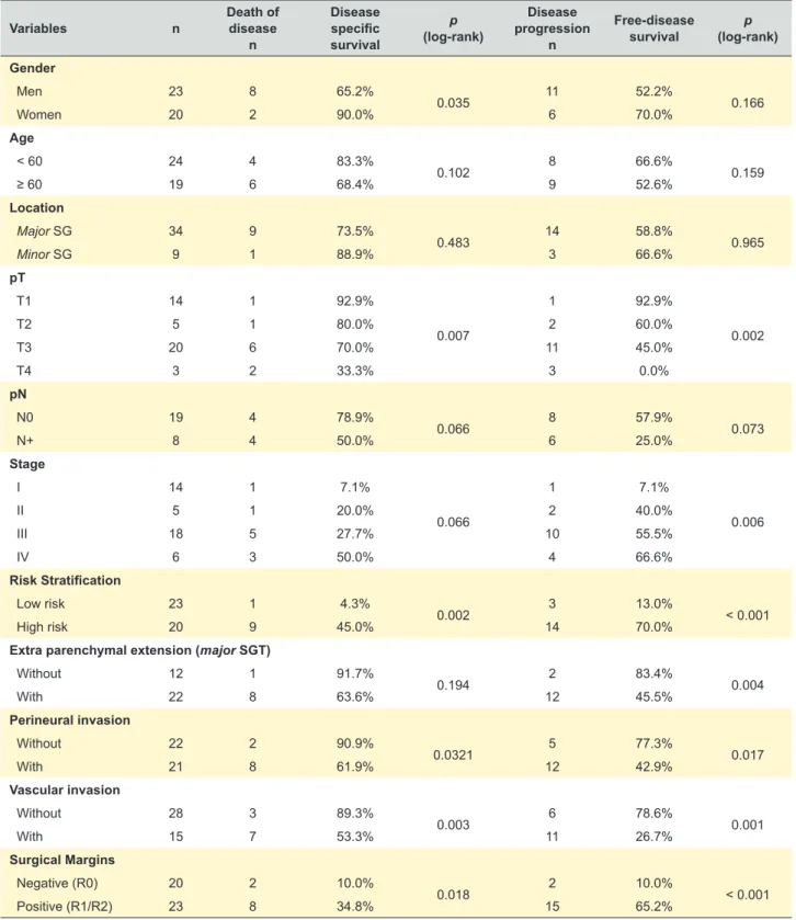
Table 2 – Univariate analysis of disease specific survival and free-disease survival in primary malignant epithelial SGT
Variables n Death of disease
n Disease specific survival p (log-rank) Disease progression n Free-disease survival (log-rank)p Gender Men 23 8 65.2% 0.035 11 52.2% 0.166 Women 20 2 90.0% 6 70.0% Age < 60 24 4 83.3% 0.102 8 66.6% 0.159 ≥ 60 19 6 68.4% 9 52.6% Location Major SG 34 9 73.5% 0.483 14 58.8% 0.965 Minor SG 9 1 88.9% 3 66.6% pT T1 14 1 92.9% 0.007 1 92.9% 0.002 T2 5 1 80.0% 2 60.0% T3 20 6 70.0% 11 45.0% T4 3 2 33.3% 3 0.0% pN N0 19 4 78.9% 0.066 8 57.9% 0.073 N+ 8 4 50.0% 6 25.0% Stage I 14 1 7.1% 0.066 1 7.1% 0.006 II 5 1 20.0% 2 40.0% III 18 5 27.7% 10 55.5% IV 6 3 50.0% 4 66.6% Risk Stratification Low risk 23 1 4.3% 0.002 3 13.0% < 0.001 High risk 20 9 45.0% 14 70.0%
Extra parenchymal extension (major SGT)
Without 12 1 91.7% 0.194 2 83.4% 0.004 With 22 8 63.6% 12 45.5% Perineural invasion Without 22 2 90.9% 0.0321 5 77.3% 0.017 With 21 8 61.9% 12 42.9% Vascular invasion Without 28 3 89.3% 0.003 6 78.6% 0.001 With 15 7 53.3% 11 26.7% Surgical Margins Negative (R0) 20 2 10.0% 0.018 2 10.0% < 0.001 Positive (R1/R2) 23 8 34.8% 15 65.2%
crude mortality rate was of 80.0%. On the other hand, no recurrence or disease progression was observed in the 11 low-grade mucoepidermoid carcinomas. Other classical pathologic prognostic factors evaluated (risk stratification, stage of disease, tumor stage, resection margins, perineural invasion and vascular invasion) displayed significant prognostic value for disease-free survival of patients and/or disease specific survival (Table 2).
DISCUSSION
In summary, we found similar prevalence of SGT in both genders and a wide age distribution. Primary benign epithelial tumors were the most frequent [77.3% (n = 228)], predominantly pleomorphic adenomas and Warthin tumors. Primary malignant epithelial tumors accounted for 14.8% (n = 43) of all tumors, most frequently mucoepidermoid, adenoid cystic and acinic cell carcinomas. Primary epithelial tumors were more frequently malignant in minor (33.3%)
ARTIGO ORIGINAL further specification) histotypes. Fonseca I et al9 reported a study (1991) on pediatric epithelial SGT in a Portuguese oncology institute (South of Portugal). Our results are limited in this age group, but indicate a similarity in the proportion (3.2%) and of the most common histotypes (e.g. pleomorphic adenoma and mucoepidermoid carcinoma) in pediatric epithelial SGT of both studies.
Oliveira F et al10 reported a study (2013) of parotid tumors (n = 153) diagnosed in a Portuguese oncology institute (South of Portugal). In our study (n = 242) we found a higher frequency of benign tumors in the parotid (82.5% vs 72.4%, respectively). Noteworthy, we found differences in the frequency of malignant histotypes, even considering that our series comprises a slightly lower number of cases: mucoepidermoid carcinoma being more frequent in our study (26.2% vs 18.92%, respectively), and myoepithelial carcinoma as well as carcinoma ex-pleomorphic adenoma being less frequent (4.7% vs 16.2%; and 4.7% vs 13.5%, respectively). We also observed a lower rate of relapse (28.1% vs 40.5%, respectively). These differences may, at least in part, indicate diverse diagnostic inclusion criteria and referral bias in both studies. Regrettably, the report by Oliveira F et al does not provide staging and survival data to compare with our results.
Monteiro LS et al11 reported a study on the prognostic factors in primary malignant SGT (n = 136, from 1992 to 2002) from another Portuguese oncology institute (Nortern Portugal). Noteworthy, compared to our study, we identified a higher proportion of primary malignant tumors in major salivary glands (79.1% vs 65.4%, respectively) and of mucoepidermoid carcinoma (37.2% vs 21.3%, respectively), similar proportion of acinic cell carcinoma (11.6% vs 8.1%, respectively), and lower frequency of adenoid cystic carcinoma (14.0% vs 25.0%, respectively). Notably, we observed lower proportion of tumors diagnosed in stage IV (14% vs 39%, respectively), which might explain differences in the 5-year overall survival rates observed in both studies (76.9% vs 71%, respectively). Again, these differences may be probably related to the fact that our hospital is not a reference center for the management of patients with advanced SGT. Yet, it would be interesting to clarify if the time lag between both studies (2005 - 2015 vs 1992 - 2002, respectively), despite referral issues aforementioned, might reflect epidemiological differences of SGT diagnosed and treated in patients in Northern Portugal or not.
CONCLUSION
Salivary gland tumors are rare tumors and represent 0.14% of all the patients diagnosed with cancer at Centro Hospitalar São João, Porto, Portugal. Importantly, these tumors represent an increasing complex area in pathology, with many different histotypes and distinct prognosis. Our results fit with age, gender, histological subtype, frequency and clinical behavior of salivary gland tumors reported in European series. Notably, we identified some dissimilarity of our results compared with Portuguese series, than in major (13.9%) salivary glands. Although infrequent,
salivary carcinomas are prone to significant morbidity and mortality. We found local recurrence in 30.2% and distant metastases in 25.6%. The mean disease-free interval was 26 (± 37.5) months; most metastases were in the lung and central nervous system. The 5 and 10 year disease-free survival rates were 63.4% and 50.1%, respectively; the 5 and 10 year disease-specific survival rates were 76.9% and 57.9%, respectively.
Primary salivary tumors included also lymphomas [n = 8 (2.7%)] and soft tissue tumors [n = 5 (1.7%)]. Secondary tumors included metastases of carcinomas [n = 7 (2.4%)] and involvement by lymphoma [n = 1 (0.3%)].
Demographic data is helpful for understanding the clinical and pathological features of salivary gland tumors. The rarity of SGT and their complexity, along with the occurrence of distinct histological subtypes, make their study an increasingly challenging diagnostic and therapeutic area.
Despite having been performed in a large tertiary hospital in Northern Portugal, based on a 11 year time period, the limitations of this study include referral bias of patients, restricted number of these uncommon malignant epithelial tumors, and lack of longer follow-up time, especially concerning SGT with reported less aggressive behavior. Even so, the results of our study are in line with those reported in other European countries.1,2,7,16–18 Indeed, we observed a high frequency of benign epithelial tumors (adenomas), being pleomorphic adenoma the most common. Notably, we found a high frequency of Warthin tumors in the male gender, which may probably be related to smoking habits being more common in men in the past.19 The proportion of primary malignant tumors is slightly lower than reported by others,1,2 which might be related with referral bias of our series. Nevertheless, the most frequent subtypes correspond with those reported in other studies. The disease-specific survival rate appears to be similar to what has been previously reported in other European studies,20,21 and seems to be influenced by the histological subtype, grade and tumor stage at diagnosis of patients. Indeed, the presence of 53.4% of low-risk and low stage malignant tumors are in line with the calculated disease-specific and overall survival curves above the median observed in the patients of our study.
Interestingly, concerning secondary tumors, we notice the lack of melanoma metastasis in our series, which is probably related to the fact that our hospital is not a reference center for the management of patients with advanced melanomas.
There are few published Portuguese series assessing malignant oral cancers,8 epithelial SGT of children and adolescents,9 tumors in the parotid gland10 and prognostic factors in malignant SGT.11
We cannot compare our results with those reported by Tavares C et al8 because the histotypes of malignant SGT are not clearly stated. Actually in their report there is only evidence of mucoepidermoid and adenocarcinoma (without
ARTIGO ORIGINAL which might indicate different inclusion diagnostic criteria,
clinical referral and time lag variation among studies. It would be important to add our data with data from other referral hospitals in Northern Portugal, in a larger study, to clarify the behavior of different SGT subtypes, namely those with reported characteristic molecular features, in order to improve management of patients harboring these tumors.
PROTECTION OF HUMANS AND ANIMALS
The authors declare that the Ethics Committee of APDP-Diabetes granted ethical approval of the study and that the procedures followed the regulations established by
the Helsinki Declaration of the World Medical Association.
DATA CONFIDENTIALITY
The authors declare having followed the protocols in use at their working center regarding patients’ data publication.
CONFLICTS OF INTEREST
All authors report no conflict of interest.
FUNDING SOURCES
No subsidies or grants contributed to this work. REFERENCES
1. Lukšić I, Virag M, Manojlović S, Macan D. Salivary gland tumours: 25 years of experience from a single institution in Croatia. J Craniomaxillofac Surg. 2012;40:e75-81.
2. Eveson JW, Cawson R. Salivary gland tumours. A review of 2410 cases with particular reference to histological types, site, age and sex distribution. J Pathol. 1985;146:51–8.
3. Ito FA, Ito K, Vargas PA, de Almeida OP, Lopes MA. Salivary gland tumors in a Brazilian population: a retrospective study of 496 cases. Int J Oral Maxillofac Surg. 2005;34:533–6.
4. Tian Z, Li L, Wang L, Hu Y, Li J. Salivary gland neoplasms in oral and maxillofacial regions: a 23-year retrospective study of 6982 cases in an eastern Chinese population. Int J Oral Maxillofac Surg. 2010;39:235–42. 5. Subhashraj K. Salivary gland tumors: a single institution experience in
India. Br J Oral Maxillofac Surg. 2008;46:635–8.
6. Adebiyi KE, Emmanuel MM. Neoplastic salivary gland lesions: a retrospective analysis of 135 cases from Lagos State University Teaching Hospital, Ikeja, Lagos, Nigeria. West Afr J Med. 2014;33:206– 10.
7. Bradley PJ, McGurk M. Incidence of salivary gland neoplasms in a defined UK population. Br J Oral Maxillofac Surg. 2013;51:399–403. 8. Tavares C, Guimarães J, Lopes O, Felino A, Coimbra F. Epidemiological
profile of malignant oral cancers in a population of northern Portugal. Rev Port Estomatol Med Dent Cir Maxilofac. 2016;57:229–35. 9. Fonseca I, Martins G, Soares J. Epithelial salivary gland tumors of
children and adolescents in southern Portugal. A clinicopathologic study of twenty-four cases. Oral Surg Oral Med Oral Pathol. 1991;72:696– 701.
10. Oliveira F, Costa E, Pereira S, Pacheco R, Magalhães M. Tumores das glândulas parótidas - Casuística dos últimos 10 anos do serviço de ORL do IPO de Lisboa. Rev Port Otorrinol Cirur Cerv Fac. 2013;51:157-60.
11. Monteiro LS, Bento MJ, Antunes L, Lopes C. Fatores de prognóstico em neoplasias malignas de glândulas salivares. Rev Port Estomatol Med Dent Cir Maxilofac. 2012;53:199–205.
12. El-Naggar AK, Chan J, Takata T, Grandis J, Blootweg P, editors. WHO classification of head and neck tumours. 4th ed. Lyon: IARC Press; 2017.
13. Edge SB, Byrd DR, Compton CC, Fritz AG, Greene FL, Trotti A, editors. AJCC cancer staging manual. 7th ed. France: Springer; 2010.
14. Raja R. Seethala. Histologic grading and prognostic biomarkers in salivary gland carcinomas. Adv Anat Pathol. 2011;18:29–45.
15. Seethala RR. An update on grading of salivary gland carcinomas. Head Neck Pathol. 2009;3:69–77.
16. Ascani G, Pieramici T, Messi M, Lupi E, Rubini C, Balercia P. Salivary glands tumours: a retrospective study of 454 patients. Minerva Stomatol. 2006;55:209–14.
17. Debets JM, Munting JD. Parotidectomy for parotid tumours: 19-year experience from the Netherlands. Br J Surg. 1992;79:1159–61. 18. Rodríguez Paramás A, Lendoiro Otero C, González García JA, Souviron
Encabo R, Scola Yurrita B. Tumores malignos de la glándula parótida. Acta Otorrinolaringol Española. 2005;56:211–4.
19. Sadetzki S, Oberman B, Mandelzweig L, Chetrit A, Ben-Tal T, Jarus-Hakak A, et al. Smoking and risk of parotid gland tumors: Aa nationwide case-control study. Cancer. 2008;112:1974–82.
20. Bjørndal K, Krogdahl A, Therkildsen MH, Overgaard J, Johansen J, Kristensen CA, et al. Salivary gland carcinoma in Denmark 1990-2005: outcome and prognostic factors: Results of the Danish Head and Neck Cancer Group (DAHANCA). Oral Oncol. 2012;48:179–85.
21. Wahlberg P, Anderson H, Biörklund A, Möller T, Perfekt R. Carcinoma of the parotid and submandibular glands - a study of survival in 2465 patients. Oral Oncol. 2002;38:706–13.