RMRP
Is a Non-Coding RNA Essential for Early Murine
Development
Joseph Rosenbluh1,2, Deepak Nijhawan1,2, Zhao Chen1, Kwok-Kin Wong1, Kenkichi Masutomi3*, William C. Hahn1,2*
1Department of Medical Oncology, Dana–Farber Cancer Institute and Departments of Medicine, Brigham and Women’s Hospital and Harvard Medical School, Boston, Massachusetts, United States of America,2Broad Institute of Harvard and Massachusetts Institute of Technology (MIT), Cambridge, Massachusetts, United States of America,3Division of Cancer Stem Cell, National Cancer Center Research Institute, Tokyo, Japan
Abstract
RMRPis a non-coding RNA that is ubiquitously expressed in both humans and mice.RMRPmutations that lead to decreased RMRPlevels are found in the pleiotropic syndrome Cartilage Hair Hypoplasia. To assess the effects of deletingRMRP, we
engineered a targeting vector that contains loxP sequences flankingRMRPand created hemizygous mice harboring this
engineered allele (RMRPconditional). We found that insertion of this cassette suppressedRMRPexpression, and we failed to obtain viable mice homozygous for theRMRPconditional allele. Furthermore, we were unable to obtain viable homozygous
RMRPnull mice, indicating thatRMRPis essential for early embryonic development.
Citation:Rosenbluh J, Nijhawan D, Chen Z, Wong K-K, Masutomi K, et al. (2011)RMRPIs a Non-Coding RNA Essential for Early Murine Development. PLoS ONE 6(10): e26270. doi:10.1371/journal.pone.0026270
Editor:Saverio Bellusci, Childrens Hospital Los Angeles, United States of America
ReceivedJune 10, 2011;AcceptedSeptember 23, 2011;PublishedOctober 19, 2011
Copyright:ß2011 Rosenbluh et al. This is an open-access article distributed under the terms of the Creative Commons Attribution License, which permits unrestricted use, distribution, and reproduction in any medium, provided the original author and source are credited.
Funding:This research was supported by a US National Institutes of Health postdoctoral fellowship to J.R. F32GM090437-01, and R01 AG23145 (WCH). The funders had no role in study design, data collection and analysis, decision to publish, or preparation of the manuscript.
Competing Interests:The authors have declared that no competing interests exist.
* E-mail: [email protected] (WCH); [email protected] (KM)
Introduction
RMRP is a non-coding RNA that is highly expressed in a
wide range of human and murine tissues [1]. Mutations inRMRP have been detected in individuals afflicted with Cartilage Hair Hypoplasia (CHH) [2], a syndrome characterized by short stature, sparse hair, immunodeficiency and in a subset of patients severe combined immunodeficiency or life threatening anemia. Cell-based reporter assays have shown thatRMRPmutations result in decreased RMRP stability, which may account for the severe phenotypes seen in CHH [3]. Individuals that carry only a single RMRPmutation do not exhibit phenotypes associated with CHH
[2]; however, affected individuals harbor mutations in bothRMRP alleles [4] suggesting that these mutations inactivate the RMRP gene product.
The biological function(s) of RMRP remain incompletely understood. Biochemical studies have demonstrated that RMRP RNA binds to the mitochondrial posttranscriptional modification complex RNase MRP [5]. However, no apparent mitochondrial defects have been found in CHH patients. In addition,RMRPis also found in the nucleolus. We recently reported that together with the catalytic subunit of telomerase (hTERT),RMRPforms an RNA dependent RNA polymerase that converts single stranded RMRPRNA into double strandedRMRP[6].
To gain further insight into the biological functions ofRMRP, we generated a genetically engineered mouse that lacksRMRP.
Results and Discussion
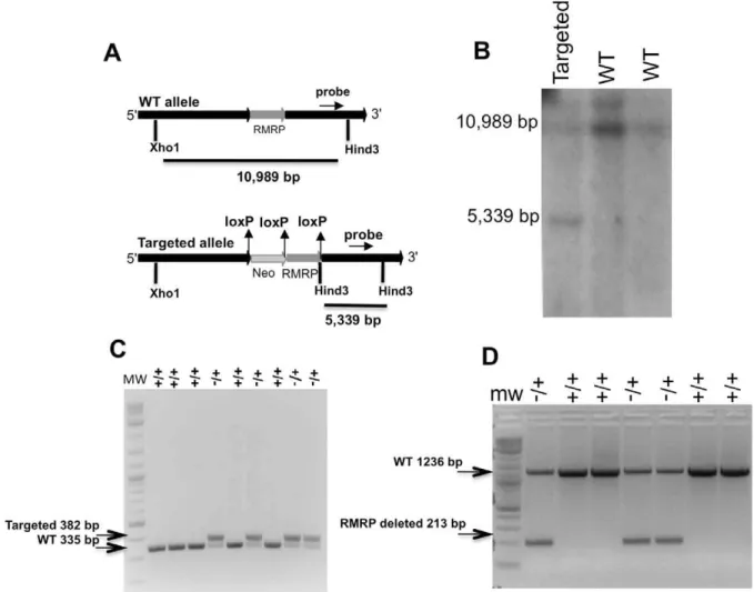
We created a targeting vector specific for murineRMRPusing the pEasyflox backbone [7]. The targeting vector contains the
RMRPgene and promoter (800 bp up stream of murineRMRP
[1]) flanked by two loxP sequences. A neomycin selectable marker flanked by two loxP sequences was placed upstream of RMRP (Figure 1A).
This targeting vector was introduced into mouse embryonic cells and individual clones containing the integrated targeting vector were selected by treatment with G418. Using southern blot analysis with a probe that can detect both the WT and targeted alleles, we found that 10% of the clones had integrated theRMRP targeting vector into the endogenous RMRP locus (Figure 1B). One of these clones was injected into female donor blastocysts producing 10 pups, 6 of which were chimeric, based on coat color. The chimeric mice were bred to FVB/N mice and the resulting pups were genotyped using a PCR based assay (Figure 1C). These mice contain theRMRPgene flanked by two loxP sequences and an insert coding for neomycin resistance upstream (RMRP conditional, RC) (Figure 1A).
We failed to obtain homozygous RC mice by crossing the hemizygous RC mice. Despite multiple attempts, we were unable to separate embryos earlier then E6.5 from the placenta. The RC mice harbor the neomycin resistance gene upstream of theRMRP gene, suggesting that insertion of DNA elements upstream of RMRP results in early embryonic lethality (Table 1). Thus, we
hypothesized that the neomycin insertion impairs critical genomic elements that are essential forRMRPexpression. Since prior work has confirmed that a subset of CHH patients harbor mutations in the RMRP promoter and these mutations decrease RMRP expression (1, 2), these observations suggest that the RMRP promoter is particularly sensitive to nucleotide changes.
hemizygous mice were crossed to a mouse that ubiquitously and constitutively expresses the Cre recombinase (CMV-Cre). Using PCR with primers that are specific for the predicted engineered RMRPallele after recombination, we confirmed that the RMRP was deleted in the offspring of the hemizygous mice (Figure 1D). Similar to what we observed in RC mice, we failed to obtain pups harboring homozygous deletion of RMRP (Table 1). These observations suggest that that insertion of exogenous DNA sequences upstream ofRMRPresults in aberrantRMRPexpression and results in embryonic lethality.
The levels of RMRP may be critical for RMRP function. Specifically, Nakashima et al. have proposed a model by which
RMRPmutations found in CHH patients leads to destabilization
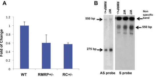
ofRMRP[3]. When we assessed total levels ofRMRPin murine embryonic fibroblasts (MEFs) obtained from RMRP or RC hemizygous mice, we found that RMRP was expressed at 50% of the level found in wild type MEFs (Figure 2A). RC andRMRP+/2 mice were monitored from birth to 18 months of age and no abnormality in size, fur or behavior was detected. This is in consonance with what has been observed in human carriers of RMRPmutations [2].
We previously found that two species ofRMRPare present in human cells: single strandedRMRPRNA and a double stranded RMRPRNA composed of a single RNA containing both the sense
and antisense strands [6]. The double stranded version ofRMRP requires the presence of the catalytic subunit of telomerase, TERT. Using Northern blot analysis with probes designed to detect sense or antisenseRMRP, we detected decreased levels of both species of RMRP in total RNA extracted from RMRP+/2 E13.5 MEFs as compared to WT MEFs (Figure 2B). TheRMRP antisense probe detects both single and double strandedRMRP and theRMRPsense probe detects only double strandedRMRP. These observations demonstrate that reduction ofRMRPreduces the function of the TERT-RMRPRdRP.
To assess the embryonic stage that requiresRMRPwe isolated embryos from breeding ofRMRP+/2 mice at several early time Figure 1. Targeting of murineRMRP.A. Murine targeting vector (MTV) B. Southern blot of ES cells following selection for alleles with integrated MTV (the southern probe is shown in figure 1a) C. PCR analysis of RC (RMRPconditional) pups D. PCR analysis of pups derived from the interbreeding of RC mice and mice expressing CMV-Cre.
doi:10.1371/journal.pone.0026270.g001
Table 1.RMRPdepletion is embryonic lethal.
Mating N (male/female) +/+ +/2 2/2
RMRP+/2
6RMRP+/2 45 (20/25) Expected 11.25 22.5 11.25 Observed 19 26 0
RC+/2
6RC+/2 47 (23/24) Expected 11.75 23.5 11.75 Observed 15 31 0
points. We failed to identify homozygous embryos as early as E6.5 (Table 2) indicating, that RMRP is required early in embryonic development.
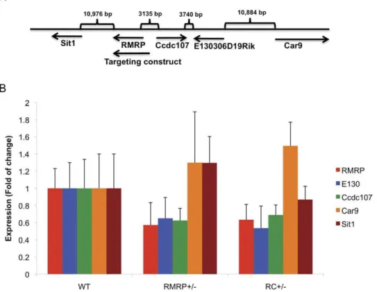
TheRMRPgene is located in close proximity to several other genes including Sit1, Ccdc107, E130 and Car9 (Figure 3A). To assess whether the expression of these genes are affected by RMRP depletion, we measured the expression of these genes in
E13.5 MEFs derived from WT, RMRP+/2 or RC+/2 mice. We confirmed that RMRP levels were decreased by 50% in both RMRP+/2and RC+/2MEFs. We also found that the expression of Ccdc107and E130were also decreased inRMRP+/2and RC+/2 MEFs while the expression of Sit1 and Car9 was not affected (Figure 3B). Although the targeting of RMRP involved the insertion of a small number of nucleotides, RMRP,Ccdc107and E130 are located closely together, and deletion ofRMRPaffects
the expression of all of these genes.
Based on these observations, we tested whetherCcdc107orE130 are essential for survival and thus may also contribute to the observed embryonic lethal phenotype. We used MEFs from E13.5 WT orRMRP+/2mice. Using RNAi, we reduced the expression of these genes to 5–20% of levels found in cells transfected with a control siRNA (Figure 4A and B). Seven days post transfection the cells were tested for viability, by monitoring the ATP content of the cells (Figure 4C). We failed to observe any alteration in cell proliferation/viability after suppression of eitherCcdc107orE130 indicating that these genes are not essential. However, we cannot eliminate the possibility that the residual low levels ofCcdc107or
E130(5–20% of control) are enough to sustain viability and when
completely deleted will lead to embryonic lethality. Due to the very close proximity between these genes it is very difficult to target RMRPwithout disruptingCcdc107andE130expression and further attempts to targetRMRPshould take this into consideration.
RMRPis a ubiquitously expressed non-coding RNA [1] that has
critical functions both in mice and humans. We found thatRMRP is essential for development at early stages of embryogenesis. We further demonstrated that insertion of DNA elements upstream of the RMRP promoter cause a decrease in RMRP expression in hemizygous mice and are lethal in homozygous mice. These observations suggest that expression ofRMRPis tightly regulated and essential for early developmental processes. We conclude that future attempts to targetRMRPmust consider the tight regulation and early requirement ofRMRP.
Materials and Methods
All laboratory animals will be cared for in the animal quarters of the Dana-Farber Cancer Institute under the direct supervision of the Dana-Farber Cancer Institute Animal Care and Use Committee (ACUC) under assurance number A3023-01. The Dana-Farber Cancer Institute is an Association for Assessment and Accreditation of Laboratory Animal Care International (AAALAC) accredited institution meeting or exceeding all standards for animal care and use. This work presented herein is has been approved by the ACUC under animal protocol 10-004.
Construction ofRMRPmouse targeting vector
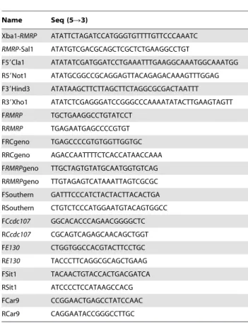
The pEasyflox vector [7] was used as a backbone to create the murine targeting vector. A 1097 bp fragment corresponding to RMRPand 800 bp of the upstream promoter sequence was PCR
amplified from RP23-207P5 (http://bacpac.chori.org) using Xba1-RMRP and RMRP-Sal1 primers (Table 3). The PCR product was digested with Xba1 and Sal1 and ligated to the same sites in the pEasyflox to create pEasyflox-RMRP. Next a 4064 bp fragment upstream of the RMRP gene was PCR amplified from RP23-207P5 using F59Cla1 and R59Not1 primers (Table 3). Following digestion with Not1 and Cla1 the PCR product was ligated to the same sites in pEasyflox-RMRPto create pEasyflox-59-RMRP. The downstream sequence of the targeting vector was amplified from RP23-207P5 using F39Hind3 and Figure 2. RMRPdepletion leads to reduced levels ofRMRPtranscript. Total RNA was produced from E13.5 MEFs andRMRPlevel was
measured by A. qRT-PCR B. Northern blot using either a sense or antisenseRMRPprobe. Error bars represent SD of three replicas. doi:10.1371/journal.pone.0026270.g002
Table 2.RMRPdepletion is lethal in early stage embryos.
N +/+ +/2 2/2
E13.5 27 Expected 6.75 13.5 6.75
Observed 11 16 0
E10.5 8 Expected 2 4 2
Observed 1 7 0
E6.5 20 Expected 5 10 5
Observed 4 16 0
doi:10.1371/journal.pone.0026270.t002
Figure 3. The effect ofRMRPdepletion on neighboring genes.A. Map of the genomic structure and genes surroundingRMRP. B. Expression of RMRP,Sit1,Ccdc107,E130andCar9in E13.5 MEFs from the indicated genotypes. Error bars represent SD of three replicas.
doi:10.1371/journal.pone.0026270.g003
Figure 4. Genes nearRMRPare not essential for cellular viability.MEFs from E13.5 mice of either A. WT or B.RMRP+/2were transfected with siRNAs targetingCcdc107orE130. Three days later RNA was extracted from the cells and qRT-PCR was preformed using primers forRMRP,Ccdc107or E130. C. The same cells as in A and B were plated (5000 cells/well) in a 96 well plate and 7 days post transfection cell number was assessed by Cell titer glow. Error bars represent SD of three replicas.
R39Xho1 primers (Table 3) and the 4029 bp fragment was digested with Hind3 and Xho1 and ligated into the same sites in pEasyflox-59-RMRPto create theRMRPmouse targeting vector. The sequence of the vector was confirmed by direct sequencing.
Northern blot
RNA was extracted from MEFs using an RNeasy kit (Qiagene). 10ml of sample buffer (95% formamide 5% bromophenolblue) was added to 5mg of total RNA in a total volume of 20ml. The samples were applied to a 6% TBE-urea acrylamide gel (invitrogen cat number: EC6865BOX) and then blotted onto a Hybond-N+membranes (GE Healthcare). Following cross-linking with a UV crosslinker the membrane was pre-incubated for 1 hour at 60uC with hybridization buffer (0.5 M NaHPO4PH = 7.2, 1 mM EDTA,
7% SDS). A radiolabeled RNA probe corresponding to the full length sense or antisense murineRMRPwas added and incubated with the membrane overnight at 60uC. Following 4 washes with 26SSC the gel was exposed and visualized.
Southern blot
DNA was prepared from ES cells targeted with the murine RMRPtargeting vector using the Qiaamp kit (Qiagene). 10mg of
DNA was digested with Hind3, loaded onto a 1% agarose fomaldhyde gel and blotted onto a Hybond-N+membranes (GE Healthcare). Following UV crosslinking the membrane was pre-incubated for 1 hour at 65uC with hybridization buffer (0.5 M NaHPO4PH = 7.2, 1 mM EDTA, 7% SDS). Next the probe was
added to the blot and allowed to incubate overnight at 65uC. Following 3 washes with 16SSC the membrane was exposed. The probe used for detection of targeted cells was generated by PCR amplification from a BAC clone (RP23-207P5 (http://bacpac. chori.org)) using FSouthern and RSouthern primers (Table 3).
Quantitative RT-PCR
Total RNA extracted from MEFs was reverse transcribed using the advantage RT-PCR kit (Clontech). Following reverse tran-scription the quantity of the transcript was determined using specific primers (Table 3) and syber green PCR mix (applied biosystems).
Genotyping
Tails were clipped from three week old mice and DNA was prepared. Genotyping was doneRMRPnull mice were genotyped using FRMRPgeno and RRMRPgeno (Table 3) and RC mice were genotyped using FRCgeno and RRCgeno (Table 3).
siRNA transfection and viability assay
For silencing the expression of Ccdc107 or E130, we used three independent siRNA duplexes targeting each gene. The siRNAs were purchased from IDTCcdc107cat number: MMC.RNAI.N001037913. 12.5, MMC.RNAI.N001037913.12.2, MMC.RNAI.N001037913. 12.1 andE130cat number: MMC.RNAI.N001013377.12.8, MMC. RNAI.N001013377.12.3, MMC.RNAI.N001013377.12.1. For the negative control Ambion siRNA negative control 1 was used (cat number AM4635) 100,000 MEFs were plated in a 6 well plate and were transfected with 20 ng of the duplex mix. Three days later RNA was extracted from a fraction of the transfected MEFs and expression was assessed by qRT-PCR. The same cells were plated onto a 96 well plate (5000 cells/well, 4 wells/sample) and 7 days post transfection viability was measured using cell titer glow (Promega) 15ml/well.
Author Contributions
Conceived and designed the experiments: JR DN ZC KKW KM WCH. Performed the experiments: JR ZC. Analyzed the data: JR DN KKW WCH. Contributed reagents/materials/analysis tools: JR ZC KKW. Wrote the paper: JR KKW KM WCH.
References
1. Hermanns P, Bertuch AA, Bertin TK, Dawson B, Schmitt ME, et al. (2005) Consequences of mutations in the non-codingRMRPRNA in cartilage-hair hypoplasia. Hum Mol Genet 14: 3723–3740.
2. Ridanpaa M, van Eenennaam H, Pelin K, Chadwick R, Johnson C, et al. (2001) Mutations in the RNA component of RNase MRP cause a pleiotropic human disease, cartilage-hair hypoplasia. Cell 104: 195–203.
3. Nakashima E, Tran JR, Welting TJ, Pruijn GJ, Hirose Y, et al. (2007) Cartilage hair hypoplasia mutations that lead toRMRPpromoter inefficiency or RNA transcript instability. Am J Med Genet A 143A: 2675–2681.
4. Hermanns P, Tran A, Munivez E, Carter S, Zabel B, et al. (2006)RMRP mutations in cartilage-hair hypoplasia. Am J Med Genet A 140: 2121–2130. 5. Chang DD, Clayton DA (1987) A mammalian mitochondrial RNA processing
activity contains nucleus-encoded RNA. Science 235: 1178–1184.
6. Maida Y, Yasukawa M, Furuuchi M, Lassmann T, Possemato R, et al. (2009) An RNA-dependent RNA polymerase formed by TERT and theRMRPRNA. Nature 461: 230–235.
7. Schenten D, Gerlach VL, Guo C, Velasco-Miguel S, Hladik CL, et al. (2002) DNA polymerase kappa deficiency does not affect somatic hypermutation in mice. Eur J Immunol 32: 3152–3160.
Table 3.primers used in this study.
Name Seq (5R3)
Xba1-RMRP ATATTCTAGATCCATGGGTGTTTTGTTCCCAAATC
RMRP-Sal1 ATATGTCGACGCAGCTCGCTCTGAAGGCCTGT
F59Cla1 ATATATCGATGGATCCTGAAATTTGAAGGCAAATGGCAAATGG
R59Not1 ATATGCGGCCGCAGGAGTTACAGAGACAAAGTTTGGAG
F39Hind3 ATATAAGCTTCTTAGCTTCTAGGCGCGACTAATTT
R39Xho1 ATATCTCGAGGGATCCGGGCCCAAAATATACTTGAAGTAGTT
FRMRP TGCTGAAGGCCTGTATCCT
RRMRP TGAGAATGAGCCCCGTGT
FRCgeno TGAGCCCCGTGTGGTTGGTGC
RRCgeno AGACCAATTTTCTCACCATAACCAAA
FRMRPgeno TTGCTAGTGTATGCAATGGTGTCAG
RRMRPgeno TTGTAGAGTCATAAATTAGTCGCGC
FSouthern GATTTCCCATCTACTACTTACACTGA
RSouthern CTGTCTCCCATGGAATGTACAGTGGCC
FCcdc107 GGCACACCCAGAACGGGGCTC
RCcdc107 CGCAGTCAGAGCAACAGCTGGT
FE130 CTGGTGGCCACGTACTTCCTGC
RE130 TACCCTTCAGGCGCAGCTGAAG
FSit1 TACAACTGTACCACTGACGATCA
RSit1 ATCCCCTCCATAAGCCACG
FCar9 CCGGAACTGAGCCTATCCAAC
RCar9 CAGGAATACCGGGCCTTGC
doi:10.1371/journal.pone.0026270.t003