Pathogenesis
Xinghong Yang1*, Theresa Thornburg1, Zhiyong Suo2, SangMu Jun1,3, Amanda Robison1, Jinquan Li1, Timothy Lim1, Ling Cao1, Teri Hoyt1, Recep Avci2, David W. Pascual1,3
1Department of Immunology & Infectious Diseases, Montana State University, Bozeman, Montana, United States of America,2Imaging and Chemical Analysis Laboratory, Department of Physics, Montana State University, Bozeman, Montana, United States of America,3Department of Infectious Diseases and Pathology, University of Florida, Gainesville, Florida, United States of America
Abstract
Flagella are cell surface appendages involved in a number of bacterial behaviors, such as motility, biofilm formation, and chemotaxis. Despite these important functions, flagella can pose a liability to a bacterium when serving as potent immunogens resulting in the stimulation of the innate and adaptive immune systems. Previous work showing appendage overexpression, referred to as attenuating gene expression (AGE), was found to enfeeble wild-typeSalmonella. Thus, this approach was adapted to discern whether flagella overexpression could induce similar attenuation. To test its feasibility, flagellar filament subunit FliC and flagellar regulon master regulator FlhDC were overexpressed inSalmonella enterica
serovar Typhimurium wild-type strain H71. The results show that the expression of either FliC or FlhDC alone, and co-expression of the two, significantly attenuates Salmonella. The flagellated bacilli were unable to replicate within macrophages and thus were not lethal to mice. In-depth investigation suggests that flagellum-mediated AGE was due to the disruptive effects of flagella on the bacterial membrane, resulting in heightened susceptibilities to hydrogen peroxide and bile. Furthermore, flagellum-attenuatedSalmonellaelicited elevated immune responses toSalmonellapresumably via FliC’s adjuvant effect and conferred robust protection against wild-typeSalmonellachallenge.
Citation:Yang X, Thornburg T, Suo Z, Jun S, Robison A, et al. (2012) Flagella Overexpression AttenuatesSalmonellaPathogenesis. PLoS ONE 7(10): e46828. doi:10.1371/journal.pone.0046828
Editor:Gunnar F. Kaufmann, The Scripps Research Institute and Sorrento Therapeutics, Inc., United States of America
ReceivedJuly 31, 2012;AcceptedSeptember 5, 2012;PublishedOctober 3, 2012
Copyright:ß2012 Yang et al. This is an open-access article distributed under the terms of the Creative Commons Attribution License, which permits unrestricted use, distribution, and reproduction in any medium, provided the original author and source are credited.
Funding:This study was supported by National Institutes of Health Grants R21 AI080960, R01 AI041123, and P20 RR020185, an equipment grant from the M. J. Murdock Charitable Trust, and the Montana State University Agricultural Experiment Station. It was also supported in part by Office of Navy Research Award N00014-10-1-0946. The funders had no role in study design, data collection and analysis, decision to publish, or preparation of the manuscript.
Competing Interests:The authors have declared that no competing interests exist.
* E-mail: [email protected]
Introduction
Salmonella enterica are Gram-negative pathogens capable of infecting humans and animals causing salmonellosis [1]. Although more than 2,500 S. enterica serovars have been identified to date [2], only a few cause the majority of infections [3]. S. enterica serovar Typhi causes typhoid fever, which annually accounts for 16 million cases worldwide [4], while serovar Typhimurium causes ,1.3 billion cases of non-typhoid fever globally each year [1,5]. The most common S. Typhimurium isolated from humans is definitive phage type DT104, which has acquired multiple drug resistance [6]. DT104 is widely distributed in food animals and has been implicated in increased morbidity and mortality when compared with pan-susceptible Salmonella [7]. Therefore, a protective and cost-effective vaccine against typhoid and non-typhoid fevers would constitute an important control measure.
Currently, two typhoid vaccines are commercially available: attenuated S. Typhi strain Ty21a and the purified capsular polysaccharide of S. Typhi antigen Vi [8,9]. Live oral vaccine Ty21a is well-tolerated, but modestly immunogenic, requiring 3–4 consecutive doses to achieve moderate levels of protection [10,11]. Intramuscular vaccine Vi is protective but commonly associated with injection site reactions [12]. Thus, although Ty21a is licensed in 56 countries and Vi is licensed in more than 92 countries [13],
neither has been widely adopted in public health programs in countries where typhoid and non-typhoid fevers are endemic [8,9]. During the past few decades, efforts have been devoted to define virulence gene interruption to develop live Salmonella vaccines. However, none of these mutants is yet licensed for human application, which implies the conventional method for attenuating wild-type (wt) Salmonella to generate a live vaccine remains problematic. Therefore, the quest for new strategies to attenuate bacterial pathogens may expedite the development of Salmonellavaccines.
down-regulated inside the host [24]. The expression of flagella is very likely tightly regulated to prevent flagella-associated vulnerabilities from being exposed to the host. To test the hypothesis that heterologous or attenuating gene expression (AGE) can be used to enfeeble Salmonella [25], flagella were overexpressed inS. Typhimurium. The results show that flagella overexpression dramatically attenuates Salmonella virulence both in vitroandin vivo and is capable of conferring protection against salmonellosis.
Results
Overexpression of Flagella InhibitsSalmonellaGrowth
Plasmids pTP2fliC, pTP2flhDC, and pTP2DC+C were constructed using E. coli H681 (Figure 1A). They were transformed to Dasd S. Typhimurium P1 [25] to obtain three new strains: P1-pTP2fliC, -pTP2flhDC, and -pTP2DC+C. The previously constructed strain P1-pY, which recovers full virulence to its parental strain H71 [25], was used as a control. To assess the expression levels of FliC by each construct, a Western blot analysis was performed to measure sheared and cell-associated FliC. Depicted in ascending order (Figure 1B), P1-pTP2fliC, -pTP2flhDC, and -pTP2DC+C showed enhanced FliC expression relative to -pY strain. FESEM analysis confirmed the Western blot results. P1-pTP2DC+C cells were entangled with dense flagella, -pTP2flhDC and -pTP2fliC cells showed less, and -pY cells exhibited sparse flagella (Figure 1C). These results show that the overexpression of either fliC or flhDC alone enhances flagellum expression relative to P1-pY, whereas co-expression offliCandflhDCfurther promotes greater flagella expression.
The growth rates of the four strains, P1-pTP2fliC, -pTP2flhDC, -pTP2DC+C, and -pY, were then compared in LB at 37uC. As early as 1.5 hrs post-inoculation, P1-pTP2fliC and -pTP2DC+C grew significantly more slowly than -pY, and -pTP2flhDC grew significantly more slowly than -pY at two hrs post-inoculation (Figure 1D). In the logarithmic growth phase, P1pTP2fliC, -pTP2flhDC, and -pTP2DC+C displayed slower growth rates than -pY. The strain expressingfliCalone, P1-pTP2fliC, had a growth rate similar to that of the strain expressing flhDC alone, -pTP2flhDC, and both grew faster than -pTP2DC+C. Hence, this result suggests that the overexpression of fliC, flhDC, or both represses bacterial growth.
Overexpression of Flagella InactivatesSalmonella Viability
To determine whether the overexpressed flagella produced any impact on the viability of the salmonellae, a viability assay was conducted. The results showed that at nearly all sampling time points, regardless of temperature (4uC, 23uC, or 37uC) or medium (Milli-Q water or sterile phosphate-buffered saline (sPBS)), the survival rates of P1-pTP2fliC, -pTP2flhDC, and -pTP2DC+C were significantly less than that of control -pY (Figure 2). In general, the survival rates of all tested strains were diminished in Milli-Q water, although this was not always the case when they were in sPBS. The harshest condition for all tested strains was Milli-Q water at 37uC, in which cells from all strains died quickly (Figure 2C). All strains survived better in sPBS than in Milli-Q water. This was particularly true when the temperature was 23uC or 37uC. This indicates that a balanced osmotic condition is favorable for maintaining the salmonellae viability. Overall, overexpression of flagella results in the decreased viability of salmonellaeex vivo.
Overexpression of Flagella AttenuatesSalmonella Virulence
To assess their susceptibility to killing by macrophages, these four strains were tested for survival in RAW264.7 cells. At every infection ratio, P1-pTP2fliC, -pTP2flhDC, and -pTP2DC+C were less capable of infecting macrophages than -pY shortly after onset of infection (t = 0 hr) (Figure 3A). After infection, P1pTP2fliC, -pTP2flhDC, and -pTP2DC+C were unable to survive in
Figure 1. Characterization of flagellatedSalmonellastrains.(A)
Schematic maps of plasmids (1) pTP2fliC, (2) pTP2flhDC, (3) pTP2DC+C,
and (4) pY. The term ‘‘pmt’’ indicates fusion promoter of PtetA,PphoP.
(B) Detection of flagellin expression via Western blot. (1–4) The
extracellular flagella sheared from cell surfaces were detected. (19-49)
The intracellular flagellin contained within bacterial cells was detected.
Approximately 7.26108CFU of each strain were loaded into SDS-PAGE
wells. (C) Flagellum observation via FESEM. Cells depicted for each
strain possessed approximately the average number of flagellum
filaments for that strain. (D) Overexpression of flagella hinders bacterial
growth. RecombinantSalmonellastrains were analyzed for growth rate
indexed by OD600value, and the statistical differences were indicated:
**P,0.01 and ***P,0.001 for P1-pTP2fliC vs. -pY; ""P
,0.01 for
-pTP2flhDC vs. -pY; and {
P,0.05, {{
P,0.01, and {{{
P,0.001 for
-pTP2DC+C vs. -pY. Depicted are the mean of triplicate samples6SEM
(n = 3 experiments).
doi:10.1371/journal.pone.0046828.g001
macrophages, regardless of infection dose, as their bacterial burdens were diminished at 8 and 24 hrs post-infection. In contrast, at all of the tested infection doses, control P1-pY was able to reproduce within macrophages, since its bacterial colony forming units (CFUs) at 24 hrs were greater than its initial CFUs (t = 0). This is similar to what we observed previously [25]. These results suggest that overexpression of flagella limits the recombi-nantSalmonella’scapacity for infection, survival, and multiplication within macrophages.
Since the genetic manipulations were performed in wt Salmonella, we queried whether these recombinant strains would be attenuatedin vivo. Groups of mice were orally infected with one of each strain, and four days later, spleens, Peyer’s patches, and livers were evaluated for extent of colonization. No differences were discerned for the splenic weights among the four groups, but significant differences were found in the Peyer’s patches and liver weights between P1-pTP2fliC, -pTP2flhDC, or -pTP2DC+C, and -pY (P,0.01). The splenic bacterial burdens for P1pTP2fliC, -pTP2flhDC, and -pTP2DC+C were 11,002-, 41,256-, and 13,752-fold, respectively, less than those of -pY (Figure 3B). In the Peyer’s patches, the bacterial burdens for P1pTP2fliC, -pTP2flhDC, and -pTP2DC+C were 4,612-, 179-, and 6,918-fold, respectively, less than those of -pY. In livers, the P1-pTP2flhDC load was 6,370-fold less than -pY-infected mice, while no salmonellae were detected in -pTP2fliC- or -pTP2DC+C-infected mice. These findings explicitly show that overexpression of flagella attenuatesSalmonella’s virulencein vivoand thus results inSalmonella being less inflammatory to the host.
To assess whether these recombinant strains are lethal, groups of mice were orally infected and monitored for 4 weeks. All mice administered P1-pTP2fliC or -pTP2DC+C survived (9/9 and 9/9,
respectively), one third of mice administered -pTP2flhDC survived (3/9), but all mice dosed with -pY succumbed to infection (6/6) (Figure 3C). Lethality to mice seems to be associated with liver colonization, since both P1-pTP2flhDC and -pY are able to colonize liver (Figure 3B), and mice from these two groups died. In contrast, P1-pTP2fliC and -pTP2DC+C are unable to colonize the liver, and all mice in these two groups survived. This result further indicates that the overexpression of flagella greatly attenuates Salmonella in vivo and that the expression of main filament subunit FliC alone is able to vitiate the lethal capacity of Salmonella.
Mechanism of Flagellum-mediatedSalmonella Attenuation
To understand the mechanism by which overexpression of flagella causes Salmonella attenuation, the flagellated salmonellae were subjected to antimicrobial assays (Figure 4). Polymyxin B (PMB), a peptide antibiotic, is lethal to gram-negative bacteria via binding lipid A [26]. To test whether the flagellated strains exhibit an increased susceptibility to PMB, we measured the minimum inhibitory concentration (MIC) of PMB for these four strains [25]. The results showed that the PMB MICs for P1pTP2fliC, -pTP2flhDC, and -pTP2DC+C were all significantly less than that of control -pY (Figure 4A). Because PMB exerts elevated detrimental effects on bacteria with compromised outer mem-branes [27], this result implies that the cell memmem-branes of flagellated salmonellae are permeabilized relative to control, P1pY. Expectedly, the survival rates of strains P1pTP2flhDC and -pTP2DC+C were significantly lower than that of -pY after cells were treated with hydrogen peroxide (Figure 4B), and the survival rates of -pTP2fliC, -pTP2flhDC, and -pTP2DC+C were all
Figure 2. Overexpression of flagella reducesSalmonellaviability.RecombinantSalmonellastrains were analyzed forex vivoviability in Milli-Q
water (A,B,C) and sPBS (D,E,F) at 4uC (A,D), 23uC (B,E), and 37uC (C,F). The percentages of live cells were analyzed on a daily base. The statistical
differences were calculated to be *P,0.05, **P,0.01, and ***P,0.001 for P1-pTP2fliC, -pTP2flhDC, or -pTP2DC+C vs. -pY. Depicted are the mean6
significantly less than that of -pY when grown on LB plus 1% bile salt (Figure 4C). These results suggest that overexpression of flagella disrupts the salmonellae membrane integrity, making them more susceptible to antimicrobial agents, i.e., hydrogen peroxide and bile. Altering their barrier function might be the reason these strains exhibited reduced viability ex vivo (Figure 2), reduced virulence in vitro (Figure 3A), and diminished virulence in vivo (Figure 3B and C).
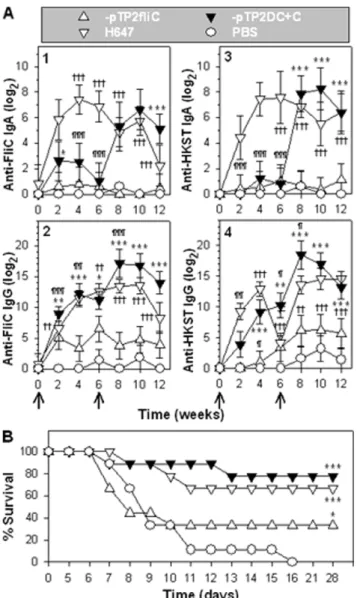
The Flagellum-attenuatedSalmonella Confer Protection against wtSalmonella Challenge
Since P1-pTP2fliC and -pTP2DC+C are not lethal, we queried whether these strains could be used as vaccines for Salmonella. Groups of mice were vaccinated with P1-pTP2fliC, -pTP2DC+C, DaroA S. Typhimurium (strain H647), or sPBS. Mice were monitored biweekly for anti-FliC and anti-HKST (heat-killed SalmonellaTyphimurium) antibody (Ab) responses. At 6 weeks post-primary immunization, mice were boosted with an additional oral dose. The vaccine strain H647-dosed mice [25] was used as a positive vaccination control, and the sPBS-dosed mice provided the negative control group. P1-pTP2DC+C induced stronger anti-FliC copro-IgA titers than those from -pTP2fliC-vaccinated mice, but less than H647 during the primary immunization phase (Figure 5A-1). After boosting, P1-pTP2DC+C induced much stronger anti-FliC titers than those of -pTP2fliC and H647. P1-pTP2DC+C and H647 induced greater anti-FliC serum IgG titers than -pTP2fliC, and after boost immunization, it showed the greatest serum IgG titers than any of the groups (Figure 5A-2). P1-pTP2DC+C and -pTP2fliC induced weaker anti-HKST copro-IgA titers than H647 during the priming phase (Figure 5A-3). After boosting, P1-pTP2DC+C induced similar titers to H647, but greater than -pTP2fliC. P1-pTP2DC+C and H647 induced similar anti-HKST serum IgG titers, but greater than -pTP2fliC during the priming phase (Figure 5A-4). Likewise after boosting, P1-pTP2DC+C induced similar anti-HKST titers to H647, but greater than -pTP2fliC. These results suggest that booster immunization is essential for P1-pTP2DC+C to elicit robust immune Ab titers. P1-pTP2DC+C is more immunogenic than -pTP2fliC regarding both the HKST and the flagellin antigens. These results suggest that flagella overexpression enhances Ab responses to FliC and HKST.
To evaluate its efficacy against wt strain challenge, these mice were subsequently orally challenged with wtS. Typhimurium H71 (Figure 5B). While the sPBS-dosed mice all succumbed to infection, mice vaccinated with P1-pTP2DC+C conferred 77.8% (7/9), H647 66.7% (6/9), and -pTP2fliC 33.3% (3/9) protection. These results show that P1-pTP2DC+C conferred the best protection against wtSalmonellainfection.
Discussion
In addition to being a virulence factor [28], flagella are involved in biofilm formation [18], accelerate bacterial invasion of host cells [29], are essential for multiplication in vivo [30], confer an advantage in the early stage of infection in animals [31], and activate the host innate immune system while inactivating apoptosis of epithelial cells [32]. Flagella also exhibit adjuvant properties acting as a TLR5 agonist [33], which render this structure a protective antigen against salmonellosis [34]. Hence, the exquisite regulation of flagella is crucial for Salmonella when interacting with the animal host. Flagellum expression has the potential to turn on to provide a great advantage in the early stage of infection [31], and then turn off to minimize host recognition
Figure 3. Overexpression of flagella attenuates Salmonella
virulence. (A) Recombinant Salmonella strains were analyzed for virulence in RAW264.7 macrophages. At all of the three bacteriatomacrophage infection ratios of 1:1, 10:1, and 100:1, P1pTP2fliC,
-pTP2flhDC, and -pTP2DC+C were not able to reproduce in
macrophag-es, while -pY did. The statistical differences in bacterial CFUs were
calculated to be ***P,0.001 for P1-pTP2fliC vs. -pY;"
P,0.05,""
P,0.01,
and"""P
,0.001 for -pTP2flhDC vs. -pY; and{{{P
,0.001 for -pTP2DC+C
vs. -pY. Depicted are the mean6SD (n = 3 independent experiments).
(B) Recombinant Salmonella strains were evaluated for ability to
colonize spleen, Peyer’s patches, and liver 4 days after oral infection. The statistical differences in tissue weights and CFUs were calculated to
be *P,0.05, **P,0.01, and ***P,0.001 for P1pTP2fliC, pTP2flhDC, or
-pTP2DC+C vs. -pY; ND: not detectible. A total of five to seven mice were
used in each group, and the depicted results are the mean6SEM. (C)
Survival fractions of the mice orally infected with 16109CFUs of
P1-pTP2fliC, -pTP2flhDC, or -pTP2DC+C were compared with that of -pY,
***P,0.001. A total of nine mice were used for P1-pTP2fliC, -pTP2flhDC,
or -pTP2DC+C, with six mice were used for -pY. Depicted are the mean
of two independent experiments. doi:10.1371/journal.pone.0046828.g003
once infection can be established [24]. Consequently, Salmonella tightly controls its flagellum expression.
Since bacterial self-preservation relies on the precise regula-tion of flagella, the absence of control of flagellum expression has formidable consequences. First, the constitutive expression of flhDC imposes a heavy metabolic burden on the cells as previously observed [35], which is evidenced by the slow growth rates of P1-pTP2flhDC versus (vs.) -pY (Figure 1D). This is because a number of pathways or genes are under the control of FlhDC [22], and as a result, their continued ‘‘on’’ status consumes much of the substrates and energy. This is further supported by the observation that co-expression of fliC and flhDC in strain P1-pTP2DC+C results in an elevated flagellin yield impeding cell growth (Figure 1D). Second, the constitutive expression of flhDC causes salmonellae to be unable to survive in water or sPBS, since the population of P1-pTP2flhDC abates more rapidly when compared to the virulence-restored control strain, -pY (Figure 2). In contrast, co-expression of flhDC and fliC in strain P1-pTP2DC+C improves survival relative to -pTP2flhDC. The reason for this outcome is not immediately clear. However, since P1-pTP2DC+C produces more flagellin than -pTP2flhDC, we may deduce that FliC has some protective effects on the -pTP2flhDC-damaged membrane. Although there were no significant differences in their sensitiv-ities to PMB, significant differences in their sensitivsensitiv-ities to hydrogen peroxide treatment were observed as evidenced by the survival rates for P1-pTP2DC+C and -pTP2flhDC being 31.0% vs. 23.0% (P,0.01), respectively, and when grown on LB agar plus 1% bile salt, the survival rates for -pTP2DC+C and -pTP2flhDC were 74.8% vs. 65.9% (P,0.05). These differences in susceptibilities to antimicrobials suggest FliC may confer
some protection. Consequently, P1-pTP2flhDC and
-pTP2DC+C are greatly attenuated as analyzed in vitro and in vivo. Third, both strains’ capabilities of infecting, surviving, and replicating within the macrophages are diminished when compared to the virulence-restored control strain, P1-pY. Likewise, their abilities to colonize mouse tissues are also compromised (Figure 3). However, P1-pTP2DC+C is nonlethal
to mice, while -pTP2flhDC still retains some virulence implicating FliC has a role in this attenuation.
FliC expression alone is sufficient to attenuate wtSalmonella. The overexpression offliCin P1-pTP2fliC imposes growth restrictions, diminishes viability in water and sPBS, reduces its capacity to infect and replicate in macrophages, and reduces its capacity to colonize mouse tissues relative to control -pY strain. In fact, all mice dosed with P1-pTP2fliC survived (Figure 3C). It seems that the AGE effects of FliC differ from FlhDC, which relates to the milieu they encounter. When evaluatedex vivo, P1-pTP2fliC had greater viability in both water and sPBS than -pTP2flhDC. When evaluated during infection, P1pTP2fliC was less viable than -pTP2flhDC as evidenced by the fewer CFUs at 8 and 24 hrs post-infection of macrophages.In vivo, P1-pTP2fliC was less virulent than -pTP2flhDC and may be linked to the former inability to colonize the liver while the latter can. Although both of these strains appeared attenuated, at least by the criterion in RAW264.7 macrophages, unlike P1-pTP2fliC, -pTP2flhDC was lethal to mice. The fact that P1-pTP2flhDC remained lethal to mice suggests that this strain may regain its virulence in vivo. This restored virulence may be attributed to enhanced degradation of FlhDCin vivo, resulting in the salmonellae recovering part of their virulence. In fact, the rapid degradation of FlhDC has been previously observed inProteus mirabilis[36].
Constitutive to its antimicrobial defense, host phagocytic cells produce hydrogen peroxide to defend against bacterial infections [37]. Secreted bile provides another antimicrobial barrier inter-fering with the bacterial membrane and being bactericidal [38]. The increased susceptibilities by the flagellatedSalmonellastrains to antimicrobial attack may account for the underlying mechanisms that these strains are attenuated.
Attenuation of wtSalmonellavia AGE rather than the virulence gene deletion has been previously demonstrated to be a useful method [25,39]. AGE-impaired strains have been found to elicit robust immune responses againstSalmonellaand confer substantial protection [25]. In this study, we further showed that overexpres-sion of a homologous antigen, FliC, is also capable of attenuating wtSalmonellato generate live vaccine, e.g., P1-pTP2DC+C, which
Figure 4. FlagellatedSalmonellastrains show enhanced sensitivity to treatments with polymyxin B (PMB), hydrogen peroxide, and bile salt.The sensitivities of the flagellatedSalmonellastrains to (A) PMB, (B) H2O2, and (C) bile salt were determined. The PMB MIC, the survival rates
after H2O2treatment or in the presence of bile salt were statistically calculated and are indicated: *P,0.05, **P,0.01, and ***P,0.001 for P1-pTP2fliC,
-pTP2flhDC, or -pTP2DC+C vs. -pY. Depicted are the mean6SEM (n = 3).
provided the best protection against wt Salmonella challenge. Hence, using AGE as a method described in this study demonstrates one alternative to impair Salmonella and other Gram-negative bacteria as an approach to generate live bacterial vaccines.
Materials and Methods
Ethics Statement
All animal care and procedures were in accordance with institutional policies for animal health and well-being and approved by Montana State University Institutional Animal Care and Use Committee under protocol 2009-30. After challenge, mice were observed for symptoms twice daily for 4 weeks. An alternate source of water such as a sterile water gel was provided if they showed retardation in reaching the water. However, if they became moribund (difficult to move to reach food and water, and combined with serious fur ruffling) they were euthanized by CO2
and recorded as death.
Bacterial Strains, Media, Plasmids, Primers and Growth Conditions
Dasd Salmonella Typhimurium strain P1 (Table 1) was trans-formed withasd+
plasmids carrying heterologous genes, and the recombinant strains were stocked at280uC. Bacterial organisms in the logarithmic growth phase were harvested from liquid lysogeny broth (LB) medium, and the cell optical density at 600 nm (OD600) was adjusted to ,0.05 with LB medium for growth rate analysis using BioScreen C at a continuous agitation of 150 rpm at 37uC for 5.5 hrs [39]. The growth rate of each strain was measured in triplicate per experiment, and each experiment was repeated three times. The susceptibilities of the recombinant Salmonellato hydrogen peroxide (2.5 mM) and bile salt (1%) were determined according to a protocol previously described [39]. The MIC of PMB was analyzed as previously described [25]. The logarithmic growth phase cells were used for field emission scanning electron microscopy (FESEM) observation for detection of flagellum expression [40]. The salmonellae were harvested after overnight growth on LB agar and resuspended in sPBS for oral immunizations, and morphological evaluations via FESEM. For each strain, 20 cells were imaged, and a representative example with the average amount of flagella is depicted.
To clone the flagellar major component encoding genefliC[41] and the master regulator encoding genesflhDC[42] either singly or co-expressed in wt Dasd Salmonella, fliC and flhDC of S. Typhimurium strain H71 were amplified by polymerase chain reaction (PCR) with primers fliC-F+fliC-R and flhDC-F+flhDC-R, respectively (Table 1). Then fliC and flhDC were inserted into pV55 [43] to generate plasmids pTP2fliC and pTP2flhDC, respectively. To co-express fliC and flhDC, fliC from pTP2fliC was inserted into pTP2flhDC to construct plasmid pTP2DC+C. Thus,fliC in pTP2fliC,flhDC in pTP2flhDC, and bothfliCand flhDC in pTP2DC+C are all under the control of the hybrid promoter of PtetA,PphoP [43]. Plasmids pTP2fliC, pTP2flhDC, and pTP2DC+C were transformed to S. Typhimurium P1 to generate P1-pTP2fliC, -pTP2flhDC, and -pTP2DC+C, respec-tively. Strain P1-pY was used as a control [25].
Western Blot Analysis
After overnight growth at 37uC on LB agar, P1pTP2fliC, -pTP2flhDC, -pTP2DC+C, and -pY cells were harvested and flagella were sheared from cells by rigorous vortexing [44]. The mouse anti-FliC monoclonal Ab (clone 6H11, Santa Cruz Biotechnology, Santa Cruz, CA) was used as the primary Ab, and the goat anti-mouse IgG HRP-conjugated Ab was used as the secondary Ab (Southern Biotechnology Associates, Birmingham, AL). The procedure was as previously described [43,45].
Figure 5. The flagellum-attenuated Salmonella strains confer protective immunity against wtSalmonellachallenge.(A) ELISA titration of anti-FliC and anti-HKST Ab endpoint titers. BALB/c mice (4–5
individuals/group) were orally immunized with 16109 CFU of
P1-pTP2fliC, -pTP2DC+C, H647, and sPBS at weeks 0 and 6. The copro-IgA
(1, 3) and serum IgG (2, 4) specific for FliC (1, 2) and HKST (3, 4) Ab titers were measured biweekly, and the significant differences are indicated:
*P,0.05, **P,0.01, and ***P,0.001 for P1-pTP2DC+C vs. -pTP2fliC;
"P
,0.05, ""P
,0.01, and """P
,0.001 for -pTP2DC+C vs. H647; and
{P
,0.05, {{P
,0.01, and {{{P
,0.001 for -pTP2fliC vs. H647. Each
experiment was repeated twice, and depicted are the means6SEM.
(B) Protective efficacy assay. Immunized mice (A) were orally
challenged with 56107CFUs of wtS. Typhimurium H71, and survival
fractions obtained from P1-pTP2fliC (n = 9 mice), -pTP2DC+C (n = 9
mice), or H647-dosed mice (n = 9 mice) were compared with
sPBS-dosed (n = 9 mice) mice and significances are indicated: ***P,0.001 for
P1-pTP2DC+C, -pTP2fliC, or H647 vs. sPBS. The depicted are the mean of
two independent experiments. doi:10.1371/journal.pone.0046828.g005
Assessment of the Flagellated Salmonellae Viability
A previous study showed that water is able to exacerbate the autolysis of E. coli [46]. Thus, using Milli-Q water to treat flagellated salmonellae may reveal whether the membranes of the flagellated salmonellae are impaired. Sterile PBS, on the other hand, is an osmotically balanced buffer and is widely used as a vehicle for oral delivery [43]. Thus, sPBS was used as a control for Milli-Q water. Additionally, three temperatures, 4uC mimicking food (produce) chilling, room temperature (,23uC) for gross maintenance [47], and 37uC, temperature mimicking normal human temperature, were used for the ex vivo viability assay.
After overnight growth on LB agar at 37uC, P1pTP2fliC, -pTP2flhDC, -pTP2DC+C, and -pY cells were diluted in Milli-Q water or sPBS to a final concentration of,3 CFUs/ml. One-ml
aliquots of each suspension were maintained at 4uC, 23uC, and 37uC, respectively, for five days. Each day, 10ml of the cell suspension was plated onto LB agar, with six repeats (10ml66) per sample. Bacterial CFUs were enumerated, and the daily survival percentages were calculated in comparison to the corresponding initial populations on day 0.
Evaluation of Infection and Replication of Flagellated Salmonellae in Macrophages
RAW264.7 macrophages (American Type Culture Collection) were used for evaluating the infection and replication of P1-pTP2fliC, -pTP2flhDC, -pTP2DC+C, and -pY. Infections, macrophage lysis, and bacterial CFU enumeration were conduct-ed as previously describconduct-ed [25].
Mouse Studies
Pathogen-free female BALB/c mice (National Cancer Insti-tute, Frederick Cancer Research Facility) 7–9 weeks of age were used throughout this study. All mice were maintained at the Montana State University Animal Resource Center under
pathogen-free conditions in individually ventilated cages under HEPA-filtered barrier conditions and were fed sterile food and water ad libitum. To assess the virulence of flagellated salmonellae, 16109 CFUs of P1pTP2fliC, pTP2flhDC, -pTP2DC+C, and -pY contained in 200ml of sPBS were fed orally by gavage to BALB/c mice previously treated with a 100ml 50% saturated sodium bicarbonate solution 30 min prior to challenge [48]. At four days post-infection, the spleens, Peyer’s patches, and livers were aseptically removed for determination of tissue weights and bacterial burden. Another group of mice dosed with the same protocol and procedure were monitored for survival for four weeks after infection, and the experiment was repeated twice. For challenge study, a dose of 56107CFUs of wt H71 contained in 200ml sPBS was given via oral gavage.
Statistical Analysis
The Tukey Kramer multiple comparisons test was used for analyzing differences among experimental parameters. The Kaplan-Meier method (GraphPad Prism, GraphPad Software, Inc., La Jolla, CA) was utilized to obtain the mouse survival fractions after mice were administered with recombinantSalmonella strains. Using the Mantel-Haenszel log rank test, theP-values for statistical differences between attenuated Salmonella strains and control wtSalmonellastrain-dosed mice were discerned at the 95% confidence interval. This method was also used for evaluating the protective efficacy after mice were challenged with wtSalmonella H71.
Acknowledgments
We appreciate Ms. Lois Avci and Ms. Nancy Kommers for their assistance in preparation of this manuscript.
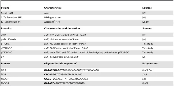
Table 1.Bacterial strains, plasmids, and primers used in this study.
Strains Characteristics Sources
E. coliH681 Dasd [49]
S. Typhimurium H71 Wild-type strain [49]
S. Typhimurium P1 Dasd::kanRH71 [25,50]
Plasmids Characteristics and derivation Sources
pV55 asd+,lcrVunder control of PtetA
,PphoP [43]
pJGX15C-asd+ asd+
,cfa/I under control of PtetA [49]
pTP2fliC asd+,
fliCunder control of PtetA,PphoP This study
pTP2flhDC asd+,flhDCunder control of PtetA
,PphoP This study
pTP2DC+C asd+
, bothflhDCandfliCunder control of PtetA,PphoP, derived from pTP2flhDC This study
pY asd+
, derived from pJGX15C-asd+
[25]
Primers Oligonucleotide sequences1 Enzyme sites
fliC-F GATATCGAGCTCGGAGGAAAAGATCATGGCACAAG EcoRI,SacI
fliC-R CTCGAGGCTCCGGAATTAAAAAAGG XhoI
flhDC-F GAGCTCGGAGGTTATTCTGGATGGGAACA SacI
flhDC-R GATATCAAGCTTACCGCTGCTGGAGTG EcoRI
Author Contributions
Conceived and designed the experiments: XY DWP. Performed the experiments: XY TT ZS SJ AR JL TL LC TH RA. Analyzed the data: XY
DWP. Contributed reagents/materials/analysis tools: DWP RA XY. Wrote the paper: XY DWP.
References
1. Coburn B, Grassl GA, Finlay BB (2007)Salmonella, the host and disease: a brief review. Immunol Cell Biol 85: 112–118.
2. Bell RL, Gonzalez-Escalona N, Stones R, Brown EW (2011) Phylogenetic evaluation of the ‘Typhimurium’ complex ofSalmonellastrains using a seven-gene multi-locus sequence analysis. Infect Genet Evol 11: 83–91.
3. CDC (2007) Preliminary FoodNet data on the incidence of infection with pathogens transmitted commonly through food–10 states, 2006. MMWR Morb Mortal Wkly Rep 56: 336–339.
4. Pang T, Levine MM, Ivanoff B, Wain J, Finlay BB (1998) Typhoid fever– important issues still remain. Trends Microbiol 6: 131–133.
5. Kennedy M, Villar R, Vugia DJ, Rabatsky-Ehr T, Farley MM, et al. (2004) Hospitalizations and deaths due toSalmonellainfections, FoodNet, 1996–1999. Clin Infect Dis 38 Suppl 3: S142–148.
6. Threlfall EJ (2000) EpidemicSalmonella typhimuriumDT 104 - a truly international multiresistant clone. J Antimicrob Chemother 46: 7–10.
7. Varma JK, Greene KD, Ovitt J, Barrett TJ, Medalla F, et al. (2005) Hospitalization and antimicrobial resistance in Salmonella outbreaks, 1984– 2002. Emerg Infect Dis 11: 943–946.
8. Fraser A, Paul M, Goldberg E, Acosta CJ, Leibovici L (2007) Typhoid fever vaccines: Systematic review and meta-analysis of randomised controlled trials. Vaccine 25: 7848–7857.
9. Fraser A, Goldberg E, Acosta CJ, Paul M, Leibovici L (2009) Vaccines for preventing typhoid fever. Cochrane Database of Systematic Reviews: 1–54. 10. Levine MM, Ferreccio C, Abrego P, Martin OS, Ortiz E, et al. (1999) Duration
of efficacy of Ty21a, attenuatedSalmonella typhilive oral vaccine. Vaccine 17 Suppl 2: S22–27.
11. Viret JF, Favre D, Wegmuller B, Herzog C, Que JU, et al. (1999) Mucosal and systemic immune responses in humans after primary and booster immunizations with orally administered invasive and noninvasive live attenuated bacteria. Infect Immun 67: 3680–3685.
12. Marcus LC, Froeschle JE, Hill DR, Wolfe MS, Maus D, et al. (2007) Safety of typhim Vi vaccine in a postmarketing observational study. J Travel Med 14: 386–391.
13. Namgyal P (2011) Typhoid Vaccines - Ad-hoc consultation on typhoid vaccine introduction and typhoid surveillance, Bangkok Meeting on Typhoid. April 18– 20, 2011: World Health Organization.
14. Ibarra JA, Steele-Mortimer O (2009)Salmonella–the ultimate insider.Salmonella virulence factors that modulate intracellular survival. Cell Microbiol 11: 1579– 1586.
15. Schmitt CK, Ikeda JS, Darnell SC, Watson PR, Bispham J, et al. (2001) Absence of all components of the flagellar export and synthesis machinery differentially alters virulence ofSalmonella entericaserovar Typhimurium in models of typhoid fever, survival in macrophages, tissue culture invasiveness, and calf enterocolitis. Infect Immun 69: 5619–5625.
16. Franchi L, Amer A, Body-Malapel M, Kanneganti TD, Ozoren N, et al. (2006) Cytosolic flagellin requires Ipaf for activation of caspase-1 and interleukin 1ß in Salmonella-infected macrophages. Nat Immunol 7: 576–582.
17. Miao EA, Alpuche-Aranda CM, Dors M, Clark AE, Bader MW, et al. (2006) Cytoplasmic flagellin activates caspase-1 and secretion of interleukin 1ß via Ipaf. Nat Immunol 7: 569–575.
18. Santos PG, Santos PA, Bello AR, Freitas-Almeida AC (2011) Association of Aeromonas caviae polar and lateral flagella with biofilm formation. Lett Appl Microbiol 52: 49–55.
19. Shimoyama T, Kato S, Ishii S, Watanabe K (2009) Flagellum mediates symbiosis. Science 323: 1574.
20. Wang Q, Suzuki A, Mariconda S, Porwollik S, Harshey RM (2005) Sensing wetness: a new role for the bacterial flagellum. EMBO J 24: 2034–2042. 21. Kawagishi I, Imagawa M, Imae Y, McCarter L, Homma M (1996) The
sodium-driven polar flagellar motor of marineVibrioas the mechanosensor that regulates lateral flagellar expression. Mol Microbiol 20: 693–699.
22. Anderson JK, Smith TG, Hoover TR (2010) Sense and sensibility: flagellum-mediated gene regulation. Trends Microbiol 18: 30–37.
23. Heuner K, Brand BC, Hacker J (1999) The expression of the flagellum of Legionella pneumophilais modulated by different environmental factors. FEMS Microbiol Lett 175: 69–77.
24. Sano G, Takada Y, Goto S, Maruyama K, Shindo Y, et al. (2007) Flagella facilitate escape ofSalmonellafrom oncotic macrophages. J Bacteriol 189: 8224– 8232.
25. Yang X, Suo Z, Thornburg T, Holderness K, Cao L, et al. (2012) Expression of Escherichia colivirulence usher protein attenuates wild-typeSalmonella. Virulence 3: 29–41.
26. Hancock RE (1997) Peptide antibiotics. Lancet 349: 418–422.
27. Sikora AE, Lybarger SR, Sandkvist M (2007) Compromised outer membrane integrity inVibrio choleraeType II secretion mutants. J Bacteriol 189: 8484–8495. 28. Winter SE, Thiennimitr P, Nuccio SP, Haneda T, Winter MG, et al. (2009) Contribution of flagellin pattern recognition to intestinal inflammation during Salmonella enterica serotype Typhimurium infection. Infect Immun 77: 1904– 1916.
29. van Asten FJ, Hendriks HG, Koninkx JF, van Dijk JE (2004) Flagella-mediated bacterial motility accelerates but is not required forSalmonellaserotype Enteritidis invasion of differentiated Caco-2 cells. Int J Med Microbiol 294: 395–399. 30. Cogan TA, Jorgensen F, Lappin-Scott HM, Benson CE, Woodward MJ, et al.
(2004) Flagella and curli fimbriae are important for the growth ofSalmonella entericaserovars in hen eggs. Microbiology 150: 1063–1071.
31. Robertson JM, McKenzie NH, Duncan M, Allen-Vercoe E, Woodward MJ, et al. (2003) Lack of flagella disadvantagesSalmonella entericaserovar Enteritidis during the early stages of infection in the rat. J Med Microbiol 52: 91–99. 32. Vijay-Kumar M, Wu H, Jones R, Grant G, Babbin B, et al. (2006) Flagellin
suppresses epithelial apoptosis and limits disease during enteric infection. Am J Pathol 169: 1686–1700.
33. Smith KD, Andersen-Nissen E, Hayashi F, Strobe K, Bergman MA, et al. (2003) Toll-like receptor 5 recognizes a conserved site on flagellin required for protofilament formation and bacterial motility. Nat Immunol 4: 1247–1253. 34. Bergman MA, Cummings LA, Alaniz RC, Mayeda L, Fellnerova I, et al. (2005)
CD4+
-T-cell responses generated during murine Salmonella enterica serovar Typhimurium infection are directed towards multiple epitopes within the natural antigen FliC. Infect Immun 73: 7226–7235.
35. Glick BR (1995) Metabolic load and heterologous gene expression. Biotechnol Adv 13: 247–261.
36. Claret L, Hughes C (2000) Rapid turnover of FlhD and FlhC, the flagellar regulon transcriptional activator proteins, duringProteusswarming. J Bacteriol 182: 833–836.
37. Douglass II WC (2003) Hydrogen Peroxide: Medical Miracle, Rhino Publishing, S.A.; New Edition edition (June 26, 2003). Page 19.
38. Merritt ME, Donaldson JR (2009) Effect of bile salts on the DNA and membrane integrity of enteric bacteria. J Med Microbiol 58: 1533–1541. 39. Cao L, Lim T, Jun S, Thornburg T, Avci R, et al. (2012) Vulnerabilities in
Yersinia pestis cafoperon are unveiled by aSalmonellavector. PLoS One 7: e36283. 40. Suo Z, Yang X, Avci R, Deliorman M, Rugheimer P, et al. (2009) Antibody
selection for immobilizing living bacteria. Anal Chem 81: 7571–7578. 41. Inoue YH, Kutsukake K, Iino T, Yamaguchi S (1989) Sequence analysis of
operator mutants of the phase-1 flagellin-encoding gene, fliC, in Salmonella typhimurium. Gene 85: 221–226.
42. Tomoyasu T, Takaya A, Isogai E, Yamamoto T (2003) Turnover of FlhD and FlhC, master regulator proteins forSalmonellaflagellum biogenesis, by the ATP-dependent ClpXP protease. Mol Microbiol 48: 443–452.
43. Yang X, Hinnebusch BJ, Trunkle T, Bosio CM, Suo Z, et al. (2007) Oral vaccination with Salmonellasimultaneously expressingYersinia pestisF1 and V antigens protects against bubonic and pneumonic plague. J Immunol 178: 1059– 1067.
44. Bahrani FK, Johnson DE, Robbins D, Mobley HL (1991)Proteus mirabilisflagella and MR/P fimbriae: isolation, purification, N-terminal analysis, and serum antibody response following experimental urinary tract infection. Infect Immun 59: 3574–3580.
45. Cao L, Suo Z, Lim T, Jun S, Deliorman M, et al. (2012) Role of overexpressed CFA/I fimbriae in bacterial swimming. Phys Biol 9: 036005.
46. Leduc M, van Heijenoort J (1980) Autolysis ofEscherichia coli. J Bacteriol 142: 52– 59.
47. Kinsella KJ, Rowe TA, Blair IS, McDowell DA, Sheridan JJ (2006) Survival and recovery ofSalmonella entericaserovar Typhimurium DT104 at low temperature and water activity in a broth system. Foodborne Pathog Dis 3: 375–383. 48. Yang X, Thornburg T, Holderness K, Suo Z, Cao L, et al. (2011) Serum
antibodies protect against intraperitoneal challenge with enterotoxigenic Escherichia coli. J Biomed Biotechnol 2011: 632396.
49. Wu S, Pascual DW, VanCott JL, McGhee JR, Maneval DR, Jr., et al. (1995) Immune responses to novelEscherichia coliandSalmonella typhimuriumvectors that express colonization factor antigen I (CFA/I) of enterotoxigenicE. coliin the absence of the CFA/I positive regulatorcfaR. Infect Immun 63: 4933–4938. 50. Suo Z, Avci R, Yang X, Pascual DW (2008) Efficient immobilization and
patterning of live bacterial cells. Langmuir 24: 4161–4167.