in Human, Mouse and Rat T Lymphocytes
Suresh K. Mendu., Amol Bhandage., Zhe Jin, Bryndis Birnir*
Molecular Physiology and Neuroscience, Department of Neuroscience, Uppsala University, Uppsala, Sweden
Abstract
c-aminobutyric acid (GABA) is the most prominent neuroinhibitory transmitter in the brain, where it activates neuronal GABA-A receptors (GABA-A channels) located at synapses and outside of synapses. The GABA-A receptors are primary targets of many clinically useful drugs. In recent years, GABA has been shown to act as an immunomodulatory molecule. We have examined in human, mouse and rat CD4+and CD8+T cells which subunit isoforms of the GABA-A channels are
expressed. The channel physiology and drug specificity is dictated by the GABA-A receptor subtype, which in turn is determined by the subunit isoforms that make the channel. There were 5, 8 and 13 different GABA-A subunit isoforms identified in human, mouse and rat CD4+ and CD8+ T cells, respectively. Importantly, the c2 subunit that imposes
benzodiazepine sensitivity on the GABA-A receptors, was only detected in the mouse T cells. Immunoblots and immunocytochemistry showed abundant GABA-A channel proteins in the T cells from all three species. GABA-activated whole-cell transient and tonic currents were recorded. The currents were inhibited by picrotoxin, SR95531 and bicuculline, antagonists of GABA-A channels. Clearly, in both humans and rodents T cells, functional GABA-A channels are expressed but the subtypes vary. It is important to bear in mind the interspecies difference when selecting the appropriate animal models to study the physiological role and pharmacological properties of GABA-A channels in CD4+and CD8+T cells and when
selecting drugs aimed at modulating the human T cells function.
Citation:Mendu SK, Bhandage A, Jin Z, Birnir B (2012) Different Subtypes of GABA-A Receptors Are Expressed in Human, Mouse and Rat T Lymphocytes. PLoS ONE 7(8): e42959. doi:10.1371/journal.pone.0042959
Editor:Kjetil Tasken, University of Oslo, Norway
ReceivedJanuary 31, 2012;AcceptedJuly 16, 2012;PublishedAugust 21, 2012
Copyright:ß2012 Mendu et al. This is an open-access article distributed under the terms of the Creative Commons Attribution License, which permits unrestricted use, distribution, and reproduction in any medium, provided the original author and source are credited.
Funding:Human pancreatic lymph nodes were obtained from The Nordic network for Clinical Islet Transplantation, supported by the Swedish national strategic research initiative EXODIAB (Excellence of Diabetes Research in Sweden) and the Juvenile Diabetes Research Foundation. The authors thank the Swedish Research Council, Swedish Diabetes Research Foundation, The Ernfors Foundation, Uppsala University and EXODIAB for financial support. ZJ held a postdoctoral fellowship sponsored by EXODIAB and The Swedish Association for Medical Research (SSMF). The funders had no role in study design, data collection and analysis, decision to publish, or preparation of the manuscript.
Competing Interests:The authors have declared that no competing interests exist.
* E-mail: [email protected]
.These authors contributed equally to this work.
Introduction
The GABA-A receptor is an ion channel that opens whenc -aminobutyric acid (GABA) binds to its binding site on the receptor complex. In the brain, GABA is the most important neuroinhi-bitory transmitter. It is released from neuronal presynaptic terminals and activates the GABA-A channels located at the postsynaptic site [1]. GABA in low, submicromolar concentrations is also present around the neurons where it activates high-affinity GABA-A channels located outside of synapses [1,2]. These channels are called extrasynaptic GABA-A channels [3]. Although the focus over the years has been on the GABA-A channels in the brain, there is growing evidence indicating a significant, physio-logical function of GABA and GABA-A channels in a number of non-neuronal tissue [4]. Components of the GABA signaling systems have also been identified in cells of the immune system [5,6,7,8,9,10,11,12,13].
The GABA-A ion channel is a pentameric chloride channel that is commonly made of three different types of subunits. To-date 19 different mammalian GABA-A subunits have been cloned (a1–6,
b1–3,c1–3,d,e,p,h,r1–3) [14]. The subunits can combine in many different arrangements to form the pentameric channel. This is important as the specific subunits in the channel complex
determine the pharmacological specificity of the channel [14]. It has been shown that benzodiazepine-site ligands can differentiate between GABA-A channel subtypes based on the type ofaandc
subunits present in the channel complex [1]. Other drugs like the general anaesthetics and even GABA have been used to extend the list of subtypes further as their affinity is also related to the subunit composition of the channels [1]. In immune cells, the GABA-A channels are not located in a synapse and are only exposed to low concentrations of GABA similar to extrasynaptic GABA-A channels in neurons.
GABA is present in blood in submicromolar concentration [15,16] and may be produced by the immune cells themselves [7,17]. Immune cells such as CD4+
, CD8+
chemo-taxis [7,8,11,13,18,19,20]. As the functional and pharmacological properties are dictated by the pentameric subunit-composition of the GABA-A channel complex, it is important to know the subtypes in the cells when selecting drugs to act on the channels. A number of drugs modulating the functional properties of GABA-A channels are widely used in clinical settings. These drugs include the general anesthetics and benzodiazepines that differ in their selectivity at GABA-A channels. In order to minimize side-effects involving the immune system when these drugs are used, it is essential to know which GABA-A subtypes are formed in the immune cells. Furthermore, when selecting the appropriate experimental animal model, it is desirable that it expresses similar channel subtypes to their human counterpart. T lymphocytes have a central role in the cell-mediated immune response of the adaptive immune system. We have previously shown that 10 of the 19 GABA-A channel subunit mRNAs (a1,a2,a3,a4,a6,b3,c1,
d, r1, r2) are expressed in both CD4+
and CD8+
T cells from biobreeding (BB) rats [18]. Here we examined the interspecies variability in terms of expression of the 19 GABA-A channel subunit isoforms in CD4+
and CD8+
T cells from humans, rats and mice. It is the first time all potential GABA-A channel subtypes in native T cells have been examined and compared for the three species. The results show that different combinations of GABA-A channel subunit isoforms are expressed in T cells from humans, rats and mice.
Results
Differential expression of GABA-A channel subunit mRNAs in human, rat and mouse CD4+and CD8+T cells and Jurkat cells
Here we examined whether the 19 different GABA-A channel subunit mRNAs were present in CD4+and CD8+T cells from rats
(Wistar), mice (C57BL/6J) and humans as well as in a human CD4+T cell line, the Jurkat cells. Primers specific for each
GABA-A channel subunit (Table 1) were verified using the appropriate brain cDNA samples from humans, rats or mice.
In both CD4+
and CD8+
T cells isolated from mesenteric lymph nodes from Wistar rats, 13 GABA-A channel subunit mRNAs were detected (Fig. 1A and Table 2). There was an abundant expression of the a1, a2, a3, a4, a6, b3, c1 and r2 subunits, modest expression of theh,r1 andr3 subunits and low expression of theb2 andpsubunits. The mRNA levels of these 13 GABA-A channel subunits did not differ between the CD4+and CD8+T
cells (Fig. 1A). In CD4+
and CD8+
T cells isolated from mesenteric lymph nodes from C57BL/6J mice (Fig. 1B and Table 2), 8 GABA-A channel subunit mRNAs were consistently detected in both cell types. There was abundant expression of thea2,a3,a5,c2 andd
subunits, modest expression of the b2 andc1 subunits and low expression of theb3 subunit. The mRNA levels of these 8 subunits did not differ between the CD4+
and CD8+
T cells (Fig. 1B). Interestingly, we detected only 5 GABA-A channel subunit mRNAs in both CD4+
and CD8+
T cells isolated from human pancreatic lymph nodes (Fig. 1C and Table 2). These were thea1,
a5,b1,p and r2 subunits and the mRNA levels did not differ between the CD4+
and CD8+ T cells (Fig. 1C). Using the same human GABA-A primer sets and reference gene (Table 1), 9 GABA-A channels subunit mRNAs including thea1,a3,a5,a6,
b1,b2,b3,p andr2 subunits were detected in Jurkat cells, the human T cell lymphoblast-like cell line (Fig. 1D and Table. 2). The mRNA expression levels for theb1 andr2 subunits in Jurkat cells were significantly lower than those in the CD4+
or CD8+
human T cells (Kruskal-Wallis one-way ANOVA on ranks, post hoc Tukey test, p,0.05)
Thec2 subunit confers specific pharmacology on the GABA-A channel complex and was only detected in samples from the mouse T cells where it was abundantly expressed (Fig. 1B). We, therefore, designed additional primer pairs specific for the rat or the humanc2 subunit (Table 1) to further examine whether thec2 subunit could be detected in the rat and human T cells. These primers were designed to amplify all splice variants ofc2 transcript and targeted different regions of thec2 transcript from those used for the quantitative PCR. These additional primers were also verified using the rat and human brain samples, respectively. However, noc2 subunit mRNA transcript was detected in the rat and the human T cells or the Jurkat cells with these additional primer sets (data not shown).
GABA-A channel subunit proteins in the native lymphocytes and the Jurkat cell line
Western blot analysis was used to confirm the presence of the GABA-A channel subunit proteins in rat and mouse CD4+and
CD8+
T cells and in the Jurkat cell line (Fig. 2). Experiments where the primary antibody was omitted served as negative controls and resulted in no specific bands at the predicted molecular weights in the blots. Protein extracts from rat and mouse brains served as positive controls whereas b-actin served as a control for protein loading.
In rat CD4+
and CD8+
T cells, the mRNAs of several alpha subunits (a1, a2, a3, a4 and a6) were detected by RT-qPCR. Therefore, we first used an antibody, GABA-A a1–6, that is reported to recognize all GABA-A channelsaisoforms of mouse, rat and human origins. This polyclonal antibody was raised against amino acids 157–456 near the C-terminus of the human GABA-A a1 protein. This region is highly conserved: in rat GABA-Aa1 (100%),a2 (80%),a3 (71%),a4 (76%),a5 (71%) and
a6 (55%) proteins. The predicted molecular weight for the rata
GABA-A subunits ranges from 51 to 62 kDa. In a Western blot several bands were detected within the expected molecular weight range using protein extracts from rat brain (4 bands), rat CD4+(2
bands) and rat CD8+
(2 bands) T cells (Fig. 2A). Surprisingly only a faint band was detected in the brain sample in the expected molecular range for the a1 subunit, which is the most highly expressedasubunit in the brain. We, therefore, further examined the a1 isoform using an a1-specific antibody that was raised against amino acids 28–43 of the rat GABA-Aa1 protein. This antigen peptide sequence is also conserved in the human GABA-A
a1 protein. Fig. 2B shows that a single band was detected at the expected molecular weight of,52 kDa in protein extracts from
rat CD4+, CD8+T cells and Jurkat cells as well as in protein
extracts from rat brain that served as a positive control. However, some nonspecific bands were observed in the blot as well (data not shown).
The GABA-A b3 subunit mRNA was the most abundant b
subunit isoform in rat CD4+and CD8+T cells as determined by
the RT-qPCR. By using a GABA-Ab3 antibody, a single band was detected at the expected molecular weight of ,54 kDa in
protein extracts from rat CD4+, CD8+ T cells and Jurkat cells
(Fig. 2C). The molecular weight of the three differentbsubunits ranges from 54.1 to 54.6 kDa and all b-subunit antibodies available to-date show some cross reactivity for the three isoforms. In the protein extracts from rat brain there were 2 bands (,54 kDa), which may indicate different b subunits isoforms,
Table 1.Human, rat and mouse primers list for quantitative real-time RT-PCR.
Gene name Forward primer (59-39) Reverse primer (59- 39)
Product size (bp)
Human
a1 (GABRA1) GGATTGGGAGAGCGTGTAACC TGAAACGGGTCCGAAACTG 66
a2 (GABRA2) GTTCAAGCTGAATGCCCAAT ACCTAGAGCCATCAGGAGCA 160
a3 (GABRA3) CAACTTGTTTCAGTTCATTCATCCTT CTTGTTTGTGTGATTATCATCTTCTTAGG 102
a4 (GABRA4) TTGGGGGTCCTGTTACAGAAG TCTGCCTGAAGAACACATCCA 105
a5 (GABRA5) CTTCTCGGCGCTGATAGAGT CGCTTTTTCTTGATCTTGGC 105
a6 (GABRA6) ACCCACAGTGACAATATCAAAAGC GGAGTCAGGATGCAAAACAATCT 67
b1 (GABRB1) CCAGGTCGACGCCCACGGTA GTGGCCTTGGGGTCGCTCAC 102
b2 (GABRB2) GCAGAGTGTCAATGACCCTAGT TGGCAATGTCAATGTTCATCCC 137
b3 (GABRB3) CCGTTCAAAGAGCGAAAGCAACCG TCGCCAATGCCGCCTGAGAC 105
c1 (GABRG1) CCTTTTCTTCTGCGGAGTCAA CATCTGCCTTATCAACACAGTTTCC 91
c2 (GABRG2) CACAGAAAATGACGGTGTGG TCACCCTCAGGAACTTTTGG 136
c2 (GABRG2)-2* AGCAACCGGAAACCAAGC TCCATTTTGGCAATGCGG 269/245
c2 (GABRG2)-3 AACATGGTGGGGAAAATCTG GGCAGGAGTGTTCATCCATT 196
e(GABRE) TGGATTCTCACTCTTGCCCTCTA GGAGTTCTTCTCATTGATTTCAAGCT 107
h(GABRQ) CCAGGGTGACAATTGGCTTAA CCCGCAGATGTGAGTCGAT 63
p(GABRP) GGCCTTGCTAGAATATGCAGTTG CTTTGTTGTCCCCCTATCTTTGG 76
r1 (GABRR1) Hs00266687_m1 from AppliedBiosystem Hs00266687_m1 from AppliedBiosystem 94
r2 (GABRR2) CCTAGAAGAGGGCATAGACATCG TCCAGTAGCTGCTGCATTGTTTG 99
r3 (GABRR3) TGATGCTTTCATGGGTTTCA CGCTCACAGCAGTGATGATT 111
B2M CCTGCCGTGTGAACCATGTGACT GCGGCATCTTCAAACCTCCATGATG 94
Rat
a1 (Gabra1) CTCCTACAGCAACCAGCTATACCC GCGGTTTTGTCTCAGGCTTGAC 113
a2 (Gabra2) AAGAGAAAGGCTCCGTCATG GCTTCTTGTTTGGTTCTGGAGTAG 134
a3 (Gabra3) ACAAGCACCACCTTCAACATAG AGGTCTTGGTCTCAGCAGGA 174
a4 (Gabra4) GATGTCAACAGCAGAACTGAGGTG TTGTGCCAGATCCAGAAGGTGGTG 345
a5 (Gabra5) GCCTTGGAAGCAGCTAAAATC GAAGTCTTCTCCTCAGATGCTCT 178
a6 (Gabra6) CACTCTGACTCCAAGTACCATCTG GTACACAAGGTTGAATCCTG 221
b1 (Gabrb1) CCCTCAGAAAAAAGGAGCGA TCACGGCTGCTCAGTGGTTT 231
b2 (Gabrb2) GCCTGGATGTCAACAAGATGGACC CTAGGCAACCCAGCTTTCCGATAC 169
b3 (Gabrb3) CCTACTAGCACCGATGGATGTT GATGCTTCTGTCTCCCATGTAC 163
c1 (Gabrg1) TGACACGTTCTTCAGGAACTCAA AACCCTTCCATCACTCCATATCC 91
c2 (Gabrg2) CGGAAACCAAGCAAGGAT TCTCTTGAAGGTGGGTGGCA 134
c2(Gabrg2)-2 CGGCCCGACATAGGAGTGAAACC TCGTGATCCAGTGAGCATCCGC 250
c2 (Gabrg2)-3 CGCTCTACCCAGGCTTCACTAGC TCGGGCCGAAGTTTGTTGTCGT 157
e(Gabre) CAGATGGCTCTCATCCATAAGGA GCTAGAGAAAGACAGAGGGCAAGA 129
h(Gabrq) TAGGACTTGGCTGGCAGAGAGTA ATGCTGGAGGAGAGCTCGAA 72
p(Gabrp) TGACAACAGTGTTGTCCATGACA TGGCCTTTATGAAGCAGTTGGT 81
r1 (Gabrr1) TGGACAGCAGCTACAGTCACGG AAGCAGCTGGGAAAATGATC 209
r2 (Gabrr2) CAAGAAGCCACATTCTTCCA TTCTGGAAGATATAGAGTCC 133
r3 (Gabrr3) GGTGTGAGCGCCTCTATGC GGGAGCTGACCCACATGTACA 70
Hprt CTCATGGACTGATTATGGACAGGAC GCAGGTCAGCAAAGAACTTATAGCC 123
Mouse
a1 (Gabra1) AAA AGC GTG GTT CCA GAA AA GCT GGT TGC TGT AGG AGC AT 84
a2 (Gabra2) GCTACGCTTACACAACCTCAGA GACTGGCCCAGCAAATCATACT 117
a3 (Gabra3) GCCGTCTGTTATGCCTTTGTATTT TTCTTCATCTCCAGGGCCTCT 118
a4 (Gabra4) AGAACTCAAAGGACGAGAAATTGT TTCACTTCTGTAACAGGACCCC 118
a5 (Gabra5) GATTGTGTTCCCCATCTTGTTTGGC TTACTTTGGAGAGGTGGCCCCTTTT 100
a6 (Gabra6) GGTGACCGGGCATCCCAGTGA TGTTACAGCACCCCCAAATCCTGGC 197
The GABA-Ac2 subunit mRNA was detected only in mouse T cells. To examine whether the GABA-A c2 subunit protein is present in mouse T cells, a GABA-Ac2 specific antibody raised against amino acids 39–67 of the mouse GABA-Ac2 protein was used. This antigen peptide sequence is also 100% conserved in the human and rat GABA-A c2 protein. Fig. 2 shows that a single major band was detected at the molecular weight of,49 kDa in
protein extracts from mouse brain (Fig. 2D and E) and mouse CD4+
and CD8+
T cells (Fig. 2D, E) but some minor nonspecific bands could also be detected (Fig. 2E). The predicted molecular weight of mouse GABA-A c2 protein is 54 kDa, however, the cleavage of the signal peptide (the first 38 amino acids) leads to a 49 kDa c2 protein. The specific band was abolished by pre-absorption of the antibody with the synthetic GABA-A c2 immunogenic peptide (Fig. 2E and F).
Localization of GABA-A subunit proteins in native T lymphocytes and the Jurkat cell line
The Western blot results confirmed the presence of several GABA-A subunit proteins in T cells and Jurkat cells. We further examined the cellular localization of the GABA-A subunits by immunocytochemistry. Negative controls where the primary antibody was omitted were devoid of immunostaining (data not shown).
In rat CD4+
and CD8+
T cells somewhat punctate immuno-fluorescent staining of the GABA-A a1, a2 and b subunit was observed (Fig. 3A and 3B). Similar staining patterns were observed for the GABA-Aa1 subunit in human CD4+
and CD8+
T cells (Fig. 3C) and for the GABA-Aa2 and thec2 subunits in mouse CD4+
and CD8+
T cells (Fig. 3E). The GABA-A c2 specific immunostaining in mouse T cells was blocked by pre-absorption of the antibody with c2 immunogenic peptide (Fig. 3F). In Jurkat cells (Fig. 3D), the immunostaining for the GABA-Aa1 andb3 appeared more diffuse throughout the cytoplasm.
In order to examine the cellular location of these GABA-A channel subunits, we co-labeled the membrane of rat CD4+
and Jurkat cells with a lipophilic dye (DiI red). The dye labeled the
plasma membrane for both cell-types and in some cells, gained entry into the cells (Fig. 4A and B). The b3-antibody and a 1-antibody subunit labeling was present in the plasma membrane of rat CD4+
and Jurkat cells demonstrating that GABA-A receptors do reach the plasma membrane (Fig. 4A and B).
GABA-activated currents in T lymphocytes
We further examined if functional GABA-A channels could be detected in the T cells and Jurkat cells using the patch-clamp technique. We recorded whole-cell GABA-activated transient or tonic currents from rat CD4+(n = 6) and CD8+(n = 5) T cells and
Jurkat cells (n = 6) (Fig. 5). At a negative holding potential (280 mV) in chloride solutions (ECl=220 mV), 1mM or 1 mM
GABA application to the cells resulted in an inward, transient current where the peak amplitude ranged from21.2 to26.8 nA (n = 7, holding potential =280 mV, Fig. 5 A–D). Surprisingly, from each cell we often only recorded one current response. Subsequent applications of GABA resulted in no current activation indicating an extensive run-down of the response in the cells. The currents were outward at positive holding potentials and blocked by 100mM picrotoxin (+40 mV, Fig. 5E, n = 2) or 100mM bicuculline (n = 1, not shown). Extrasynaptic GABA-A channels generate small amplitude but long-lasting currents in neurons that is termed tonic current. We examined if we could activate tonic currents in the T cells. Tonic currents were activated with 1mM or 1 mM GABA and inhibited with 100mM SR95531 (holding potential =+40 mV, Fig. 5F) in rat CD4+(n = 2) and CD8+(n = 3)
T cells or 100mM bicuculline (Jurkat cells, holding poten-tial =260 mV, n = 2, 5G). The whole-cell current results show that T cells and Jurkat cells express functional GABA-A channels that respond with transient or tonic currents when exposed to GABA.
Discussion
Our results demonstrate that human, mouse and rat CD4+and
CD8+
T cells express GABA-A channel mRNAs that are translated into proteins that form functional channels in the
Table 1.Cont.
Gene name Forward primer (59-39) Reverse primer (59- 39)
Product size (bp)
b2 (Gabrb2) GCTGGTGAGGAAATCTCGGTCCC CATGCGCACGGCGTACCAAA 70
b3 (Gabrb3) GAGCGTAAACGACCCCGGGAA GGGACCCCCGAAGTCGGGTCT 100
c1 (Gabrg1) ATCCACTCTCATTCCCATGAACAGC ACAGAAAAAGCTAGTACAGTCTTTGC 100
c2 (Gabrg2) ACTTCTGGTGACTATGTGGTGAT GGCAGGAACAGCATCCTTATTG 147
c3 (Gabrg3) ATTACATCCAGATTCCACAAGATG CAC AGG TGT CCT CAA ATT CCT 149
d(Gabrd) TCAAATCGGCTGGCCAGTTCCC GCACGGCTGCCTGGCTAATCC 147
e(Gabre) ACTGCGCCCTGGCATTGGAG AGGCCCGAGGCTGTTGACAA 70
h(Gabrq) GCTGGAGGTGGAGAGCTATGGCT CCCCAGGTACGTGTACTGAGGGA 115
p(Gabrp) TCGGTGGTGACCCAGTTCGGAT TCTGTCCAACGCTGCCGGAG 115
p(Gabrp) TCGGTGGTGACCCAGTTCGGAT TCTGTCCAACGCTGCCGGAG 115
r1 (Gabrr1) CCATCTAGGAAAGGCAGCAG GAGCTTCGTCTCAGGATTGG 92
r2 (Gabrr2) GCTGCCTGTTGCATCATAGA ATACAAATGGCTTGGCTTGG 153
r3 (Gabrr3) CAACTCAACAGGAGGGGAAA TCCACATCAGTCTCGCTGTC 101
b-actin(Actb) GTCCACACCCGCCACCAGTTCG ATGCCGGAGCCGTTGTCGAC 71
plasma membrane of the cells. CD4+
and CD8+
T cells express the same GABA-A subunits but the specific isoforms differ between the species. Five, eight and thirteen different types of GABA-A subunits were detected in the T cells from humans, C57BL/6J mice and Wistar rats, respectively. Each species expressed at least two a subunit isoforms, one or more types of b subunits but differed widely in what other types of subunits were expressed. The different profile of subunit isoforms expressed in the cells from humans, mice and rats is highly significant as it demonstrates that the GABA-A channel subtypes will vary according to species. The Western blots and immunostaining images demonstrated that the GABA-A channel proteins are abundant and located in the plasmalemma and throughout the cytoplasm of the CD4+
and the CD8+
T cells. The staining pattern often appeared punctate, possibly indicating clustering of channels in vesicles or the plasma membrane. GABA evoked transient and tonic currents in the cells, somewhat similar to what is recorded in neurons.
There have been relatively few studies that have examined the expression of the GABA-A channels in immune cells [5]. The GABA-A transcripts are often present in the cells but it varies
which subunits have been detected.a1,a2,b1,b2,c3 anddwere identified in CD4+
T cells from the type-1 diabetic NOD (non-obese diabetic) mice [11] whereas in CD4+
T cells from an experimental autoimmune encephalomyelitis (EAE) mouse model
a1,b1,c2 andewere examined but not detected [21].a1,a4,b2,
b3,c1 anddwere detected in an EAE cell line [8] and in CD4+
and CD8+
T cells from Biobreeding (BB) rats,a1,a2,a3,a4,a6,
b3,c1,d,r1 andr2 were identified [18]. In these studies, only in two cases [8,18] have all 19 subunits been examined. It is, therefore, possible that more subunit isoforms can be detected in the T cells from the mouse models. However, the NOD mouse expressed thec3 subunit that we did not detect in the C57BL/6J mouse model used in this study and the combination of subunits expressed in the BB rats differ from the Wistar rats in this study. Whether it is the strain of animals or possibly the state of activation of the cells that regulates the subunit isoforms expression pattern remains to be determined.a1,a2,b3 anddhave been detected in cultured peritoneal macrophages andb1 andefrom macrophages isolated from an EAE mouse model [21]. Human peripheral monocytes have been reported to express thea1,a3,a4,b2,b3,d Figure 1. Expression of GABA-A channel subunit mRNAs in CD4+or CD8+T cells from rats, mice and human donors and the Jurkat cell line.Expression of all 19 GABA-A receptor channel subunit isoforms was examined using RT-qPCR in T cells. Subunit isoforms that were detected are shown in A–D. In CD4+(open bar) and CD8+(grey bar) T cells from mesenteric lymph nodes of Wistar rats (A, n = 4) or C57BL/6J mice (B, n = 3), 13 and 8 different GABA-A channel subunit mRNAs were detected, respectively. The mRNA expression level for each subunit did not differ between the CD4+and the CD8+T cells. (C) In both CD4+(open bar) and CD8+(grey bar) T cells isolated from human pancreatic lymph nodes (4 different donors) 5 different GABA-A channel subunit mRNAs were detected. (D) In Jukart cells, 9 different GABA-A channel subunit mRNAs were detected. The mRNA level of each subunit was normalized to reference genes, calculated as 22DCt and presented as mean with SEM. The reference genes were hypoxanthine phophoribosyltransferase (Hprt) for rat T cells,b-actin (Actb)for mouse T cells, andb2-microglobin (B2M) for human T cells and Jurkat cells.
andesubunits [6,22] or only theb2 subunit [13]. Dionisio et al. (2011) also examined human periperal monocytes and consistently detected thea1, dand r2 subunits. Clearly, immune cells from humans, mice and rats do have the necessary building blocks to form GABA-A ion channels but what determines which subtype of the GABA-A channel is expressed and whether the expression varies with the state of the activation of the T cells or even among different subtypes of T cells (e.g. naı¨ve T, Treg, TH, TCM, TEM
cells) remains to be clarified.
The physiology and pharmacology of GABA-A channels (GABA-A receptor) is determined by the subunit composition of the channel [1]. In human T cells, the pentameric GABA-A channels may be formed byabsubunits alone or in combination with ther2 and even thep subunits. TheabGABA-A channel subtype does exist in the brain but represents a minority of neuronal GABA-A channels [14,23]. The functional and phar-macological properties of theabchannels have been extensively studied in heterologous expression systems. These channels are sensitive to drugs like barbiturates (pentobarbital [24]), intrave-nous (propofol, etomidate [25]) and volatile anaesthetics (isoflur-ane, haloth(isoflur-ane, chloroform [26]) but not to the benzodiazepines (diazepam, [27,28]) that require the incorporation of the c2 subunit in the channel complex [1,27]. Less is known about channels containing ther2 and thepsubunits [1]. Ther2 subunit can also form pentameric channels alone [29,30]. These homomeric channels have in the past been referred to as the GABA-C channels but today they are considered a subtype of the GABA-A channels [1].
Precaution needs to be taken when selecting model animals for studies. Although, both rats and mice can serve as good models for
studying various functional properties of the GABA-A channels in immune cells and how the channels influence the function of the immune system, the GABA-A channel subtypes in humans and rodents CD4+
and CD8+
T cells differ. The b subunits have limited influence on the pharmacology of the channels whereas the
a subunits and the potential 3rd-type of subunit in the channel complex, e.g. c, d, r, can have dominating effects [1]. The expression profile of subunit isoforms is similar for the native human T cells and the Jurkat cell line. Importantly, the highest expressing a subunit is the same (a1) and so are the non-a, b
subunits,r2 andp. As a model system for examining pharma-cological properties of human T cells, the Jurkat cell-line appears to be a possible alternative.
Perhaps the most significant difference among the CD4+
and CD8+
T cells from humans, mice and rats is that only in mice is the c2 subunits expressed. As the human GABA-A CD4+
and CD8+T cells do not contain thec2 subunits, those channels will
not be modulated by anaesthetics such as diazepam but may be modulated by intravenous and volatile anaesthetics that only requireaandbsubunits in the channels complex to enhance the GABA-A channel function [1,25,26]. Our results support the hypothesis put forth by Wheeler et al. (2011) that side-effects of anaesthetics on immune function via GABA-A channels some-times observed in intensive care units may potentially be decreased by selecting drugs that do not modulate e.g. GABA-A function in T cells.
GABA can affect a wide variety of functional properties of immune cells like cytokine secretion, cell proliferation, phagocytic activity and chemotaxis [5] but we know relatively little about the mechanism of how GABA influences these processes. In the T cells not only are the GABA-A channels transcripts present but the channel proteins are abundantly expressed and form functional channels. The cells respond with large amplitude, fast decaying currents or low amplitude, long lasting currents when exposed to GABA. These current responses are reminiscent of currents observed in neurons where they regulate neuronal excitability. By modulating the membrane potential, the GABA-A channels in T cells may affect a number of processes that take place in the cells. Changes in the membrane potential will affect the open probability of other channels present in the plasma membrane such as voltage-gated Ca2+ channels. A shift in the membrane
potential also affects the driving force on ions crossing the cell membrane though specific channels such as the CRAC (Ca2+
release-activated Ca2+
) channel. These processes will influence e.g. the intracellular calcium concentration.
In conclusion, our study demonstrates the presence of multiple, functional GABA-A channels in CD4+
and CD8+
T cells from humans, mice and rats. The subtype profile differs between the species. It is still not known whether these interspecies differences in T cells also exist in other subpopulation of immune cells. Depending on the subunit composition of GABA-A channels in the immune cell, the response to drugs acting at GABA-A channels may vary widely and therefore differentially influence the immune cells. This is important to bear in mind when selecting the appropriate animal models to study the role of GABA and the GABA-A channels in the immune cells.
Materials and Methods
Ethics Statement
Animal and Human Tissue. Wistar rats and C57BL/6J mice at the age of 65 days and 142–146 days, respectively, of both sexes were used in the study. All animals were housed and bred in specific pathogen free conditions. Animals were kept in 12 h light
Table 2.GABA-A channel subunit mRNA expression in rat, mouse and human T cells (both CD4+and CD8+) and Jurkat
cells.
GABA-A subunits Rat Mouse Human Jurkat cells
a1 ++ 2 ++ +
a2 ++ ++ 2 2
a3 ++ ++ 2 +
a4 ++ 2 2 2
a5 2 ++ + (+)
a6 ++ 2 2 (+)
b1 2 2 ++ ++
b2 (+) + 2 ++
b3 ++ (+) 2 ++
c1 ++ + 2 2
c2 2 ++ 2 2
c3 2 2 2 2
d 2 ++ 2 2
e 2 2 2 2
h + 2 2 2
p (+) 2 (+) (+)
r1 + 2 2 2
r2 ++ 2 ++ +
r3 + 2 2 2
++: Abundant expression;+: Modest expression; (+): Low expression:2: No expression.
and 12 h dark cycle with pellet food and water. The animals were sacrificed in accordance with local ethical guidelines, and the animal care protocols (approval number C244/11) were specifi-cally approved by the Uppsala Djurfo¨rso¨kiska Na¨md, Sweden (the animal ethics committee for Uppsala University). Adipose tissue containing lymph nodes from cadaver donors in Ringer solution was obtained from the Nordic Islet Transplantation Program (www.nordicislets.org) by the courtesy of prof. Olle Korsgren, Uppsala University. The research program and written informed consent for the original human work that produced the tissue samples were approved of by regional etikpro¨vningsna¨mnden Uppsala (the ethics committee for Uppsala University) and was given the approval number 2009/298.
Tissue Collection and Cell Isolation Procedure. The procedure has been described in detail [18]. In brief, mesenteric lymph nodes were collected from rats or mice and placed in ice-cold 16PBS (phosphate buffered saline). The lymph nodes were
then minced to release the cells. The tissue mix was filtered and a pure single cell suspension obtained. Human pancreatic lymph nodes were collected from pancreatic adipose tissue from deceased human donors and followed the same procedure to get the single cell suspension.
Separation of rat, mouse and human CD4+and CD8+T cell. To separate rat or mouse CD4+
and CD8+
T cells, the cell suspensions were labeled and incubated for 15 min at 4–8uC with rat CD8a or CD4 Microbeads or mouse CD8a(ly-2) or CD4 (L3T4) Microbeads (Miltenyi Biotec, Germany), respectively. To separate human CD4+
and CD8+
T cells, the cell suspensions were labeled and incubated for 10 min at 4–8uC with primary
non-CD8+
or non-CD4+
T cell biotin antibodies cocktail, and further incubated with secondary anti-biotin Microbeads for additional 15 min at 4–8uC. The respective labeled cells were washed with MACS buffer containing PBS pH 7.2, 2 mM EDTA, 0.5% BSA (MiltenyiBiotec, Germany) and centrifuged for 10 min at 3006g at 4uC. The cell pellets were resuspended in MACS buffer. The magnetically labeled cells suspended in the MACS buffer were separated by using manual MACS cell separation set-up with LS columns and magnet (Miltenyi Biotech, Germany). The positive labeled cells of rat or mouse CD4+and CD8+T cell fractions were
collected independently. The negative selection protocol was used to separate human CD4+
and CD8+
T cell fractions. The separated CD4+and CD8+T cells were assessed for viability using
trypan blue exclusion method and counted.
Cell Line Cultures. Jurkat E6. 1 cells (human leukaemic T cell lymphoblast) obtained from ECACC (Salisbury, UK) were seeded in RPMI-1640 (2 mM glutamine, 25 mM HEPES, 10% heat inactivated FBS, 100 U/ml penicillin, 10mg/ml streptomy-cin, 5mMb-mercaptoethanol) at a density of 56105cells/ml and every two days the cells grew to 80% confluence. Once confluent Jurkat cells were collected for experiments.
RNA isolation and RT-qPCR
The methods have been described in detail [18,31]. Briefly, total RNA was extracted from the magnetically separated CD4+
and CD8+
T cells from rat, mice, humans and Jurkat E6. 1 cell line using the GenElute total RNA miniprep (Sigma). The gene-specific primer pairs (primer sequences shown in Table 1) were designed using NCBI’s Primer-BLAST or Primer Express
Figure 2. Detection of GABA-A channel subunit proteins in CD4+and CD8+T cells from rats, mice and the Jurkat cell line.
(A–C). Rat CD4+and CD8+T cell protein extracts immunoprecipitated with anti-a1–6 (A, n = 6), anti-a1 (B, n = 3) or anti-b3 antibody (C, n = 3) and bands at the correct molecular weight were identified. Thea1 andb3 GABA-A channel subunits proteins were also identified in extracts from Jurkat cells (B and C). (D) Thec2 GABA-A channel subunit protein was detected in mouse CD4+and CD8+T cells (n = 4). Protein extracts from rat and mouse brains served as positive controls. In A–D, the blots withb-actin served as loading controls. (E–F) The bands specific forc2 subunit were absent in the presence of thec2 blocking peptide in protein extracts from mouse brain (E) and mouse CD4+and CD8+T cells (F). The amounts of proteins loaded were: (A)
20mg for all lanes; (B) 60mg for rat CD4+, CD8+T cells and Jurkat cells, 15mg for rat brain; (C) 30mg for all lanes; (D, E, F) 60mg for mouse CD4+and
CD8+
T cells; 10mg for mouse brain. Molecular weight in kDa is given in Table S1.
Software version 3.0 (Applied Biosystems) and further verified using the appropriate species brain cDNA. SYBR Green chemistry was used to detect target genes. Reference genes were selected from commonly used ‘‘housekeeping’’ genes. In humans we examined beta-actin (ACTB), beta-2-microglobulin (B2M), 18S rRNA (eukaryotic 18S ribosomal RNA), glyceraldehyde-3-phos-phate dehydrogenase (GAPDH) and selected B2M in accordance with the literature [32,33,34]. In rats we then examined the level of expression of GAPDH, hypoxanthinephosphoribosyl-transfer-ase (HPRT), cyclophilin A (PPIA), 18S rRNA and selected the HPRT based on appropriate level of expression and minimal variability [35,36,37]. In mouse we used ACTB [34,38].
Western Blot Analysis
For extraction of proteins from T cells and Jurkat cells, cell lysis buffer [12.5 mM Tris base, 12.5 mM NaCl, 1% Triton X-100 and protease inhibitor cocktail tablet (Roche Applied Science), pH 7.4] was used while brain proteins were extracted by homogenizing total brain in tissue homogenization buffer (12.5 mM Tris base, 37.5 mM NaCl, 1 mM MgCl2, 0.125 mM EDTA, 2% Triton
X-100 and protease inhibitor cocktail tablet, pH 7.4). The sample was incubated for 10–20 min at 4uC, homogenized with a sterile pestle, and then passed through a syringe twice followed by centrifugation at 150006 g for 20 min. The supernatant was
collected and the protein concentration was measured in System multiscan instrument using the RC DCTM protein assay kit
(Bio-Figure 3. Immunolabeling of GABA-A channel subunits in rats, mice and human T cells and the Jurkat cell line.(A) Rat CD4+ (n = 85) and CD8+(n = 186) T cells,b3 GABA-A channel subunit immunolabeling is observed in most cells. Insert in (A) represent the cell identified with the arrowhead. (B) Rat CD4+
and CD8+
T cells,a1 (n = 44; n = 43),a2 (n = 53; n = 55) GABA-A subunit immunolabeled. The representative images show the punctate labeling pattern (C) Human CD4+(n = 34) and CD8+(n = 43) T cells,a1 GABA-A subunit immunolabeled. (D) Jurkat cells,a1 (n = 104) orb3 (n = 65) GABA-A subunit immunolabeled. (E) Mouse CD4+
and CD8+
T cells,a2 (n = 42; n = 53) orc2 (n = 36; n = 54) GABA-A subunit immunolabeled. (F) In mouse CD4+T cells, thec2 GABA-A subunit immunolabeling was absent in the presence of the blocking peptide (n = 35). Subunit colour-labeling:a1 green;a2 green,b3 red;c2 green. The nuclei were stained with DAPI (blue). Scale bars in A and F = 5mm, in insert in A and in B–E = 2mm.
Rad, USA). Protein samples (10–60 g) subjected to SDS-PAGE using 10% polyacrylamide gels and transferred to PVDF membranes (Amersham Biosciences). The membranes were blocked with 5% fat-free milk in Tris buffered saline containing 0.1% Tween (TBS-T) for 1 h and incubated overnight at 4uC with the following primary antibodies: rabbit anti-GABAAR a1–6
(1:200, Santa Cruz Biotechnology, Germany), rabbit anti-GA-BAAR a1 (1:1000, Synaptic Systems, Germany), goat
anti-GABAARb3 (1:500, Santa Cruz Biotechnology, Germany), rabbit
anti-GABAARc2 (1:500, Synaptic Systems, Germany) and rabbit
anti-b-actin (1:1000, Biovision, USA), respectively. Experiments using the c2 blocking-peptide for rabbit anti-GABAAR c2
(Synaptic Systems, Germany) were done a similar way except that the c2 antibody was pre-incubated with 10 fold higher concentration of thec2 peptide for 1 h. After washing with TBS-T, the membranes were further incubated with horseradish peroxidase-conjugated secondary antibody (1:10000, Jackson Immunoresearch laboratories, USA) for 1 h and then the immunoreactive protein bands were visualized by enhanced chemiluminescence (ECL) detection kit (GE Healthcare, Sweden).
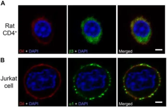
Figure 4. GABA-A channel subunits immunolabeled in the cytoplasm and in the plasma membrane.(A) Rat CD4+
T cell,b3 GABA-A channel subunit immunolabeling was observed both in the cytoplasm and in the plasma membrane (n = 41). (B) Jurkat cell, a1 GABA-A subunit immunolabeling was observed prominently in the plasma membrane as punctate pattern (n = 59). Plasma membrane labelling: DiI (red); subunit colour-labeling:b3 (green),a1 (green). The nuclei were stained with DAPI (blue). Scale bar = 2mM.
doi:10.1371/journal.pone.0042959.g004
Figure 5. GABA activates GABA-A currents in T cells.Whole-cells currents were evoked by application of 1mM or 1 mM GABA to rat CD4+T
cells (A, B, F), rat CD8+
T cells (C) or Jurkat E6. 1 cells (D, E, G). In symmetrical chloride solutions the currents were inward at negative potentials (A, B, C, D;280 mV and in G,260 mV) and outward at positive potential (E, F;+40 mV). Picrotoxin (PTX) inhibits the GABA-activated transient current. (F, G)
Tonic currents were activated by GABA (1mM, 1 mM) and inhibited by 100mM SR95531 (F) or 100mM bicuculline (G), GABA-A channel antagonists.
The difference between the dotted lines shows the amplitude of the tonic current (51 pA). Applications of drugs are indicated by the bars located above the current traces.
Immunocytochemistry and Confocal Imaging
Cells were resuspended in PBS and stained either in Falcon tubes or on poly-L-lysine or TESPA (3-triethoxysilylpropylamine) coated cover slips. Cells were fixed with 4% paraformaldehyde (PFA) in 0.1 M phosphate buffered saline (PBS) for 20 min at RT. After fixation, cells were washed with PBS, blocked with 5% bovine serum albumin (BSA) in PBS for 15 min and incubated overnight at 4uC with the following primary antibodies: rabbit anti-GABAARa1 (1:50, Novus Biologicals, USA) or rabbit
anti-GABAAR a1 (1:500, Synaptic Systems, Germany), rabbit
anti-GABAAR a2 (1:500, Synaptic Systems, Germany), rabbit
anti-GABAARc2 (1:500, Synaptic Systems, Germany) and mouse
anti-GABAARb3 (1:500, NeuroMab, USA), respectively. The control
experiment for thec2 antibody was performed in the mouse CD4+
T cells in the same way as with thec2 antibody apart from pre-incubation with 10 fold higher concentration of thec2 blocking peptide for 1 h. After washing with PBS, cells were incubated with a fluorophore-conjugated secondary antibody (Jackson Immunor-esearch Laboratories, USA) at RT for 1 h. All antibodies were diluted in 5% donkey serum in PBS. Cell nuclei were stained with DAPI and the plasma membrane was labeled with lipophilic tracer DiI (Invitrogen, USA) as indicated. The cells were immersed in the fluorescent mounting media (Dako, Sweden), and examined with a confocal microscope (LSM Meta, Carl Zeiss, Germany). The omission of primary antibodies served as negative controls.
Electrophysiological Recording
The isolated rat CD4+
and CD8+
T cells or Jurkat cells were collected (1–36106cells) and centrifuged for 2 min at 1006g. The supernatant was removed and the cell pellet was washed with the extracellular solution containing in mM: 145 NaCl, 5 KCl, 1 MgCl2, 1.8 CaCl2, 10 TES pH 7.3, 297 mOsm, centrifuged for
2 min at 1006g and then resuspended in 50–100ml extracellular solution. Nanion’s Port-a-Patch chip technology (Nanion, Ger-many) or conventional clamp methods were used to patch-clamp the cells. Using the Port-a-Patch chip technology the success rate of obtaining whole-cell recordings of native T cells was greater than with the conventional-patch-clamp method. For the Port-a-Patch, a 5ml cell suspension (106cell/ml) was dispensed into the extracellular chamber containing the recording chip, resistance 5 or 8–10 MVfor rat T cells and 2–3 or 5 MVfor Jurkat cells. The whole-cell configuration was established and currents were
recorded at holding potentials of280 mV or+40 mV. For the conventional patch-clamp recordings, the holding potential was
260 or + 40 mV. GABA (1mM or 1 mM) and GABA plus 100mM SR95531, 100mM bicuculline or 100mM picrotoxin (all drugs from Sigma, Germany) were prepared in the extracellular recording solution and perfused into the extracellular chamber at a rate of 1 ml/min. The Port-a-Patch internal recording solution contained in mM: 50 CsCl, 10 NaCl, 60 Cs-fluoride, 20 EGTA, 10 mM TES pH 7.3, 284 mOsm. The extracellular recording solution contained in mM: 80 NaCl, 3 KCl, 10 MgCl2, 35 CaCl2,
10 HEPES pH 7.3, 296 mOsm. The conventional patch-clamp pipette (internal) solution contained in mM: KCl 135, CsCl 5, CaCl2 1.8, MgCl2, TES 15 pH 7.4 282 mOsm and the extracellular solution contained in mM: NaCl 145, KCl 5, MgCl2 1, CaCl2 1.8 Ph 7.3, 297 mOsm. 5 mM EGTA was sometimes included in the pipette solution. All patch-clamp recordings were performed at room temperature (20–22uC). Currents were recorded using an Axopatch 200B amplifier, filtered at 2 kHz, digitized on-line at 10 kHz using an analogue-to-digital converter and analyzed with pClamp 10.2 software (Molecular Devices, USA).
Statistical analyses
Data are presented as mean6SEM. Differences in expression levels were analyzed using one way ANOVA or Kruskal-Wallis one way ANOVA on ranks, post hoc Tukey test. All statistical tests were performed using Sigma Plot version 11 (Systat Software, San Jose, CA, USA).
Supporting Information
Table S1 Molecular weight of GABA-A channel sub-units.Values were calculated from the amino acid sequences as given in the NCBI data base.
(DOC)
Author Contributions
Conceived and designed the experiments: SKM AB ZJ BB. Performed the experiments: SKM AB. Analyzed the data: SKM AB ZJ BB. Wrote the paper: SKM ZJ BB. Approved final version of the manuscript: SKM AB ZJ BB.
References
1. Olsen RW, Sieghart W (2008) International Union of Pharmacology. LXX. Subtypes of gamma-aminobutyric acid(A) receptors: classification on the basis of subunit composition, pharmacology, and function. Update. Pharmacol Rev 60: 243–260.
2. Birnir B, Korpi ER (2007) The impact of sub-cellular location and intracellular neuronal proteins on properties of GABA(A) receptors. Curr Pharm Des 13: 3169–3177.
3. Kullmann DM, Ruiz A, Rusakov DM, Scott R, Semyanov A, et al. (2005) Presynaptic, extrasynaptic and axonal GABAA receptors in the CNS: where and why? Prog Biophys Mol Biol 87: 33–46.
4. Erdo SL, Wolff JR (1990) gamma-Aminobutyric acid outside the mammalian brain. J Neurochem 54: 363–372.
5. Jin Z, Mendu SK, Birnir B (2011) GABA is an effective immunomodulatory molecule. Amino Acids.
6. Alam S, Laughton DL, Walding A, Wolstenholme AJ (2006) Human peripheral blood mononuclear cells express GABAA receptor subunits. Mol Immunol 43: 1432–1442.
7. Bhat R, Axtell R, Mitra A, Miranda M, Lock C, et al. (2010) Inhibitory role for GABA in autoimmune inflammation. Proc Natl Acad Sci U S A 107: 2580– 2585.
8. Bjurstom H, Wang J, Ericsson I, Bengtsson M, Liu Y, et al. (2008) GABA, a natural immunomodulator of T lymphocytes. J Neuroimmunol 205: 44–50. 9. Reyes-Garcia MG, Hernandez-Hernandez F, Hernandez-Tellez B,
Garcia-Tamayo F (2007) GABA (A) receptor subunits RNA expression in mice
peritoneal macrophages modulate their IL-6/IL-12 production. J Neuroimmunol 188: 64–68.
10. Tian J, Chau C, Hales TG, Kaufman DL (1999) GABA(A) receptors mediate inhibition of T cell responses. J Neuroimmunol 96: 21–28.
11. Tian J, Lu Y, Zhang H, Chau CH, Dang HN, et al. (2004) Gamma-aminobutyric acid inhibits T cell autoimmunity and the development of inflammatory responses in a mouse type 1 diabetes model. J Immunol 173: 5298–5304.
12. Wang Y, Feng D, Liu G, Luo Q, Xu Y, et al. (2008) Gamma-aminobutyric acid transporter 1 negatively regulates T cell-mediated immune responses and ameliorates autoimmune inflammation in the CNS. J Immunol 181: 8226–8236. 13. Wheeler DW, Thompson AJ, Corletto F, Reckless J, Loke JC, et al. (2011) Anaesthetic impairment of immune function is mediated via GABA(A) receptors. PLoS One 6: e17152.
14. Olsen RW, Sieghart W (2009) GABA A receptors: subtypes provide diversity of function and pharmacology. Neuropharmacology 56: 141–148.
15. Petty F, Sherman AD (1984) Plasma GABA levels in psychiatric illness. J Affect Disord 6: 131–138.
16. Gerber JC, Hare TA (1980) Gaba in Peripheral-Tissues - Presence and Actions in Endocrine Pancreatic Function. Brain Research Bulletin 5: 341–346. 17. Soltani N, Qiu H, Aleksic M, Glinka Y, Zhao F, et al. (2011) GABA exerts
protective and regenerative effects on islet beta cells and reverses diabetes. Proc Natl Acad Sci U S A.
rats developing diabetes compared to their congenic littermates. Mol Immunol 48: 399–407.
19. Dionisio L, Jose De Rosa M, Bouzat C, Esandi Mdel C (2011) An intrinsic GABAergic system in human lymphocytes. Neuropharmacology 60: 513–519. 20. Reyes-Garcia MG, Garcia-Tamayo F (2009) A neurotransmitter system that
regulates macrophage pro-inflammatory functions. J Neuroimmunol 216: 20– 31.
21. Bhat R, Steinman L (2009) Innate and adaptive autoimmunity directed to the central nervous system. Neuron 64: 123–132.
22. Sidorkiewicz M, Brocka M, Bronis M, Grek M, Jozwiak B, et al. (2011) The altered expression of alpha1 and beta3 subunits of the gamma-aminobutyric acid A receptor is related to the hepatitis C virus infection. Eur J Clin Microbiol Infect Dis.
23. Halonen LM, Sinkkonen ST, Chandra D, Homanics GE, Korpi ER (2009) Brain regional distribution of GABA(A) receptors exhibiting atypical GABA agonism: roles of receptor subunits. Neurochem Int 55: 389–396.
24. Birnir B, Tierney ML, Dalziel JE, Cox GB, Gage PW (1997) A structural determinant of desensitization and allosteric regulation by pentobarbitone of the GABAA receptor. J Membr Biol 155: 157–166.
25. Krasowski MD, Koltchine VV, Rick CE, Ye Q, Finn SE, et al. (1998) Propofol and other intravenous anesthetics have sites of action on the gamma-aminobutyric acid type A receptor distinct from that for isoflurane. Mol Pharmacol 53: 530–538.
26. Jenkins A, Greenblatt EP, Faulkner HJ, Bertaccini E, Light A, et al. (2001) Evidence for a common binding cavity for three general anesthetics within the GABAA receptor. J Neurosci 21: RC136.
27. Pritchett DB, Sontheimer H, Shivers BD, Ymer S, Kettenmann H, et al. (1989) Importance of a novel GABAA receptor subunit for benzodiazepine pharma-cology. Nature 338: 582–585.
28. Draguhn A, Verdorn TA, Ewert M, Seeburg PH, Sakmann B (1990) Functional and molecular distinction between recombinant rat GABAA receptor subtypes by Zn2+. Neuron 5: 781–788.
29. Wang TL, Guggino WB, Cutting GR (1994) A novel gamma-aminobutyric acid receptor subunit (rho 2) cloned from human retina forms bicuculline-insensitive homooligomeric receptors in Xenopus oocytes. J Neurosci 14: 6524–6531. 30. Alakuijala A, TalviOja K, Pasternack A, Pasternack M (2005) Functional
characterization of rat rho2 subunits expressed in HEK 293 cells. Eur J Neurosci 21: 692–700.
31. Jin Z, Bazov I, Kononenko O, Korpi ER, Bakalkin G, et al. (2012) Selective changes of GABAA channel subunit mRNAs in the hippocampus and orbitofrontal cortex but not in prefrontal cortex of human alcoholics. Front Cell Neurosci 5: 1–12.
32. Zhang X, Ding L, Sandford AJ (2005) Selection of reference genes for gene expression studies in human neutrophils by real-time PCR. BMC Mol Biol 6: 4. 33. Lupberger J, Kreuzer KA, Baskaynak G, Peters UR, le Coutre P, et al. (2002) Quantitative analysis of beta-actin, beta-2-microglobulin and porphobilinogen deaminase mRNA and their comparison as control transcripts for RT-PCR. Mol Cell Probes 16: 25–30.
34. Beyer M, Thabet Y, Muller RU, Sadlon T, Classen S, et al. (2011) Repression of the genome organizer SATB1 in regulatory T cells is required for suppressive function and inhibition of effector differentiation. Nat Immunol 12: 898–907. 35. Condamine T, Le Luduec JB, Chiffoleau E, Beriou G, Louvet C, et al. (2010)
Characterization of Schlafen-3 expression in effector and regulatory T cells. J Leukoc Biol 87: 451–456.
36. Chiffoleau E, Heslan JM, Heslan M, Louvet C, Condamine T, et al. (2007) TLR9 ligand enhances proliferation of rat CD4+ T cell and modulates suppressive activity mediated by CD4+CD25+T cell. Int Immunol 19: 193– 201.
37. Hillebrands JL, Whalen B, Visser JT, Koning J, Bishop KD, et al. (2006) A regulatory CD4+T cell subset in the BB rat model of autoimmune diabetes expresses neither CD25 nor Foxp3. J Immunol 177: 7820–7832.