RESEARCH ARTICLE / ARTIGO
Thymol and carvacrol: biotransformation and antifungal
activity against the plant pathogenic fungi
Colletotrichum
acutatum
and
Botryodiplodia theobromae
Manuel A. Numpaque1, Luis A. Oviedo1, Jesús H. Gil1,2, Carlos M. García1 & Diego L. Durango1
1Grupo de Química de los Productos Naturales y los Alimentos, Facultad de Ciencias,Escuela de Química, Universidad
Nacional de Colombia, AA 3840, Antioquia, Medellín, Colombia; 2Facultad de Ciencias Agropecuarias, Departamento de Ingeniería Agrícola y Alimentos, Universidad Nacional de Colombia, AA 3840, Antioquia, Medellín, Colombia
Author for correspondence: Diego L. Durango, e-mail: dldurango@unal.edu.co
ABSTRACT
Fungal growth in fruits may cause spoilage and result in a reduction of their quality and quantity. The aim of the current study was to investigate the antifungal activity and the metabolism of thymol and carvacrol by Colletotrichum acutatum and Botryodiplodia theobromae. The results showed that both compounds provided relatively good control against these plant pathogenic fungi. Mycelial growth of C. acutatum and B. theobromae was inhibited at 50 µg/mL and above. At 150 µg/mL, thymol and carvacrol inhibited the radial growth of fungi completely and this effect remained for 240 h. Furthermore, thymol and carvacrol were metabolized by the plant pathogenic fungi in low proportion to several compounds, including thymoquinone, thymohydroquinone, thymyl- and carvacryl acetate, thymyl- and carvacryl methyl ether. The transformations affect the structural requirements of thymol and carvacrol related to their antimicrobial activity and mode of action. The relatively high antifungal activity of thymol and carvacrol against C. acutatum and B. theobromae and the low levels of microbial transformation indicate that both compounds could be an alternative to traditional chemical fungicides for control of pre- and postharvest phytopathogenic fungi on fruits or vegetables.
Key words: Alternative control, arylpropanoids, fungal detoxification, metabolic pathways, thymoquinone. RESUMEN
Timol y carvacrol: biotransformación y actividad antifúngica contra los hongos fitopatogénicos Colletotrichum acutatum y Botryodiplodia theobromae
El crecimiento de los hongos en frutas puede causar el deterioro de los alimentos y resultar en una reducción de la calidad y la productividad. El objetivo del presente estudio fue investigar la actividad antifúngica, y el metabolismo de timol y carvacrol por Colletotrichum acutatum y Botryodiplodia theobromae. Los resultados muestran que ambos compuestos despliegan un relativo buen control contra los hongos fitopatogénicos; el crecimiento micelial de C. acutatum y B. theobromae se inhibió a concentraciones de 50 µg/mL y mayores. A 150 µg/mL, timol y carvacrol inhibieron el crecimiento radial de los hongosg/mL y mayores. A 150 µg/mL, timol y carvacrol inhibieron el crecimiento radial de los hongosµg/mL, timol y carvacrol inhibieron el crecimiento radial de los hongoscompletamente y este efecto permaneció durante 240 h. Además, los hongos fitopatogénicos metabolizaron timol y carvacrol a varios compuestos en una baja proporción, incluyendo timoquinona, timohidroquinona, timil- y carvacril acetato, timil- y carvacril metil eter, entre otros. Las transformaciones afectan los requerimientos estructurales del timol y carvacrol relacionados con su actividad antimicrobiana y modo de acción. La actividad antifúngica relativamente alta de timol y carvacrol contra C. acutatum and B. theobromae y los niveles de transformación microbiana bajos, indican que ambos compuestos pueden ser una alternativa a los fungicidas químicos tradicionales para el control de los hongos fitopatogénicos en pre- y poscosecha de frutas y vegetales.
Palabras-clave: Arilpropanóides, control alternativo, desinto�icación fúngica, rutas metabólicas, timoquinona.Arilpropanóides, control alternativo, desinto�icación fúngica, rutas metabólicas, timoquinona.
INTRODUCTION
Stem-end rot (Botryodiplodia theobromae) and anthracnose (Colletotrichum acutatum) are two serious diseases that contribute significantly to harvest and postharvest loss of tamarillo, avocado, mango, papaya, and citrus in Colombia (Afanador-Kafuri et al., 2003; Martínez et al., 2009). The use of synthetic chemicals as fungicides is the primary method of control of postharvest fungal decay caused by both diseases. However, the rapid development of tolerance to commercial fungicides by C. acutatum and
B. theobromae has led to an increase in the quantities of these compounds that have to be used. Consequently, the presence of fungicide residues on the fruits decreases their quality and can prevent their e�port to some foreign markets (Tripathi & Dubey, 2004). Furthermore, the use of synthetic chemicals to control pre- and postharvest deterioration of food commodities is restricted, due to their possible carcinogenicity, teratogenicity, acute to�icity, environmental pollution and side effects on human beings (Tripathi & Shukla, 2007). Therefore, the fruit industry urgently demands alternative pre- and postharvest treatments that are
Tropical Plant Pathology 36 (1) January - February 2011 free of synthetic fungicides and acceptable to consumers.
Given these facts, the use of essential oils and some of their constituents can be a very attractive method for pre- and postharvest disease control of fruits, due to their relative safety and wide acceptance by consumers (Ormancey et al., 2001).
Carvacrol (5-isopropyl-2-methylphenol; - “C”) and thymol (2-isopropyl-5-methylphenol; - “T”) are the main components of the essential oils of some Laminaceae members like oregano, thyme, and savory. They are produced by these plant species as a chemical defense mechanism against phytopathogenic microorganisms (Vázquez et al., 2001). Accordingly, the potent antimicrobial and fungitoxic properties of (C) and (T) against various plant pathogens have been previously documented (Sokovic et al., 2002; Falcone et al., 2005). It has been found that these agents cause alterations in the hyphal morphology and hyphal aggregates, resulting in reduced hyphal diameters and lyses of hyphal wall, as these interact with the cell membrane of the pathogen (Soylu et al., 2007). In addition, chemical modification of these phenolic compounds to various ether and ester derivatives has been reported to result in change in biological activity (Mathelaet al., 2010).
Despite their antimicrobial characteristics, Chamberlain and Dagley (1968) found a Pseudomonas strain able to degrade (T) completely and (C) partially. The authors proposed a metabolic pathway for (T) that involves meta-ring opening of a trihydric phenol, 3-hydroxythymo-1,4-quinol to 3,7-di-methyl-2,4,6-trioxo-octanoate. Hydrolysis of the latter, catalyzed by β-ketolase, yields acetate, 2-ketobutyrate and isobutyrate. Thus, biotransformation experiments provide information on the detoxification mechanism used by phytopathogenic microorganisms and give an indication of the structural modifications that may be necessary if substrates of this type are to be further developed as selective fungal control agents (Daoubi et al., 2005). The aim of the present study was to evaluate the antifungal properties against C. acutatum and B. theobromae of (T) and (C), along with their metabolism.
MATERIALS AND METHODS
Biological and chemical materials
C. acutatum and B. theobromae were isolated from infected tamarillo (Solanum betacea) and avocado (Persea americana) fruits, respectively. Fungi were characterized and kindly provided by the Laboratory of Phytopathology (Universidad Nacional de Colombia-Medellín). Pure cultures were maintained on potato dextrose agar (PDA) at 24°C, and freshly subcultured before using in the transformation experiment. The substrates for biotransformation, (T) and (C), were purchased from Abaquim Ltda. (Medellín, Colombia) and Merck KGaA (Darmstadt, Germany), respectively. Thymoquinone was obtained from Sigma-Aldrich Co. (St. Louis, MO, USA). Thymohydroquinone was prepared by reduction of thymoquinone, as described by El-Dakhakhny (1963). Methylated and acetylated derivatives of (T) and (C) were obtained according to
Mathela et al. (2010), and Smith & March (2007). The purified synthetic compounds were analyzed by 1H- and 13
C-Nuclear magnetic resonance (NMR) spectroscopy and gas chromatography-mass spectrometry (GC-MS) to confirm their identity and purity.
Antifungal bioassay
In order to investigate the antifungal activity of (T) and (C), against the microorganisms, the poisoned food technique described by Grover and Moore (1962) with slight modifications, was used. In poisoned food technique, both fungi were inoculated on Potato dextrose agar (PDA) plates and incubated at 25°C for 48 h to obtain young, actively growing colonies. A total of 30 µL of ethanolic solutions of (T) and (C) at concentrations of 0.0250, 0.0375, 0.0500, 0.0625, 0.0750 and 0.100 mg/µL were taken and mixed with 15 mL of cooled molten PDA medium (final concentrations 50, 75, 100, 125, 150 and 200 µg/mL), and allowed to solidify at room temperature for thirty minutes. Concentration at 125 µg/mL of (T) and (C) was not evaluated for B. theobromae. Subsequently, a mycelial disc (5 mm in diameter) of C.
acutatum or B. theobromae, cut with a sterile cork borer
from the periphery of 48 h old cultures, was aseptically inoculated onto the agar plates containing (T) or (C). Petri dishes with PDA plus ethanol (2 µL/mL) were used as a control. The plates were incubated at room temperature and the diameter of the mycelial growth was measured every 24 hours. Incubation was ceased when the mycelial mass of control Petri dishes had almost filled it (ca. 240 and 144 hours for C. acutatum and B. theobromae, respectively). All concentrations were tested in triplicate. The relative growth inhibition of the treatment compared to the control was calculated as percentage, using the following formula:
Inhibition (%) = {1 – [radial growth of treatment (mm)/ radial growth of control (mm)]}x100
Pre-culture of C. acutatum and B. theobromae
Fungi were transferred into 1.0 L Erlenmeyer flasks containing 500 mL of Czapeck-Dox liquid medium (Solution A: Glucose 5%, yeast extract 0.1%; Solution B: K2HPO4 0.5%, NaNO3 0.2%, MgSO4.7H
2O 0.05%, FeSO4.7H2O 0.001%).
Erlenmeyer flasks were agitated in a reciprocating shaker (Centricol series 0239) at 120 rpm and room temperature for 8 days. Mycelia were harvested by filtration, washed with sterile H2O and immediately employed in the biotransformation and time course experiments.
Biotransformation, isolation and identification of metabolic products
Tropical Plant Pathology 36 (1) January - February 2011
experiments. After incubation, culture medium and mycelia were separated by filtration. Mycelia were discarded and culture medium was saturated with NaCl and extracted with ethyl acetate (EtOAc, 3x2.0 L). Afterwards, the medium was acidified to pH 2 with 1M HCl, and extracted again with CH2Cl2 (2x2.0 L). Both organic extracts were mixed, dried over anhydrous Na2SO4 and concentrated in vacuum. Some metabolites were partially purified by column chromatography (CC), passing the extract through a silica gel 60 (mobile phase: mixtures of n-hexane: EtOAc) column, and then through a Sephadex LH-20 column (mobile phase: mixture of n-hexane:CH2Cl2: methanol, 2:1:1 v/v). The structures of the products were determined by mass spectrometry (MS), 1H and 13C NMR analysis, and comparison with the NIST 2002 computerized mass spectral library and authentic standards (Table 1).
Time-course experiment
Pre-cultured C. acutatum or B. theobromae was transferred into fourteen 500 mL Erlenmeyer flasks, each containing 125 mL of Czapeck-Dox liquid medium and substrate (T) or (C), and stirred for 14 days under the same conditions as described for preculture. Daily, the content of one flask was removed for each of 14 days, saturated with NaCl, and extracted with EtOAc (3x10 mL). The extracts obtained after solvent evaporation were analyzed by thin layer chromatography (TLC) andthin layer chromatography (TLC) and (TLC) and GC-MS. The ratios between the substrate and metabolic products were determined on the basis of GC peak areas. Control cultures in which the microorganism was grown under identical conditions, but without substrate, were carried out.
Analytical methods
TLC was performed on precoated plates (Si 60 F24, 0.25 mm, Merk) using mixtures of n-hexane:EtOAc as mobile phase. Column chromatography (CC) used silica gel 60 (0.040-0.063 mm; Merck) and Sephadex LH-20 (Sigma-Aldrich Co., St. Louis, MO, USA). Gas chromatography (GC) was performed on a Hewlett-Packard 6890 (Agilent Technologies) gas chromatograph equipped with a HP 5973 MSD (Mass selective detector-Quadrupole type). A HP-5 column (30 m x 0.25 mm i.d.; coating thickness 0.25 μm) was used. Chromatographic conditions were as follows: column temperature, 0-250°C at 10°C/min and keep for five minutes; injector temperature, 150°C; detector temperature, 280°C; carrier gas, N2 at 1 mL/min. Relative composition of the individual constituents was determined from the average area of the peaks. Electron ionization mass spectrometryElectron ionization mass spectrometry (EI-MS) measurements were obtained using GC-MS. Chromatographic conditions were the same as described earlier. Substances were identified by comparison of their mass spectra with those of reference substances and by comparison with the NIST 2002 Mass Spectral Library. NMR spectra were measured on a Bruker AMX 300
NMR spectrometer (1H NMR, 300.12 MHz; 13C NMR, 75.42 MHz). Chemical shifts, δ, are expressed in ppm units downfield from TMS and coupling constant J is in Hertz (Hz).
RESULTS AND DISCUSSION
Antifungal activity
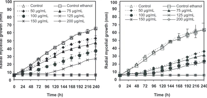
The inhibitory effects of (T) and (C) against C. acutatum and B. theobromae are shown in Figures 1 and 2. As can be seen, mycelial growth of these fungi was dependent on the concentration of (C) and (T) in the culture medium. At 10 µg/mL and above, (C) and (T) inhibited the radial growth of C. acutatum completely and this effect remained during the 240 h duration of the experiment (Figure 1). Furthermore, fungal colony development initiated only 48 and 96 hours after inoculation when C. acutatum was treated with (C) and (T) at 100 µg/mL, respectively. In addition, inhibition percentages of C. acutatum at 75 µg/mL ranged from 25-30% for (C) and 0-60% for (T). This shows a slightly higher antifungal activity of (T) in comparison to (C) against C. acutatum. In the case of B. theobromae, its growth was completely inhibited when a concentration of 10 and 200 µg/mL of (C) or (T) was used. In general, results revealed that (C) and (T) exhibited a relatively high antifungal action toward C. acutatum and B. theobromae. Some of these results are in agreement with those obtained earlier by Alzate et al. (2009). The antifungal activity of (T) and (C) against other plant pathogenic fungi has also been investigated (Tsao & Zhou, 2000; Holley & Patel, 200). In particular, (T) and (C) showed a very strong antifungal activity against Phytophthora cactorum and Cryphonectria parasitica at 28 x 10–3 mg/mL air concentration (Kim et al., 2008), and a complete growth inhibition of Alternaria arborescens and Rhizopus stolonifer (Plotto et al., 2003). Consequently, the potent antimicrobial properties of (T) and (C) against spoilage bacteria and food borne pathogens have been also reported in the literature (Valeroet al., 2006).
The mode of action for (C) and (T) is uncertain; however, activity may be linked to the hydrophobicity and consequently, the ability of both compounds to pass through the fungal cell membrane (Knobloch et al., 1988), which further affects pH homeostasis and equilibrium of inorganic ions (Ultee et al., 2000; Cristani et al., 2007) disturbing the cell structures. Furthermore, the involvement of the hydroxyl group in the formation of hydrogen bonds and the acidity of these phenolic compounds may be another important factor involved in the bioactivity. Ultee et al. (2000) additionally hypothesized that (C) acts as a proton exchanger, thereby reducing the pH gradient across the cytoplasmic membrane. This leads to the collapse of the proton motive force and depletion of the ATP pool, causing cell death. Moreover, it seems possible that both compounds may interfere with cell
Tropical Plant Pathology 36 (1) January - February 2011
TABLE 1 - GC retention time (Rt)and EI mass spectral propertiesand identification of products detected in incubation of
C. acutatum and B. theobromae with (T) and (C)
Identified by: *, proved with authentic standard and NMR; †, mass spectrum identical with literature spectrum; ‡, structure derived
from the interpretation of the mass spectrum. aNotation indicates the substrate and microorganism used: (C)-C. acutatum, CC; (C)-B. theobromae, CB; (T)-C.acutatum, TC; (T)-B. theobromae, TB. (CC1) is the same compound that (TC2), (CB2), (TB2).
7 Tropical Plant Pathology 36 (1) January - February 2011
FIGURE 1 - Effect of (C) (left) and (T) (right) on radial mycelial growth of C. acutatum. Results are shown as average
values of three replicates of mycelium diameter; bar = ±SD.
FIGURE 2 - Effect of (C) (left) and (T) (right) on radial mycelial growth of B. theobromae. Results are shown as average
values of three replicates of mycelium diameter; bar = ±SD.
wall enzymes like chitin synthase/chitinase as well as with the α- and β-glucanases of the fungus (Adams et al., 1996).
Although the difference between (T) and (C) is only in the position of the hydroxyl group, (T) was more active than (C) against C. acutatum. Growth inhibition percentages of C. acutatum achieved by (T) and (C) at 100 µg/mL for 10 days were from 86.0 (day 1) to 55.0 (day 10), and from 63.0 (day 1) to 30.5% (day 10), respectively. Different studies indicate that the position of the hydroxyl group is very important in antifungal and insecticidal activity (Kim et al., 2008; Park et al., 2008).
For B. theobromae, only slight differences in the
percentage of inhibition among (T) and (C) were found. Inhibition of B. theobromae growth by (T) at 100 µg/mL and for 6 days reached values between 97.7 (day 1) and 86.5% (day 6), whereas for (C) the values were between 93.2 (day 1) and 69.1% (day 6). As can be seen, growth inhibition percentages of C. acutatum and B. theobromae achieved by (T) and (C) at 100 µg/mL decreased with time, a fact which suggests that both fungi possess a detoxification mechanism. Similar behavior occurred when (T) and (C) were evaluated at concentrations below 150 µg/mL (data not shown).
0 10 20 30 40 50 60 70 80 90 100
0 24 48 72 96 120 144 168192 216 240 Control Control ethanol 50 μg/mL 75 μg/mL 100 μg/mL 125 μg/mL 150 μg/mL 200 μg/mL
0 10 20 30 40 50 60 70 80 90 100
0 24 48 72 96 120 144 168 192 216 240 Control Control ethanol 50 μg/mL 75 μg/mL 100 μg/mL 125 μg/mL 150 μg/mL 200 μg/mL
Radial
mycelial
growth
(mm)
Time (h)
Radial
mycelial
growth
(mm)
Time (h)
0 10 20 30 40 50 60 70 80 90 100
0 24 48 72 96 120 144
Control Control ethanol 50 μg/mL 75 μg/mL 100 μg/mL 150 μg/mL 200 μg/mL
0 10 20 30 40 50 60 70 80 90 100
0 24 48 72 96 120 144
Control Control ethanol 50 μg/mL 75 μg/mL 100 μg/mL 150 μg/mL 200 μg/mL
Radial
mycelial
growth
(mm)
Radial
mycelial
growth
(mm)
Tropical Plant Pathology 36 (1) January - February 2011 Metabolic compounds
A comparison of the TLC profiles and GC chromatograms obtained from biotransformation and the control, showed that C. acutatum transformed (C) into six major metabolites and a small amount of minor compounds not found in the control (data not shown). The structures of these metabolites were determined from the interpretation of spectral data (MS and NMR), and comparison with authentic samples (Table 1).
Two compounds (CC1 and CC2) had a molecular formula of C10H14O2 based on its mass spectrum, (M)+ =
166 amu. The molecular ion was 16 mass units higher than that of the parent compound (C), indicating the addition of a hydroxyl group to (C). Metabolite (CC1) had a fragment ion at m/z 151 (base peak) attributed to the loss of methyl group. Also, peaks of low intensity appear at m/z 123 and 43, corresponding to loss of isopropyl group and formation of isopropyl cation, respectively. From the spectral data, (CC1) was elucidated to be 2-methyl-5-isopropylhydroquinone (thymohydroquinone). Unfortunately, the very low abundance of (CC1) from the biotransformation excluded purification, enrichment and further spectroscopic characterization, and the full structural confirmation required the synthesis of this compound. The mass spectrum and retention time for (CC1) were consistent with those of synthetic thymohydroquinone obtained according to literature (El-Dakhakhny, 1963). It is noteworthy that thymohydroquinone was previously reported to be produced during biotransformation of (T) and (C) by a Pseudomonas strain (Chamberlain & Dagley, 1968). Similarly, metabolic product (CC2) gave fragment ions at m/z 43 (base peak) and 151 indicating formation of isopropyl cation and loss of methyl group. Also, (CC2) gave a peak at m/z 107 (absent in CC1) corresponding to loss of (CH3)2CO plus the radical hydrogen. This metabolite was tentatively identified as 5-(1-Hydroxy-1-methyl-ethyl)-2-methyl-phenol.
Three compounds (CC3, CC4 and CC5) presented a molecular formula C10H12O2 consistent with the molecular ion,
M+ = 164 amu. Metabolite (CC3) had fragment ions at m/z 149,
147, and 121 attributed to the loss of methyl, hydroxyl and isopropyl groups, respectively. From the spectral data, (CC3) was tentatively elucidated to be 2-Isopropenyl-5-methyl-benzene-1,4-diol. (CC4) corresponded to methyl ether derivative of carvacrol. Fragments at m/z 149 (M+−CH
3), 121 (M+−C3H7), 91 (C7H7+) and 77 confirmed
this conclusion. Also, compound CC4 was identified by comparing the retention time of GC and GC-MS with an authentic sample, and NMR data: 1H NMR: δ 1.14 (d,
6H, J = 7.0), 2.24 (s, 3H), 2.85 (sept., 1H, 7.0), 3.90 (s, 3H), 6.88 (d, 1H, J = 2.0), 7.13 (dd, 1H, J = 7.8, 2.0), 7.27 (d, 1H, J = 7.8). Mass spectrum of product (CC5) was characterized primarily by the fragment ions
at m/z 149, 136 and 121, corresponding to the loss of
a methyl group, carbon monoxide, and isopropyl group, respectively. Comparison of the mass spectrum with that reported in the NIST 2002 Mass Spectral Library indicated
that (CC5) was thymoquinone. Also, compound CC5 was identified by comparing the retention time of GC and GC-MS with an authentic sample, and NMR data: 1H NMR: δ
1.10-1.30 (d, 6H), 2.24 (s, 3H), 3.10-3.20 (m, 1H), 6.55 (s, 1H), 6.64 (s, 1H). 13C NMR: 15.4, 21.4, 26.5, 77.2, 130.4,
133.9, 145.5, 155.3, 190.1, 191.6. Thymoquinone was previously reported to be produced upon biotransformation of (T) by immobilized microalgae for 24 h (Rasoul-Amini, 2010). The authors found that (T) was hardly converted into thymoquinone and suggested that this low level of transformation may be due to (T) toxicity. The molecular formula of compound (CC6) was shown to be C
12H16O2 on
the basis of its mass spectrum (M+ 192 amu). MS analyses
gave the fragment ion (m/z 150), which corresponded to loss of the neutral molecule of ketene (M+−CH
2=C=O) and
suggests the presence of the acetyl group. The metabolite (CC6) was identified as carvacryl acetate. The structure of (CC6) was confirmed by co-injection with authentic carvacryl acetate and NMR data: 1H NMR: δ 1.32 (d, 6H, J = 7.0), 2.22 (s, 3H), 2.37 (s, 3H), 2.95 (sept., 1H, J = 7.0), 6.94 (d, 1H, J = 2.0), 7.10 (dd, 1H, J = 8.0, 2.0), 7.23 (d, 1H, J = 8.0); 13C NMR: 16.2, 21.3, 24.4, 34.0, 120.2, 124.6, 127.6, 131.4, 148.5, 149.8, 169.7.
Using (T) (80 µg/mL) as substrate and C. acutatum as biocatalyst, three major metabolites (TC1, TC2, and TC3), and many minor compounds were detected by GC analysis. The MS spectrum of metabolite (TC1) exhibited a molecular ion at m/z = 166 (M)+ and a fragmentation
pattern consistent with the loss of methyl group (M+−15;
base peak), the methyl group plus water (M+−18−15),
and the acetaldehyde plus the hydrogen radical at m/z = 121 amu. As a result, metabolite (TC1) was tentatively identified as 2-(1-Hydroxy-1-methyl-ethyl)-5-methyl-phenol. Otherwise, compound (TC2) showed a retention time (Rt) and mass spectrum indistinguishable from that obtained for an authentic sample of thymohidroquinone. In addition, metabolite (TC3) was identified by comparing the retention time of GC and GC-MS with an authentic sample of thymyl methyl ether, and NMR data: 1H NMR:
δ: 1.22 (d, 6H, J = 7.0), 2.33 (s, 3H), 3.37 (sept., 1H, J = 7.0), 3.80 (s, 3H), 6.79 (d, 1H, J = 8.0), 6.92 (dd, 1H, J = 8.0, 2.0), 7.05 (d, J = 8.0). 13C NMR: δ 21.3, 23.5, 27.6, 56.3, 112.3, 125.9, 126.6, 137.0, 137.5, 158.4. Some of these metabolites detected for C. acutatum were also found when an incubation of (C) and (T) using B. theobromae was carried out (Table 1). In fact, retention times (Rts) and mass spectra of (CC1) = (TC2), (CC2), (CC5), (CC6), and (TC1) were similar to those obtained for (CB2) = (TB2), (CB4), (CB5), (CB7), and (TB1), respectively.
9 Tropical Plant Pathology 36 (1) January - February 2011
respectively. In addition, the structure of (CB1), (CB3), and (CB6) was derived from their EI mass spectra. The fragmentation pattern of (CB1), as listed in the experimental section, was in good agreement with the proposed structure. Metabolite (CB1) had a molecular formula C10H14O2
consistent with the molecular ion, M+ = 166 amu. In
addition, this compound showed fragment ions to m/z = 135 and 105 consistent with the loss of H2CO and hydrogen radica,l and with water, ethylene and methyl radical. Thus, (CB1) was suggested to be 5-(2-hydroxy-1-methyl-ethyl)-2-methyl-phenol. Metabolite (CB3) had a molecular formula (C10H14O2; M+ = 166) and fragment ions at m/z 148 (M+−18),
133 (M+−18−15), and 105 (M+−18−28−15) attributed to the
loss of water, water + methyl radical, and water + ethylene + methyl radical, respectively. From the spectral data, (CB3) was tentatively elucidated to be 2-hydroxymethyl-5-isopropyl-phenol. Metabolic compound (CB6) had a molecular formula of C10H12O based on its mass spectrum, (M)+ = 148 amu. The molecular ion was 2 mass units (2H)
lower than that of parent compound (C), indicating the elimination of hydrogen from (C). Metabolite (CB6) had fragment ions at m/z 133 and 107 attributed, in that order, to the loss of methyl and isopropylene groups. From the spectral data, (CB6) was elucidated to be 5-Isopropenyl-2-methyl-phenol.
Upon incubation of (T) with B. theobromae, six major metabolites were detected by GC-MS analysis. The structures of (TB3), (TB5), and (TB6) were derived from their EI mass spectra. It should be emphasized that the data only tentatively suggest the identity of these metabolites. The molecular formula of compounds (TB3) and (TB5) was shown to be C10H16O2 on the basis of its mass spectrum (M+
= 168 amu). These metabolic products gave fragment ions
at m/z 153, 150, and 135 indicating the loss of methyl group,
water, and both, respectively. Also, there was a prominent peak at m/z = 126 (base peak), corresponding to a ring fission fragment. Although these data do not allow a definitive identification, they suggest a hydroxy-cyclohexenone substituted-like structure for metabolites (TB3) and (TB5). Furthermore, metabolite (TB6) had a molecular formula
C10H14O3 consistent with the molecular ion, M = 182 amu. The
mass spectrum showed fragment ions at m/z 140, 139, 111 (base peak), and 96 among others, corresponding to the loss of the ketene (M+−CH
2=C=O), ketene plus hydrogen radical, ketene
+ ethylene + hydrogen radical, and M+−C
5H10O, respectively.
(TB6) was tentatively elucidated to be 2-Hydroxymethyl-5-isopropyl-benzene-1,4-diol.
Finally, (TB4) was identified by comparing the retention times of GC-MS with those of authentic samples, and NMR data. NMR data for (TB4) were: 1H NMR: δ
1.22 (d, 6H, J = 7.0), 2.38 (s, 3H), 2.39 (s, 3H), 3.05 (sept., 1H, J = 7.0), 6.89 (d, 1H, J = 2.0), 7.11 (dd, 1H, J = 8.0, 2.0), 7.29 (d, 1H, J = 8.0); 13C NMR: 21.2, 21.3, 23.5, 27.6, 123.2, 126.9, 127.6, 137.1, 137.5, 148.4, 170.2. Also, the mass spectrum of (TB4) was consistent with that reported in the NIST 2002 Mass Spectral Library for thymyl acetate.
Time course experiment
The time course of relative concentration changes of (T) and (C) and metabolites is represented in Figures 3A-D. The starting substrate (C) was consumed slowly by both fungi. The fungus B. theobromae only transformed about 50% of (C) after 14 days, being mainly converted to (CB1) and (CB4). However, the relative abundance of both metabolites was always lower than 20%. As is shown in Figure 3A, the other metabolites reached very low concentrations.
Furthermore, C. acutatum metabolized (C) more quickly than B. theobromae. Interestingly, some metabolites produced from (C) by C. acutatum were not detected in a fungal culture of B. theobromae incubated with the same substrate. According to Figure 3B, the substrate (C) was mainly transformed by C. acutatum to (CC4) (~12%) and about 55% was consumed in 14 days. These results suggest that under the conditions used, (C) can be only slowly detoxified by the plant pathogenic fungi. Otherwise, (C) was metabolized at a faster rate than (T) by C. acutatum (Figures 3B and 3D).
As can be seen from Figure 3C, the substrate (T) was only transformed by about 55% after 14 days, using B. theobromae as biocatalyst. The major metabolic product (TB1) reached a relative abundance of ~20% after 11 days. In addition, metabolites (TB2), (TB3), (TB4), (TB5), and (TB6) presented relative abundances below 10% during the time of analysis. It seems noteworthy that (T) was consumed by B. theobromae to a greater extent than C. acutatum. In fact, analysis of extracts obtained from fungal cultures of C. acutatum incubated with (T) showed it to be weakly metabolized (<10 %) (Figure 3D). In addition, some metabolites previously detected on incubation of (T) with B. theobromae were absent in the biotransformation using C. acutatum.
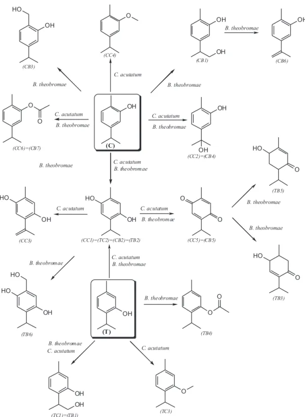
The marked difference between the metabolism of (C) and (T) by C. acutatum may reflect the higher toxicity (antifungal activity) of (T) against this pathogenic fungus. In the same way, small differences between the metabolisms of both compounds by B. theobromae are consistent with the observed similarities in the antifungal activity against this fungus. Based on the structures of the metabolites, metabolic pathways for the biotransformation of (C) and (T) by C. acutatum and B. theobromae were proposed (Figure 4).
Tropical Plant Pathology 36 (1) January - February 2011
0 10 20 30 40 50 60 70 80 90 100
0 1 2 3 4 5 6 7 8 9 10 11 1213 14
(CB1) (CB2) (CB3)
(CB4) (CB5) (CB6)
(CB7) (C)
0 10 20 30 40 50 60 70 80 90 100
0 1 2 3 4 5 6 7 8 9 10 11 12 13 14
(TB1) (TB2) (TB3)
(TB4) (TB5) (TB6)
(T)
0 10 20 30 40 50 60 70 80 90 100
0 1 2 3 4 5 6 7 8 9 10 11 12 13 14
(CC1) (CC2) (CC3)
(CC4) (CC5) (CC6)
(C)
0 10 20 30 40 50 60 70 80 90 100
0 1 2 3 4 5 6 7 8 9 10 11 12 13 14
(TC1) (TC2)
(TC3) (T)
A B
C D
Relative
abundance
(%)
Relative
abundance
(%)
Relative
abundance
(%)
Relative
abundance
(%)
Time (days) Time (days)
Time (days) Time (days)
FIGURE 3 - Time course for the microbial transformation of: (C) by B. theobromae (3a) and C. acutatum (3b); and (T)
by B. theobromae (3c) and C. acutatum (3d). (CC1) is the same compound that (TC2), (CB2), (TB2). Similarly, (CC2) = (CB4); (CC5) = (CB5); (CC6) = (CB7); and (TC1) = (TB1).
substituent, both of which may have a major influence on specific interactions with microbial cells. Veldhuizen et al. (2006) found that carvacrol methyl ether, containing a methyl ether group instead of a hydroxyl group, did not inhibit the growth of B. cereus at the concentrations tested (0 to 10 mM), whereas no growth was observed with (C) at concentrations of 0.75 mM and above. Furthermore, (TC3), (TB4), and (CC6) = (CB7) do not have the ability to release a proton and behave as a proton exchanger.
In addition, the aliphatic side chains have a direct influence on the antimicrobial activity of (C) and (T), as has been described by several researchers (Ultee et al., 2002; Veldhuizen et al., 2006). They have observed that the removal of ring substituents from (C) (using 3-isopropylphenol and o-cresol) leads to a reduction in antimicrobial activity. Therefore, the hydroxylation of the aliphatic chains of (C) and (T) may be a mechanism used by C. acutatum and B. theobromae to reduce the antifungal
11 Tropical Plant Pathology 36 (1) January - February 2011
FIGURE 4 - Possible metabolic pathways of (C) and (T) by C. acutatum and B. theobromae
We are currently evaluating the antifungal effect of the obtained metabolic compounds, and some synthetic derivatives of (T) and (C), against C. acutatum and B. theobromae. Synthetic derivatives present modifications in the aliphatic chains and benzenic ring of (T) and (C), in the same positions that were hydroxylated according to the metabolic studies.
Arylpropanoids, specifically thymol and carvacrol, which are strongly antifungal against the phytopathogenic
fungi C. acutatum and B. theobromae, are mainly metabolized via aromatic and aliphatic hydroxylation, and the methylation and acetylation of hydroxyl group over the aromatic ring. Antifungal activity of thymol was slightly higher than that observed for carvacrol against C.
acutatum.
Tropical Plant Pathology 36 (1) January - February 2011 were mainly metabolized via aromatic and aliphatic
hydroxylation, and through methylation and acetylation of hydroxyl group. These transformations affect the structural features of thymol and carvacrol associated with their antimicrobial activity and mode of action. Overall, the antifungal activity of thymol was higher than that observed for carvacrol against C. acutatum. Consequently, compared to the transformation of carvacrol, the metabolism of thymol by C. acutatum was a slower process. Similarly, the small differences in the metabolism of both compounds by B. theobromae were consistent with the observed similarities in the antifungal activity against this fungus. High antifungal activity coupled with a low level of microbial transformation makes thymol and carvacrol promising candidates to control
C. acutatum and B. theobromae.
ACKNOWLEDGEMENTS
The authors gratefully acknowledge the financial support of the Vicerrectoría de Investigación, Universidad Nacional de Colombia.
REFERENCES
Adams S, Kunz B, Weidenbörner M (1996)Mycelial deformations of Cladosporium herbarum due to the application of Eugenol and Carvacrol. Journal of Essential Oil Research8:535-540.
Afanador-Kafuri L, Minz D, Maymon M, Freeman S (2003) Characterization of Colletotrichum isolates from tamarillo, pasiflora and mango in Colombia and identification of a unique species from genus. Phytopathology 93:579-587.
Alzate DA, Mier GI, Afanador L, Durango DL, García CM (2009) Evaluación de la fitotoxicidad y la actividad antifúngica contra
Colletotrichum acutatum de los aceites esenciales de tomillo (Thymus vulgaris), limoncillo (Cymbopogon citratus), y sus componentes mayoritarios. Vitae 16:116-125.
Chamberlain EM, Dagley S (1968) The metabolism of thymol by a Pseudomonas. Biochemical Journal 110:755-763.
Cristani M, D’Arrigo M, Mandalari G, Castelli F, Sarpietro MG, Micieli D., Venuti V, Bisignano G, Saija A, Trombetta D (2007) Interaction of four monoterpenes contained in essential oils with model membranes: implications for their antibacterial activity. Journal of Agricultural and Food Chemistry 55:6300-6308. Daoubi M, Hernández-Galán R, Benharref A, Collado IG (2005) Screening study of lead compounds for natural product-based fungicides: antifungal activity and biotransformation of 6α, 7α-Dihydroxy-β-himachalene by Botrytis cinerea. Journal of Agricultural and Food Chemistry 53:6673-6677.
El-Dakhakhny M (1963) Studies on the chemical constitution of Egiptian Nigella sativa L. seeds II. The essential oil. Planta Medica 11:465-470.
Falcone PM, Speranza B, Del Nobile MA, Corbo MR, Sinigaglia M (2005) A study on the antimicrobial activity of thymol intended as a natural preservative. Journal of Food Protection 68:1664-1670.
Grover RK, Moore JD (1962) Toximetric studies of fungicides against brown rot organism. Sclerotina fruticola. Phytopathology 52:876-880.
Holley RA, Patel D (2005) Improvement in shelf-life and safety of perishable foods by plant essential oils and smoke antimicrobials. Review Food Microbiology 22:273-292.
Kim J, Lee YS, Lee SG, Shin SC, Park IK (2008) Fumigant antifungal activity of plant essential oils and components from West Indian bay (Pimenta racemosa) and thyme (Thymus vulgaris) oils against two phytopathogenic fungi. Flavour and Fragrance Journal23:272–277.
Knobloch K, Pauli A, Iberl B, Weis N, Weigand H (1988) Mode of action of essential oil components on whole cells and fungi in plate tests. In: Schreier P (Ed.) Bioflavor ’87. Berlin, New York. Walter de Gruyter Verlag. pp. 287-299.
Martínez EP, Hío JC, Osorio JA, Torres MF (2009) Identification of Colletotrichum species causing anthracnose on Tahiti lime, tree tomato and mango. Agronomía Colombiana 27:211-218.
Mathela CS, Singh KK, Gupta VK (2010) Synthesis and In Vitro
antibacterial activity of thymol and carvacrol derivatives. Acta Poloniae Pharmaceutica-Drug Research 67:375-380.
Ormancey X, Sisalli S, Coutiere P (2001) Formulation of essential oils in functional perfumery. Parfums, Cosmetiques, Actualites 157:30-40.
Park IK, Kim JN, Lee YS, Lee SG, Ahn YJ, Shin SC (2008) Toxicity of plant essential oils and their components against
Lycoriella ingenua (Diptera: Sciaridae). Journal of Economic Entomology 101:139-144.
Plotto A, Roberts DD, Roberts RG (2003) Evaluation of plant essential oils as natural postharvest disease control of tomato (Lycopersicon esculentum). Acta Horticulturae 628:737-745. Rasoul-Amini S, Fotooh-Abadi E, Ghasemi Y (2010) Biotransformation of monoterpenes by immobilized microalgae. Journal of Applied Phycology 22:1-7.
Smith MB, March J (2007) March’s Advanced Organic Chemistry: Reactions, mechanisms, and structure. 6th ed. Hoboken NJ. John Wiley & Sons, Inc.
Sokovic M, Tzakou O, Pitarakoli D, Couladis M (2002) Antifungal activities of selected aromatic plants growing wild in Greece. Molecular Nutrition and Food Research 46:317-320.
Soylu S, Yigitbas H, Soylu EM, Kurt S (2007) Antifungal effects of essential oils from oregano and fennel on Sclerotinia sclerotiorum. Journal of Applied Microbiology 103:1021-1030.
Tripathi P, Dubey NK (2004) Exploitation of natural products as an alternative strategy to control postharvest fungal rotting of fruit and vegetables. Postharvest Biology and Technology 32:235-245. Tripathi P, Shukla AK (2007) Emerging non-conventional technologies for control of post harvest diseases of Perishables. Fresh Produce 1:111-120.
Tsao R, Zhou T (2000) Antifungal activity of monoterpenoids against postharvest pathogens Botrytis cinerea and Monilinia fructicola. Journal of Essential Oil Research 12:113-121.
13 Tropical Plant Pathology 36 (1) January - February 2011
Ultee A, Kets EPW, Alberda M, Hoekstra FA, Smid EJ (2000) Adaptation of the food-borne pathogen Bacillus cereus to carvacrol. Archives of Microbiology 174:233-238.
Valero D, Valverde JM, Martinez-Romero D, Guillen F, Castillo S, Serrano M (2006) The combination of modified atmosphere packaging with eugenol or thymol to maintain quality, safety and functional properties of table grapes. Postharvest Biology and Technology 41:317-327.
Vázquez BI, Fente C, Franco CM, Vázquez MJ, Cepeda A (2001) Inhibitory effects of eugenol and thymol on Penicillium citrinum
TPP 226 - Received 10 December 2010 - Accepted 15 March 2011 Section Editor: Marciel J. Stadnik
strains in culture media and cheese. International Journal of Food Microbiology 67:157-163.
Veldhuizen EJA, Tjeerdsma-Van Bokhoven JLM, Zweijtzer C, Burt SA, Haagsman HP (2006) Structural requirements for the antimicrobial activity of carvacrol. Journal of Agricultural and Food Chemistry 54:1874-1879.