Effect of cilostazol on neointimal hyperplasia in iliac arteries of
Effect of cilostazol on neointimal hyperplasia in iliac arteries of
Effect of cilostazol on neointimal hyperplasia in iliac arteries of
Effect of cilostazol on neointimal hyperplasia in iliac arteries of
Effect of cilostazol on neointimal hyperplasia in iliac arteries of
pigs after transluminal angioplasty
pigs after transluminal angioplasty
pigs after transluminal angioplasty
pigs after transluminal angioplasty
pigs after transluminal angioplasty
Efeito do cilostazol na hiperplasia neointimal em artérias ilíacas de suínos
Efeito do cilostazol na hiperplasia neointimal em artérias ilíacas de suínos
Efeito do cilostazol na hiperplasia neointimal em artérias ilíacas de suínos
Efeito do cilostazol na hiperplasia neointimal em artérias ilíacas de suínos
Efeito do cilostazol na hiperplasia neointimal em artérias ilíacas de suínos
submetidas à angioplastia transluminal
submetidas à angioplastia transluminal
submetidas à angioplastia transluminal
submetidas à angioplastia transluminal
submetidas à angioplastia transluminal
JOEL ALEX LONGHI1; ADAMASTOR HUMBERTO PEREIRA1
A B S T R A C T A B S T R A C T A B S T R A C T A B S T R A C T A B S T R A C T
Objective Objective Objective Objective
Objective: to evaluate whether systemic administration of cilostazol reduces neointimal hyperplasia in iliac arteries of pigs submitted to balloon catheter angioplasty. MethodsMethodsMethodsMethodsMethods: twenty pigs underwent angioplasty with a 6x40 mm balloon catheter in the right common iliac artery, guided by Doppler ultrasound. The animals were randomized into two groups: group 1 (n=10), which received 50mg cilostazol twice a day, and group 2 (n=10), control. After 30 days, the animals were killed and the iliac arteries prepared for histological analysis. The histological sections were digitized and analyzed by digital morphometry. Statistical analysis was performed using the Student t and Mann-Whitney tests. ResultsResultsResultsResults: when comparing the iliac arteries submitted toResults angioplasty with those not subjected to angioplasty, there was significant neointimal hyperplasia (0.228 versus 0.119 mm2;
p=0.0001). In arteries undergoing angioplasty, there was no difference between group 1 (cilostazol) and group 2 (control) as for the lumen area (2.277 versus 2.575 mm2; p=0.08), the tunica intima (0.219 versus 0.237 mm2; p=0.64), the tunica media (2.262
vs. 2.393 mm2; p=0.53) and the neointimal occlusion percentage (8.857 vs. 9.257 %; p=0.82). ConclusionConclusionConclusionConclusion: the use of cilostazolConclusion
50mg administered in two daily doses did not reduce neointimal hyperplasia in iliac arteries of pigs submitted to balloon angioplasty catheter.
Key words Key words Key words Key words
Key words: : : : : Neointima. Hyperplasia. Phosphodiesterase Inhibitors. Angioplasty. Iliac Artery.
1. Programa de Pós-Graduação em Medicina- Ciências Cirúrgicas, Universidade Federal do Rio Grande do Sul - RS - Brazil.
INTRODUCTION
INTRODUCTION
INTRODUCTION
INTRODUCTION
INTRODUCTION
P
eripheral arterial disease has a prevalence of up to 20% in patients over 70 years old and is an important cause of morbidity1. When surgical intervention is indicated, theendovascular technique is the initial treatment for most anatomical stages in different arterial sites1,2 and in various
clinical conditions. The endovascular treatment initial success rate is high, exceeding 90% in the aortoiliac segment, but the patency falls over time due to restenosis3,4.
Drugs have been used in attempting to reduce the risk of restenosis after percutaneous vascular procedures. Cilostazol has antiplatelet, vasodilatory and antiproliferative effects5-7. It is a selective phosphodiesterase III inhibitor and
promotes increase of adenosine 3',5'-cyclic monophosphate in platelets and smooth muscle cells. The sequence of events stimulates the rapid regeneration of endothelial cells, which inhibits neointimal formation by two mechanisms: blockage of the abnormal growth of vascular smooth muscle cells (VSMC) and endothelial function improvement5,6,8-12. These
mechanisms may be responsible for reducing restenosis after coronary stent insertion observed in clinical trials and metanalyses13,14.
There are studies with small numbers of patients analyzing the use of cilostazol after peripheral endovascular procedures, especially in the femoropopliteal segment, with favorable initial results15,16. However, it is not clear whether
its application can be extended to other arterial sites. The objective of this study is to evaluate whether the systemic administration of cilostazol reduces neointimal hyperplasia in iliac arteries of pigs submitted to balloon catheter angioplasty.
METHODS
METHODS
METHODS
METHODS
METHODS
We performed the procedures in 20 Large White pigs, from different familial origins, with average eight weeks of age, from the Unidade de Experimentação Animal of Hospi-tal de Clínicas of Porto Alegre (HCPA), accompanied by a veterinarian and a nurse. The study was approved by the Comi-tê de Ética of the Grupo de Pesquisa e Pós-Graduação of HCPA and conducted according to the protocol of the Unidade de Experimentação Animal of HCPA. Project number 09-150.
bleeding; death of the animal before the deadline of arte-rial segments withdrawal; technical glitches in tissues preparation or processing.
The pigs were pre-medicated with ketamine (15mg/kg) and midazolam (0.8mg/kg) intramuscularly. After ten minutes, we held an access in the cephalic vein for administration of 0.9% saline (5ml/kg/h). During the procedure, the animals received 100% oxygen through a face mask, which, coupled with the muzzle, allowed monitoring of respiratory rate. We also monitored heart rate and the anterior leg withdrawal reflex. We kept the animals in deep sedation by the administration of a propofol “bolus”. Infiltration was performed with lidocaine (4mg/ mg) in the site of the femoral artery access. Before the procedure, we administered the analgesic tramadol (2mg/ kg) and the antimicrobial cefazolin (20mg/kg).
The right common femoral artery was surgically exposed. Through direct puncture of the common femoral artery with a 18G needle, we introduced the 0.035 inches hydrophilic guide wire and inserted the 6 Fr, 11cm sheath. Under ultrasound control, was directed the guide wire to the abdominal aorta. At this point, the animals received intravenous heparin (Heptar, Eurofarma) at a dose of 100 IU/ kg. We performed doppler ultrasonography (TITAN equipment, SonoSite) during surgery to measure the diameters of the right common iliac artery and distal abdo-minal aorta. Then, we positioned the 6x40 mm balloon catheter (Passeo-35, Biotronik) in the right common iliac artery, from the trifurcation of the aorta, under ultrasound control. We carried out the angioplasty for one minute with an 8atm pressure. The diameter of the balloon catheter was selected to provide the sizing 10-20% larger than the common iliac artery lumen in this age group, similar to that used in interventions on human patients. At the end of angioplasty, we performed a Doppler ultrasound again to confirm the iliac segment arterial patency. After removal of sheath and guide wire, the common femoral artery was sutured, and the groin closed with nonabsorbable sutures. There were no intraoperative complications.
We divided the animals into two groups and randomized treatment through the site http:// www.randomization.com. Group 1 – animals undergoing angioplasty and administration of cilostazol (Vasativ, Eurofarma) 50mg twice a day starting on the first day after surgery and administered until the 30th day; Group 2 (control) – animals submitted only to angioplasty. The animals were tagged with numbered earrings of different colors for identification.
In the postoperative period, animals were housed in specific bays, with running water and balanced chow for their age and no additional lipid supplementation. After 30 days, the animals were again anesthetized and underwent laparotomy with exposition of the abdominal aorta and iliac arteries. The surgical specimens, containing the distal ab-dominal aorta and iliac arteries, were removed en bloc after a lethal dose of 2.5% sodium thiopental.
For optical microscopy evaluation, surgical specimens were rinsed in a 0.9% NaCl solution and fixed in a neutral buffered 10% formalin solution. The common right iliac arteries (undergoing angioplasty) and left (not undergoing angioplasty) were transversally sectioned in three points measured from the aortic trifurcation: 10mm, 20mm and 30mm. These points correspond respectively to the locations 25, 50 and 75% of the used balloon length (40mm). The segments were embedded in paraffin blocks, sectioned into 4mm sections and stained with hematoxylin-eosin, Verhoeff and orcein techniques.
The images of histological sections were scanned for morphometric analysis through morphometry and image analysis with the Image-Pro Plus software version 6.0 (Me-dia Cybernetics - Silver Spring, USA) and Image (Scion Corporation - USA). The images of histological sections were digitized from the conventional optical microscopy: microscope with planachromatic lenses (Zeiss Axiostar -Germany) with phototube adjusted to 0.25 magnification, color closed circuit camera (Sony DXC-151 - Japan) and digital analog conversion board (Image-Pro Plus Capture Kit, Media Cybernetics - Silver Spring, USA), generating image files with 2560x1920 pixels. Images were scanned with 40X microscopic magnification.
The planimetry of lumen area, the tunica intima and tunica media was performed with an automated technique, without interference from the observer. The lumen area was obtained by direct measurement of the area bounded by the endothelium; the intima, subtracting the area of the lumen from the area bounded by the internal elastic lamina; the media area by subtracting the area of the lumen and the intima delimited by the external elastic lamina. The results of the morphometric measurements of the lumen, intima and media areas are presented in absolute numbers (square millimeters), using the average of the obtained areas from the three segments of each artery. Additionally, we calculated the sum of the media and intima areas and the percentage of neointimal occlusion by dividing the intima area by the area delimited by the internal elastic lamina.
For descriptive statistics, we used the average and the standard deviation for parametric variables, and the median and interquartile ranges for non-parametric variables. When comparing groups, we used the Student t test and the Mann-Whitney test. We considered p<0.05 as statistically significant.
RESULTS
RESULTS
RESULTS
RESULTS
RESULTS
three measurements) was also not different (5.07 versus 4.87 mm, p=0.39).
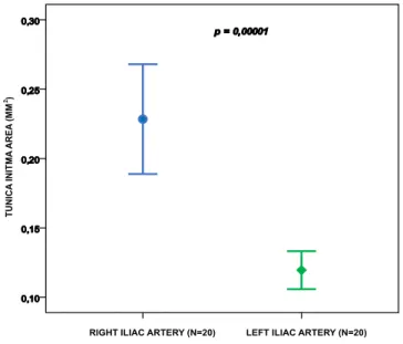
By comparing the intima area in the right common iliac artery (submitted to angioplasty) with the intima area in the left common iliac artery (not submitted to angioplasty), we observed a significant neointimal hyperplasia (0.228 versus 0.119 mm2; p=0.00001) (Figures 1 and 2). This difference
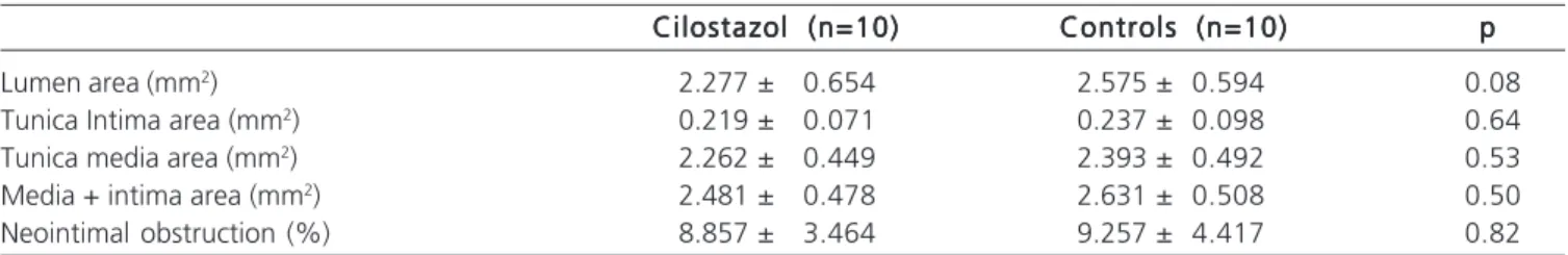
persists even when stratified by Group 1 (cilostazol) and Group 2 (control) (Figure 3). Table 1 shows the data of the digital morphometry of the right common iliac arteries subjected to balloon catheter angioplasty. There was no difference in the lumen area, the intima area, the media area, the media + intima area and the neointimal obstruction percentage between groups (Table 1 and Figure 4).
DISCUSSION
DISCUSSION
DISCUSSION
DISCUSSION
DISCUSSION
Restenosis after balloon catheter angioplasty is caused by the negative elastic remodeling and the proliferation and migration of vascular smooth muscle cells (VSMC)17. In this model, the response of the arterial wall to
the damage caused by angioplasty is the release of growth factors and other biologically active factors, which change the composition of the extracellular matrix and promote VSMC phenotypic change, from contractile to synthetic (dedifferentiation), leading to cell proliferation in the tunica media and migration to the tunica intima, forming the neointima18.
Animal models with pigs better reflect the pathophysiology of restenosis occurring in humans, with similar stages of neointimal formation19-21. In addition, pigs
present similarities in the vascular anatomy, in the coagulation system and physiology; and in medium-sized animals, one can use the same materials used with humans without the need for adjustments19.
The experimental porcine model of balloon catheter angioplasty in coronary arteries could cause significant neointimal hyperplasia only when there was rupture of the internal elastic lamina22. In rat carotid arteries,
Figure 3 Figure 3 Figure 3 Figure 3
-Figure 3 - Comparison between the intima area in the right iliac artery (submitted to angioplasty) and the left iliac artery. The value of p shows a statistically significant neointimal hyperplasia in arteries undergoing angioplasty, even when stratified by cilostazol and control groups.
Figure 2 Figure 2 Figure 2 Figure 2
-Figure 2 - Comparison between the intima area in the right iliac artery (subjected to angioplasty) and in the left iliac artery. The value of p shows a statistically significant neointimal hyperplasia in arteries undergoing angioplasty.
Figure 1 Figure 1 Figure 1 Figure 1
Figure 1 - A) Photomicrograph of the common iliac arteries of the same pig (orcein staining method, 40x original magnification); A) right common iliac artery submitted to angioplasty, with neointimal hyperplasia (arrow). B) left common iliac artery not subjected to angioplasty (normal artery).
the results are similar23. Previous work comparing versus
angioplasty versus angioplasty with stent in pigs’ iliac arteries concluded that the use of the stent causes increased neointimal hyperplasia23. These previous studies have also
balloon catheter, which might have caused minor injury to the arterial wall compared to studies using a larger oversize and multiple inflations. The option to apply the technique without a stent lies in the fact that although most studies evaluate the use of cilostazol in procedures with stent in coronary13, carotid or peripheral15,16 arteries, isolated balloon
catheter angioplasty in iliac segment remains a significant procedure in the endovascular treatment.
Experimental studies have shown that around four weeks the neointimal hyperplasia is complete, similar to that identified in humans in six to 12 months24,25. A longer
period of cilostazol administration probably would not add benefit. One limitation of this study is the assessment of the normal artery wall response. Extrapolation of the data is limited, since in the clinical setting the treated arteries present with atherosclerotic stenoses or occlusions. The adoption of an atherogenic diet does not appear to enhance
neointimal hyperplasia in response to vascular wall injury in the porcine model26.
The cilostazol dose of 50mg twice daily used in this study is similar to the maximum dose used in adults, and there were no adverse effects or change in behavioral pattern in treated pigs. Another limitation is the non-administration of other anti-platelet agents such as acetylsalicylic acid, ticlopidine and clopidogrel used in clinical trials10. However, the isolated use of cilostazol
allows evaluating the effect of the drug, since the association with other drugs can have diverse biological effects. Previous studies with animal models demonstrated that the administration of cilostazol reduced neointimal hyperplasia in animal models for the study of the coronary artery 27, the carotid artery 28 and vein grafts 29,30. Only one
study in canine model evaluated the effect of cilostazol in the iliac artery. They performed angioplasty with stent associated with embolization with coils of the common femoral artery for reduced flow in the iliac axis. The results demonstrated reduced neointimal hyperplasia and absence of thrombotic occlusion in the cilostazol group compared with controls31.
This is the first study in pigs that evaluated the use of cilostazol on neointimal hyperplasia of the iliac artery. Moreover, the work used the technique of angioplasty guided by Doppler ultrasonography, which allowed to accurately measuring arterial diameters and the consequent appropriate choice of the balloon catheter diameter.
Data from this study showed that the balloon catheter angioplasty caused significant neointimal hyperplasia in iliac arteries of pigs. The use of cilostazol 50mg twice daily for 30 days did not reduce neointimal hyperplasia in iliac arteries of pigs submitted to angioplasty. We observed no differences in the lumen area, tunica inti-ma area and tunica media area.
Acknowledgements Acknowledgements Acknowledgements Acknowledgements Acknowledgements
The authors thank the Animal Experimentation Unit and Experimental Pathology Unit of the HCPA for carrying out the experiment.
Figure 4 Figure 4 Figure 4 Figure 4
Figure 4 - Comparison between the areas of the lumen, the intima, the media and media + intima between groups. The value of p shows that there was no difference between the cilostazol and control groups.
Table 1 Table 1 Table 1 Table 1
Table 1 - Data from digital morphometry.
Cilostazol (n=10) Cilostazol (n=10) Cilostazol (n=10) Cilostazol (n=10)
Cilostazol (n=10) Controls (n=10)Controls (n=10)Controls (n=10)Controls (n=10)Controls (n=10) ppppp
Lumen area (mm2) 2.277 ± 0.654 2.575 ± 0.594 0.08
Tunica Intima area (mm2) 0.219 ± 0.071 0.237 ± 0.098 0.64
Tunica media area (mm2) 2.262 ± 0.449 2.393 ± 0.492 0.53
Media + intima area (mm2) 2.481 ± 0.478 2.631 ± 0.508 0.50
R E S U M O R E S U M O R E S U M O R E S U M O R E S U M O
Objetivo: Objetivo: Objetivo: Objetivo:
Objetivo: avaliar se a administração sistêmica de cilostazol reduz a hiperplasia neointimal nas artérias ilíacas de suínos submetidas à angioplastia com cateter balão. Métodos:Métodos:Métodos:Métodos:Métodos: vinte suínos foram submetidos à angioplastia com cateter balão 6x40 mm na artéria ilíaca comum direita, guiada por ultrassonografia com Doppler. Os animais foram randomizados em dois grupos: grupo 1 (n=10), no qual foi administrado cilostazol 50mg em duas doses diárias, e grupo 2 (n=10), considerado controle. Após 30 dias, os animais foram mortos, e as artérias ilíacas preparadas para análise histológica. Os cortes histológicos foram digitalizados e analisados por morfometria digital. A análise estatística foi realizada com o teste t de Student e o de Mann-Whitney. Resultados: Resultados: Resultados: Resultados: Resultados: comparando as artérias ilíacas submetidas à angioplastia com as artérias não submetidas à angioplastia, houve hiperplasia neointimal significativa (0,228 versus 0,119 mm2; p=0,0001). Nas artérias submetidas à angioplastia, não houve diferença entre o grupo 1 (cilostazol) e o grupo 2 (controle)
na área do lúmen (2,277 versus 2,575 mm2; p=0,08), área da íntima (0,219 versus 0,237 mm2; p=0,64), área da média (2,262 versus
2,393 mm2; p=0,53) e no percentual de obstrução neointimal (8,857 versus 9,257 %; p=0,82). Conclusão: Conclusão: Conclusão: Conclusão: Conclusão: O uso de cilostazol 50mg
administrado em duas doses diárias durante 30 dias não reduziu a hiperplasia neointimal em artérias ilíacas de suínos submetidas à angioplastia com cateter balão.
Descritores: Descritores: Descritores: Descritores:
Descritores: Neointima. Hiperplasia. Inibidores da Fosfodiesterase. Angioplastia Transluminal. Artéria Ilíaca.
REFERENCES
REFERENCES
REFERENCES
REFERENCES
REFERENCES
1. Kudo T, Chandra FA, Ahn SS. The effectiveness of percutaneous transluminal angioplasty for the treatment of critical limb ischemia: a 10-year experience. J Vasc Surg. 2005;41(3):423-35; discussion 35.
2. Norgren L, Hiatt WR, Dormandy JA, Nehler MR, Harris KA, Fowkes FG, et al. Inter-Society Consensus for the Management of Peripheral Arterial Disease (TASC II). Eur J Vasc Endovasc Surg. 2007;33 Suppl 1:S1-75.
3. Schwartz RS. Pathophysiology of restenosis: interaction of thrombosis, hyperplasia, and/or remodeling. Am J Cardiol. 1998;81(7A):14E-7E.
4. Ye W, Liu CW, Ricco JB, Mani K, Zeng R, Jiang J. Early and late outcomes of percutaneous treatment of TransAtlantic Inter-Society Consensus class C and D aorto-iliac lesions. J Vasc Surg. 2011;53(6):1728-37.
5. Rosa MP, Baroni GV, Portal VL. Cilostazol, um inibidor da fosfodiesterase III: perspectivas futuras na aterosclerose. Arq Bras Cardiol. 2006;87(5):e222-6.
6. Schror K. The pharmacology of cilostazol. Diabetes Obes Metab. 2002;4(Suppl 2):S14-9.
7. Weintraub WS. The vascular effects of cilostazol. Can J Cardiol. 2006;22(Suppl B):56B-60B.
8. Chen WJ, Chen YH, Lin KH, Ting CH, Yeh YH. Cilostazol promotes vascular smooth muscles cell differentiation through the cAMP response element-binding protein-dependent pathway. Arterioscler Thromb Vasc Biol. 2011;31(9):2106-13.
9. Jung WK, Lee DY, Park C, Choi YH, Choi I, Park SG, et al. Cilostazol is anti-inflammatory in BV2 microglial cells by inactivating nuclear factor-kappaB and inhibiting mitogen-activated protein kinases. Br J Pharmacol. 2010;159(6):1274-85.
10. Lee KM, Lee HJ, Kim MK, Kim HS, Jung GS, Hur SH, et al. Cilostazol inhibits high glucose- and angiotensin II-induced type 1 plasminogen activator inhibitor expression in artery wall and neointimal region after vascular injury. Atherosclerosis. 2009;207(2):391-8. 11. Liu Y, Shakur Y, Yoshitake M, Kambayashi Ji J. Cilostazol (pletal):
a dual inhibitor of cyclic nucleotide phosphodiesterase type 3 and adenosine uptake. Cardiovasc Drug Rev. 2001;19(4):369-86. 12. Otsuki M, Saito H, Xu X, Sumitani S, Kouhara H, Kurabayashi M,
et al. Cilostazol represses vascular cell adhesion molecule-1 gene transcription via inhibiting NF-kappaB binding to its recognition sequence. Atherosclerosis. 2001;158(1):121-8.
13. Geng DF, Liu M, Jin DM, Wu W, Deng J, Wang JF. Cilostazol-based triple antiplatelet therapy compared to dual antiplatelet therapy
in patients with coronary stent implantation: a meta-analysis of 5,821 patients. Cardiology. 2012;122(3):148-57.
14. Sakurai R, Koo BK, Kaneda H, Bonneau HN, Nagai R. Cilostazol added to aspirin and clopidogrel reduces revascularization without increases in major adverse events in patients with drug-eluting stents: a meta-analysis of randomized controlled trials. Int J Cardiol. 2013;167(5):2250-8.
15. Iida O, Yokoi H, Soga Y, Inoue N, Suzuki K, Yokoi Y, et al Cilostazol reduces angiographic restenosis after endovascular therapy for femoropopliteal lesions in the Sufficient Treatment of Peripheral Intervention by Cilostazol study. Circulation. 2013;127(23):2307-15.
16. Soga Y, Iida O, Hirano K, Suzuki K, Kawasaki D, Miyashita Y, et al. Impact of cilostazol after endovascular treatment for infrainguinal disease in patients with critical limb ischemia. J Vasc Surg. 2011;54(6):1659-67.
17. Indolfi C, Torella D, Coppola C, Stabile E, Esposito G, Curcio A, et al. Rat carotid artery dilation by PTCA balloon catheter induces neointima formation in presence of IEL rupture. Am J Physiol Heart Circ Physiol. 2002;283(2):H760-7.
18. Mongiardo A, Curcio A, Spaccarotella C, Parise S, Indolfi C. Molecular mechanisms of restenosis after percutaneous peripheral angioplasty and approach to endovascular therapy. Curr Drug Targets Cardiovasc Haematol Disord. 2004;4(3):275-87. 19. Bayes-Genis A, Kantor B, Keelan PC, Altman JD, Lubbe DF, Kang
JH, et al. Restenosis and Hyperplasia: Animal Models. Curr Interv Cardiol Rep. 2000;2(4):303-8.
20. Johnson GJ, Griggs TR, Badimon L. The utility of animal models in the preclinical study of interventions to prevent human coronary artery restenosis: analysis and recommendations. On behalf of the Subcommittee on Animal, Cellular and Molecular Models of Thrombosis and Haemostasis of the Scientific and Standardization Committee of the International Society on Thrombosis and Haemostasis. Thromb Haemost. 1999;81(5):835-43.
21. Schwartz RS. Neointima and arterial injury: dogs, rats, pigs, and more. Lab Invest. 1994;71(6):789-91.
22. Humphrey WR, Simmons CA, Toombs CF, Shebuski RJ. Induction of neointimal hyperplasia by coronary angioplasty balloon overinflation: comparison of feeder pigs to Yucatan minipigs. Am Heart J. 1994;127(1):20-31.
23. Castro Júnior C, Pereira AH, Pasa MB. Morphometric analysis of the intimal reaction after stent implantation in iliac arteries submitted to angioplasty in pigs. Acta Cir Bras. 2006;21(3):139-43. 24. De Meyer GR, Bult H. Mechanisms of neointima formation—lessons
25. Virmani R, Kolodgie FD, Farb A, Lafont A. Drug eluting stents: are human and animal studies comparable? Heart. 2003;89(2):133-8. 26. França LHG, Pereira AH, Perini SC. Self-expandable nitinol stent placement in homocysteinemic porcine aorta. Clinics. 2008;63(2):229-36.
27. Tsuchikane E, Suzuki T, Katoh O, Suzuki T. Examination of anti-intima hyperplastic effect on cilostazol-eluting stent in a porcine model. J Invasive Cardiol. 2007;19(3):109-12.
28. Ishizaka N, Taguchi J, Kimura Y, Ikari Y, Aizawa T, Togo M, et al. Effects of a single local administration of cilostazol on neointimal formation in balloon-injured rat carotid artery. Atherosclerosis. 1999;142(1):41-6.
29. Yamamoto K, Onoda K, Sawada Y, Fujinaga K, Imanaka-Yoshida K, Yoshida T, et al. Locally applied cilostazol suppresses neointimal hyperplasia and medial thickening in a vein graft model. Ann Thorac Cardiovasc Surg. 2007;13(5):322-30.
30. Malliaris SD, Munabi NC, Akelina Y, Ascherman JA. Topical cilostazol inhibits neointimal hyperplasia in a rat interposition vein graft model. Plast Reconstr Surg. 2014;134(6):895e-901e.
31. Kubota Y, Kichikawa K, Uchida H, Maeda M, Nishimine K, Makutani S, et al. Pharmacologic treatment of intimal hyperplasia after metallic stent placement in the peripheral arteries. An expe-rimental study. Invest Radiol. 1995;30(9):532-7.
Received on 10/07/2014
Accepted for publication 10/09/2014
Conflict of interest: the cilostazol used in the experiment was donated by Eurofarma Laboratory.
Source of funding: Fundo de Incentivo à Pesquisa e Eventos do Hospi-tal de Clínicas de Porto Alegre (HCPA).
Address for correspondence: Address for correspondence:Address for correspondence: Address for correspondence:Address for correspondence: Joel Alex Longhi: