The detection of KIT mutations in
acute myeloid leukemia
Detecção de mutações no gene KIT em leucemia mieloide aguda
Luis Eduardo Silva Machado1, João Renato Rebello Pinho1, Roberta Sitnik1, Nair Hideko Muto1, Elvira Deolinda Rodrigues Pereira Velloso1, Roberta Cardoso Petroni1, Paulo Vidal Campregher1
Study carried out at the Hematology Service of the Hospital Israelita Albert Einstein – HIAE, São Paulo (SP), Brazil.
1 Hematology Service, Hospital Israelita Albert Einstein – HIAE, São Paulo (SP), Brazil.
Corresponding author: Paulo Vidal Campregher – Avenida Albert Einstein, 627/701 – Morumbi – Zip code: 05651-901 – São Paulo (SP), Brazil – Phone: (55 11) 2151-5555 – E-mail: [email protected]
Received on: April 10, 2012 – Accepted on: Aug 10, 2012
Conflict of interest: none. ABSTRACT
Objective: This study describes a new method used in the clinical laboratory at Hospital Israelita Albert Einstein to detect mutations in exons 8 and 17 of the KIT gene in patients with acute myeloid leukemia. Methods: Genomic DNA extraction was performed on 54 samples of peripheral blood or bone marrow from patients with acute myeloid leukemia. The extracted DNA was amplified by polymerase chain reaction and sequenced, and the fragments were analyzed. Results: Within the analyzed samples, we detected four mutations in exon 8, two mutations in exon 17, and mutations or a double mutation in one sample. Conclusion: The tests detecting mutations in exon 8 and 17 on the KIT gene were successfully standardized. The test is now included among the routine diagnostics employed for patients at
Hospital Israelita Albert Einstein clinical laboratory.
Keywords: Leukemia, myeloid, acute; Receptor protein-tyrosine kinase; Core binding factors; Gene expression
RESUMO
Objetivo: Descrever a metodologia para detecção de mutações nos éxons 8 e 17 do gene KIT em pacientes portadores de leucemia mieloide aguda, para implementação desse teste no laboratório clínico do Hospital Israelita Albert Einstein. Métodos: Extração do DNA genômico de 54 amostras de sangue periférico ou medula óssea de pacientes com leucemia mieloide aguda para amplificação, por reação em cadeia da polimerase, sequenciamento e análise de fragmentos. Resultados: Dentre as amostras analisadas, quatro apresentaram mutação no éxon 8, duas no éxon 17 e uma amostra apresentou mutação nos dois éxons. Conclusão: A pesquisa de mutação nos éxons 8 e 17 do gene KIT foi padronizada com sucesso e o teste está em processo de inclusão no menu de exames do laboratório clínico do Hospital Israelita Albert Einstein.
Descritores: Leucemia mieloide aguda; Receptores proteína tirosina quinases; Fatores de ligação ao core; Expressão gênica
INTRODUCTION
Acute myeloid leukemia (AML) is a bone marrow malignancy characterized by the excessive proliferation of myeloid progenitor cells (blasts), which cause
neutropenia, anemia, and thrombocytopenia(1). AML is
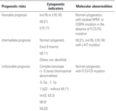
mainly diagnosed using karyotypes and by determining the molecular alterations in leukemic blasts. Using these techniques, patients are classified into groups based on their risk of relapse: high, intermediate, and low. Meanwhile, patients’ prognoses are classified based on their cytogenetic and molecular abnormalities: favorable, intermediate, or unfavorable, as depicted in table 1.
Currently, AML therapies are based solely on the patients’ risk of relapse. Thus, patients with an intermediate or high risk of relapse receive hematopoietic stem cell transplantation therapy, while low-risk patients only receive chemotherapy treatment(2).
Cytogenetic analysis classifies patients into their prognosis groups. For example, the chromosomal aberrations t(8;21)(q22;q22) and inv(16)(p13;q22) cause the genetic rearrangements RUNX1-RUNX1T1 (AML1-ETO) and CBFB-MYH11, respectively. Patients with these alterations are classified into the group with a favorable prognosis(3).
Thus, identifying the risk factors associated with the relapse of disease is critical for prognostic stratification and to determining the appropriate CBF leukemia treatment. In approximately 25% of patients with CBF AML, mutations in the proto-oncogene KIT are present(4,5).
Several independent studies have shown that KIT mutations are associated with an increased risk of relapse and higher mortality rates in CBF AML patients; however, the mechanisms behind this association
remain unknown(4-8). Therefore, developing tests to
detect KIT mutations is vital to prognostic evaluation and classification of CBF AML patients.
OBJECTIVE
The objective of this study is to develop a new, in-house test for KIT mutations in patients with AML using three complementary methodologies: DNA sequencing, fragment analysis, and conformation-sensitive gel electrophoresis (CSGE). After developing this method, we will test and subsequently implement this method
in the clinical laboratory of Hospital Israelita Albert
Einstein (HIAE).
METHODS
Samples
Bone marrow or whole blood samples of AML patients (n=54) who were previously analyzed in the HIAE
clinical laboratory were used for the cytogenetic and molecular evaluations in this study. Among the patients studied, 23 were diagnosed with CBF leukemia.
Extraction of genomic DNA
Using commercial kits, genomic DNA was extracted according to the manufacturers’ instructions: the QIAamp DNA minikits (QIAGEN, Hilden, Germany) were used for whole blood samples; the Brazol kits (LGC Biotechnology, São Paulo, Brazil) were used for bone marrow samples. The bone marrow samples were fixed in a methanol: acetic acid solution with a final elution volume of 50µL. The extracted DNA was quantified using the NanoDrop 2000 spectrophotometer (Thermo Scientific, USA) and diluted to a final concentration that ranged from 30 to 50ng/µL.
Detection of KIT gene mutations in exons 8 and 17
In CBF AML patients, there are mutations in exons 8
and 17 of the KIT gene(5); therefore, these two exons
were chosen for the current study. The mutational patterns differ between the two exons; there are point mutations in exon 17 and insertions/deletions in exon 8. Thus, to detect mutations in exon 17, we used direct sequencing of the polymerase chain reaction (PCR) product, and to detect mutations in exon 8, we used PCR with labeled primers followed by capillary electrophoresis and fragment analysis. Using primer pairs previously described in the literature(5), we studied the KIT gene. The specificity analysis of these primers
was performed using in silico PCR from [http://
genome.ucsc.edu/cgi-bin/hgPcr?command=start]. Subsequently, a new reverse primer was designed for exon 17, thereby allowing better analysis of this region.
The initial optimizations of the PCR reactions
included variations in the MgCl2 and primer
concentrations, temperature gradient tests (annealing between 51 and 71.5°C) and the addition of 4% dimethylsulfoxide (DMSO). After comparing the results, the optimal reaction conditions were selected. To detect mutations in exon 8, the following techniques were used: capillary electrophoresis, direct sequencing of the PCR product, and CSGE. A description of these processes is detailed below.
Amplification and detection of mutations in exon 17
For amplification of this region, we initially used
the primers described by Boissel et al.(5), which
produced a 294-bp product: c-Kit exon 17 FW 5’-TG GTGTACTGAATACTTTAAAACAAAA-3’; c-Kit exon 17 RV 5’-TGCAGGACTGTCAAGCAGAG-3’.
Table 1. The prognostic classifications of acute myeloid leukemia based on their cytogenetic and molecular abnormalities
Prognostic risks Cytogenetic
indicators Molecular abnormalities
Favorable prognosis inv(16) or t(16;16) Normal cytogenetics: with isolated NPM1 or CEBPA mutation in the absence of FLT3-ITD mutation t(8;21)
t(15;17)
Intermediate prognosis Normal cytogenetic t(8;21), inv(16), t(16;16): with c-KIT mutation Exon 8 trisomy
t(9;11)
Others non identified
Unfavorable prognosis Complex karyotype
(≥ 3 clonal chromosomal
abnormalities)
Normal cytogenetic: with FLT3-ITD mutation
-5, 5q-, -7,
7q-11q23 – without t(9;11)
inv(3), t(3;3)
t(6;9)
t(9;22)
Subsequently, the reverse primer was replaced to generate a 435-bp product: c-Kit exon 17 reverse2 - TAGTAATGTTCAGCATACCATGCAAATT.
The PCR reaction was performed using a cocktail containing 1x PCR buffer, 1 mM MgCl2, 0.2 mM dNTPs, 0.16µM primers c-Kit 17 FW and RV, 1 U Taq polymerase, 2µL genomic DNA (with concentrations between 30 and 50ng/µL) and sterile water qsp with a total reaction volume of 25µL. The thermocycling program consisted of an initial denaturation step at 94°C for 5 minutes, 35 cycles at 94°C for 45 seconds, 56°C for 45 seconds, 72°C for 45 seconds and a final extension at 72°C. The aliquots of the PCR product were evaluated using gel electrophoresis in 2% agarose to determine the success of the amplification.
Next, the PCR products were column purified (Illustra GFX™ DNA and Gel Purification Kit, GE
Healthcare, UK) and sequenced using the BigDye®
v3.1 (Applied Biosystems, USA) sequencing reaction kit, where each of the amplification primers were used at a concentration of 1.6µM to read both DNA strands. The sequencing was performed on an ABI PRISM 3500xL automated sequencer (Applied Biosystems, USA), and the sequences were analyzed using the
SeqScape® Software v2.7 (Applied Biosystems, USA)
with reference sequence alignment.
Amplification and detection of mutations in exon 8
For amplification of this region, we used the primers
described by Boissel et al.(5), which produced a
219-bp product: c-Kit exon 8 FW 5’-GCTGAGG TTTTCCAGCACTC-3’; c-Kit exon 8 RV 5’-AATT GCAGTCCTTCCCCTCT-3’.
The PCR reaction and the thermocycling conditions used were the same as the PCR protocol for exon 17. For capillary electrophoresis analysis, the forward primer was labeled with FAM at the 5’ end.
Initially, the PCR products were sequenced as described for exon 17. When potential mutations were detected in exon 8, these products were subsequently analyzed using CSGE. In the CSGE reaction, the PCR products were denatured for 5 minutes at 98°C followed by a reannealing step to produce heteroduplex formation, where a strand without mutations was annealed to a mutated strand for 30 minutes at 65°C. The heteroduplex was detected by identifying differential migration on a 4% agarose gel that was stained with ethidium bromide. The second strategy to detect mutations was fragment analysis using the ABI PRISM 3500xL automated sequencer (Applied Biosystems, USA) for capillary electrophoresis.
The PCR reaction was performed under the same conditions, as described above. The PCR products were diluted 30-fold in sterile water, and 1µL was used for the reaction. Next, these PCR products were added to 8.7µL of formamide and 0.3µL of the standard DNA
ladder (GeneScanTM 500 LIZ®, Applied Biosystems,
UK). The mixture was incubated for 3 minutes at 95°C and was cooled on ice for 2 minutes. The samples were applied to the sequencer and analyzed using the GeneMapper v4.1 software (Applied Biosystems, USA) to verify the fragment sizes.
Sensibility
After standardization, the sensitivity of detecting mutations in each exon was verified. For this, PCR products of a sample with approximately 50% mutated cells were diluted in a wild-type sample at equivalent DNA concentrations. Subsequently, a serial dilution was performed in the ratios of 1/1, 1/2, 1/4, and 1/8 to determine the minimum percent of mutated product that could be detected.
This work was approved by the ethics and research committee of the HIAE (process 1.663/11).
RESULTS
Detection of mutations in exons 8 and 17
Fifty-four AML patient samples were analyzed. Of these samples, 31 (AML1) were from patients that were not characterized as CBF AML, and 23 were characterized as CBF AML (AML2) patients, 16 had the chromosomal aberrations t(8;21), and 7 patients had the chromosomal aberration inv(16).
None of the AML1 group samples had mutations in the KIT gene; the samples from this group had
previously been studied for mutations in the FLT3 and
NPM1 genes. In the AML2 group, direct sequencing of
PCR products was analyzed; it was observed that one of these patients had a sequencing pattern that suggested a deletion and/or an insertion in exon 8 (Figure 1).
codons. This primer design ensured that the regions of the gene most likely to contain mutations in codons 801-828 were separated from the sequencing primers, thus improving the quality of the sequence.
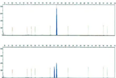
A complete analysis of the AML2 group samples revealed KIT gene mutations in seven cases. Among them, four patients had a mutation in exon 8, two patients in exon 17 (Figure 4), and one patient had mutations in both exon 8 and exon 17 (Table 2). Unfortunately, there is no information regarding the clinical outcome of these patients because this study was conducted using previously stored samples, and many of the patients did not receive follow-up care at this institution.
Figure 2. An agarose gel electrophoresis depicts the products formed after the CGSE reaction. Sample 1: patient with no sequence change without heteroduplex formation; Sample 2: patient with altered sequence with heteroduplex formation, indicating the presence of mutation in exon 8
Figure 3. The fragment analysis detected using capillary electrophoresis. (A) Single peak (218 bp): sample without mutation. (B) Double peak (215 bp and 218 bp): sample with mutation; in this case, deletion of three nucleotides
Figure 4. The electropherograms of patients 1, 2, and 3 (see Table 2) that demonstrate heterozygous mutations on exon 17
Figure 1. A depiction of the samples obtained from the AML2 Group after automated sequencing. The changes in the genomic sequence are indicated by nucleotide ambiguities at a single position in exon 8, indicated by the letters Y, W, S (IUPAC nomenclature). The sequences with red arrows belong to the same patient, who is a carrier of an exon 8 mutation in the KIT gene
Table 2. A comparison of the KIT gene mutations in patients with acute myeloid leukemia according to the exon involved
Patient Karyotype Exon 17 Exon 8
1 t(8;21) D816Y Absence of mutation
2 t(8;21) D820G Deletion of 3 nucleotides
3 t(8;21) N822K Absence of mutation
4 t(8;21) Absence of mutation Deletion of 3 nucleotides
5 inv(16) Absence of mutation Insertion of 6 nucleotides
6 inv(16) Absence of mutation Deletion of 6 nucleotides
7 inv(16) Absence of mutation Deletion of 3 nucleotides
Sensitivity
DISCUSSION
Detecting KIT gene mutations is essential for the diagnostic evaluation of CBF AML patients. Previous studies have identified these mutations in approximately
25% of CBF AML patients(4,5).
While patients with CBF AML or a wild-type KIT gene have a favorable prognosis when treated with
chemotherapy alone(9,10), the presence of KIT mutations
in this group of patients is associated with decreased disease-free survival(11) and overall survival(7,8). This fact led to the inclusion of KIT mutation detection in the prognostic stratification of patients by the National Comprehensive Cancer Network (NCCN). At present, patients with CBF AML and mutated KIT are receiving hematopoietic stem cell transplantations during their first complete remission(12).
In the present study, the detection of exon 8 and exon 17 mutations in the KIT gene was standardized. For this purpose, primers already described in the literature were used, and new primers were designed, which allowed a better evaluation of the sequencing product. Three different techniques detected the mutations, and two of the techniques were standardized for routine laboratory use. The KIT gene mutation was detected in 7 out of 23 patients with CBF AML at the HIAE laboratory. However, additional studies during early clinical stages and with a larger number of patients are necessary to estimate the actual frequency of these mutations in the population. In accordance with the literature, all of the mutations were detected in patients
from the AML2 group of CBF AML patients(13), and no
mutations were found in the KIT gene in other types of AML. However, mutations were detected in either exon 8 or exon 17, and one patient showed mutations in both exons. Although archived samples were used in this study, this test allows the patients in this study to be reclassified from a low relapse risk group to a high relapse risk group, thereby improving their prognostic stratification.
As described herein(13), the mutations in exon 8 are characterized by insertions and deletions, while in exon 17, there are point mutations. Therefore, the former are detectable using fragment analysis, while the latter require detection using sequencing.
Finally, according to previous reports, the four insertions or deletions detected in exon 8 occur in multiples of three nucleotides, thereby maintaining
the reading frame(14). The sensitivity of the tests (5%
for fragment analysis and 12.5% for sequencing) is consistent with previous data and is suitable for diagnostic sample analyses in samples that by definition contain at least 20% neoplastic cells.
However, for KIT gene mutations to detect the minimal residual disease, assays that are more sensitive are required, such as allele-specific PCR.
Several of the patients in this study had been
assessed in a previous study for mutations in their FLT3
and NPM1 genes(15). None of the patients with these
mutations showed concomitant mutation in the KIT gene. The research of KIT mutations is included in the diagnosis and treatment algorithm of the NCCN, and therefore this test will be incorporated into the tests performed at the HIAE clinical laboratory. The present study emphasizes the need to incorporate new genetic marker detections into routine diagnostics to facilitate the correct prognostic stratification and the best approach to treat acute leukemia.
CONCLUSION
In the present study, we describe the HIAE laboratory’s development and standardization of a new test to detect mutations in the KIT gene. KIT mutations were detected in seven CBF AML patients. The availability of this diagnostic test represents an advance in the treatment of CBF AML patients by providing better prognostic stratification and identifying the most appropriate treatment.
REFERENCES
1. Estey E, Döhner H. Acute myeloid leukaemia. Lancet. 2006;368(9550):1894-907. 2. Döhner H, Estey EH, Amadori S, Appelbaum FR, Büchner T, Burnett AK, Dombret H, Fenaux P, Grimwade D, Larson RA, Lo-Coco F, Naoe T, Niederwieser D, Ossenkoppele GJ, Sanz MA, Sierra J, Tallman MS, Löwenberg B, Bloomfield CD; European LeukemiaNet. Diagnosis and management of acute myeloid leukemia in adults: recommendations from an international expert panel, on behalf of the European LeukemiaNet. Blood. 2010;115(3):453-74. 3. Grimwade D, Walker H, Oliver F, Wheatley K, Harrison C, Harrison G, et al. The
importance of diagnostic cytogenetics on outcome in AML: analysis of 1,612 patients entered into the MRC AML 10 trial. The Medical Research Council Adult and Childrens Leukaemia Working Parties. Blood. 1998;92(7):2322-33. 4. Care RS, Valk PJ, Goodeve AC, Abu-Duhier FM, Geertsma-Kleinekoort WM, Wilson GA, et al. Incidence and prognosis of c-KIT and FLT3 mutations in core binding factor (CBF) acute myeloid leukaemias. Br J Haematol. 2003; 121(5):775-7.
5. Boissel N, Leroy H, Brethon B, Philippe N, de Botton S, Auvrignon A, Raffoux E, Leblanc T, Thomas X, Hermine O, Quesnel B, Baruchel A, Leverger G, Dombret H, Preudhomme C; Acute Leukemia French Association (ALFA); Leucémies Aiguës Myéloblastiques de l’Enfant (LAME) Cooperative Groups. Incidence and prognostic impact of c-Kit, FLT3, and Ras gene mutations in core binding factor acute myeloid leukemia (CBF-AML). Leukemia. 2006;20(6):965-70. 6. Cairoli R, Beghini A, Grillo G, Nadali G, Elice F, Ripamonti CB, et al. Prognostic
impact of c-KIT mutations in core binding factor leukemias: an Italian retrospective study. Blood. 2006;107(9):3463-8.
clinical outcome in patients with acute myeloblastic leukemia harboring t(8;21)(q22;q22). Leukemia. 2005;19(8):1361-6.
12. Gupta V, Tallman MS, Weisdorf DJ. Allogeneic hematopoietic cell transplantation for adults with acute myeloid leukemia: myths, controversies, and unknowns. Blood. 2011;117(8):2307-18.
13. Malaise M, Steinbach D, Corbacioglu S. Clinical implications of c-Kit mutations in acute myelogenous leukemia. Cur Hematol Malign Rep. 2009;4(2):77-82. 14. Gari M, Goodeve A, Wilson G, Winship P, Langabeer S, Linch D, et al. c-kit
proto-oncogene exon 8 in-frame deletion plus insertion mutations in acute myeloid leukaemia. Br J Haematol. 1999;105(4):894-900.
15. Velloso ED, Motta CH, Furtado JB, Bacal NS, Silveira PA, Moyses CB, et al. Molecular and cytogenetic abnormalities in acute myeloid leukemia: review and case studies. Einstein. 2011;9(2 Pt1):184-9.
T, Perrotti D, Vardiman JW, Carroll AJ, Kolitz JE, Larson RA, Bloomfield CD; Cancer and Leukemia Group B. Adverse prognostic significance of KIT mutations in adult acute myeloid leukemia with inv(16) and t(8;21): a Cancer and Leukemia Group B Study. J Clin Oncol. 2006;24(24):3904-11.
9. Byrd JC, Ruppert AS, Mrózek K, Carroll AJ, Edwards CG, Arthur DC, et al. Repetitive cycles of high-dose cytarabine benefit patients with acute myeloid leukemia and inv(16)(p13q22) or t(16;16)(p13;q22): results from CALGB 8461. J Clin Oncol. 2004;22(6):1087-94.
10. Byrd JC, Dodge RK, Carroll A, Baer MR, Edwards C, Stamberg J, et al. Patients with t(8;21)(q22;q22) and acute myeloid leukemia have superior failure-free and overall survival when repetitive cycles of high-dose cytarabine are administered. J Clin Oncol. 1999;17(12):3767-75.