SUDDEN SENSORINEURAL HEARING LOSS
EVALUATION OF CO-MORBIDITIES AND POTENTIAL CLINICAL ASSOCIATIONS
SUDDEN SENSORINEURAL HEARING LOSS
EVALUATION OF CO-MORBIDITIES AND POTENTIAL CLINICAL ASSOCIATIONS
Dissertação de Mestrado em Investigação Clínica
Clinical Research Master’s Dissertation
Prof. Doutor Paulo Vera-Cruz (orientador)
(Marinha de Guerra Portuguesa/ CEDOC – Centro de Estudos de Doenças Crónicas/ Faculdade de Ciências Médicas da Universidade Nova de Lisboa)
Prof. Doutor Pedro Caetano (orientador)
(CEDOC – Centro de Estudos de Doenças Crónicas/ Faculdade de Ciências Médicas da Universidade Nova de Lisboa)
ABSTRACT
Sudden sensorineural hearing loss (SSHL) is characterized by abrupt, mostly unilateral loss of hearing, frequently associated to aural fullness, tinnitus and vertigo. It affects 5-20/100.000 people/year (particularly working adults in the 40ths), with huge impact on quality of life. Possible causes include infectious, circulatory, traumatic, immunologic, metabolic, neoplastic, neurologic, toxic and unidentified cochlear diseases. Nevertheless, SSHL’s etiology remains unknown in most cases (80%), giving rise to controversial (and frequently ineffective) treatments. Available therapies range from corticosteroids to antivirals, vasodilators, antiaggregants, anticoagulants, vitamins and hyperbaric oxygen (HBO).
Given the lack of data concerning SSHL’s etiology and physiopathology, we intend to evaluate clinical evolution of such patients treated with HBO in the Underwater and Hyperbaric Medical Center (UHMC) at Lisbon from 2000 to 2005 during a minimum period of 5 years, in an attempt to identify eventual risk factors or clinical associations to SSHL.
The intended retrospective study is based on the review of patients’ medical charts from UHMC and confidential follow-up questionnaires applied telephonically both to patients (study group) and patients’ spouse/partner/close family member (control group), focusing past and present medical history.
A preliminary study of 20 subjects (10 of each group) was performed to anticipate difficulties and to estimate the required logistics. The identified difficulties were: 1) selection of subjects with valid phone numbers and complete medical charts (with initial and final audiograms); 2) telephonic contact with subjects from the study and control group; 3) human logistics required.
RESUMO
A surdez súbita (SS) caracteriza-se por uma perda abrupta de audição, mais frequentemente unilateral e associada a sensação de preenchimento aural, acufenos e vertigem. Afecta 5-20/100.000pessoas/ano (sobretudo adultos em fase activa na década de 40), com grande impacto na qualidade de vida. Possíveis causas incluem doenças infecciosas, circulatórias, traumáticas, imunológicas, neoplásicas, neurológicas, tóxicas e cocleares. No entanto, a causa da SS permanece desconhecida na maioria dos casos (80%), o que origina tratamentos controversos e frequentemente ineficientes. Os tratamentos disponíveis variam desde corticosteróides a antivirais, vasodilatadores, anti-agregantes, anticoagulantes, vitaminas e oxigénio hiperbárico (OHB).
Atendendo a falta de informação relativa à etiologia e fisiopatologia da SS, pretendemos avaliar a evolução clínica dos doentes com SS tratados com OHB no Centro de Medicina Subaquática e Hiperbárica (CMSH) de Lisboa entre 2000 e 2005, durante um período mínimo de 5 anos, na tentativa de identificar eventuais factores de risco ou noxas clínicas com a SS.
O estudo retrospectivo proposto baseia-se na revisão de processos clínicos do CMSH e na aplicação telefónica de questionários médicos de “follow-up” confidenciais – tanto a doentes (grupo de estudo), como aos respectivos esposos/companheiros/membros próximos da família (grupo de controlo) –, com particular ênfase nos antecedentes médicos e história clínica actual.
Um estudo preliminar de 20 pessoas (10 doentes e 10 controlos) foi efectuado para antecipar dificuldades e estimar as necessidades logísticas. As dificuldades identificadas foram: 1) selecção dos doentes com números de telefone válidos e processos clínicos completos (com audiograma inicial e final); 2) contacto telefónico com os participantes de ambos os grupos (de estudo e controlo); 3) recursos humanos requeridos.
CONTENTS
Introduction
Sudden Sensorineural Hearing Loss ………...….. 2
Pathophysiology ………. 2
Diagnosis ……… 4
Treatment ………... 5
Prognosis ……… 7
Hyperbaric Oxygen ……… 9
Biochemical effects of O2…..….………... 9
Hyperoxia and hyperoxygenation ……….. 9
Physiological and therapeutic effects of O2………. 10
Clinical indications of HBO ………... 12
Objectives ……….. 14
Material and Methods ……….. 16
Results ………..…. 19
Discussion ………. 25
Conclusion ……… 29
References ………. 31
Annexes Annex 1 ……… 38
Annex 2 ……… 43
Annex 3 ……… 50
Annex 4 ……… 54
Annex 5 ……… 56
INTRODUCTION
Sudden Sensorineural Hearing Loss
Sudden sensorineural hearing loss (SSHL) is characterized by new onset of unilateral or bilateral sensorineural hearing loss that occurs within minutes or hours. It is defined by the US National Institute for Deafness and Communication Disorders (NIDCD) as an idiopathic loss of hearing of at least 30 decibels (dB) affecting at least 3 consecutive frequencies occurring within 3 days (compared to the opposite ear’s thresholds)1. The term “idiopathic” is applied when no known cause is identified despite adequate investigation (including clinical, laboratory and imaging tests) – which happens in 85-90% of the cases2.
Estimates of the overall incidence of SSHL range from 5 to 20 per 100.000 persons per year2, affecting particularly working adults in the 40ths, with equal sex distribution. It is likely though that its incidence is increasing and also underestimated due to a high rate of spontaneous remission or recovery3-5.
This relatively common otologic condition owns its special place in the otolaryngology literature due to its unclear etiology and its consequently controversial (and frequently ineffective) treatment. Despite its 1st early description, in 1944 (De Klein)6, the entity is still poorly understood and significant uncertainty subsists among otolaryngologists concerning the choice of what seems to be the more effective treatment regimen.
Pathophysiology
SSHL is believed to occur as a direct damage to the cochlea, auditory nerve or, less frequently, higher aspects of central auditory perception or processing. Four main theories have been proposed to explain its mechanisms:
1. Viral
patients visiting their otolaryngologist without hearing loss). According to this theory, viral infection of the cochlea or the cochlear nerve and/or reactivation of a neutropic virus (such as herpesvirus) in the internal ear would be responsive for the hearing loss5,7-8. This theory is supported by higher rates of seroconversion for the herpesvirus family found in the population of patients with sudden hearing loss, and cochlear damages consistent with viral injuries found in temporal bone histopathologic studies of such patients9-10. Another supportive fact to the viral theory is the seasonal pattern of SSHL’s occurrence: higher in April and October, compatible with seasonal peaks of infection with rhinovirus (April, October), influenza, measles, rubella and respiratory syncytial’ viruses (March-May). In addition, 30 to 40% of the patients that experience SSHL relate acute respiratory infection in the precedent week.
Neverheless, the identified patterns of cochlear damage (loss of hair cells and supporting cells, atrophy of the tectorial membrane and the stria vascularis, and neuronal loss) can also be observed in other pathologies, and no virus, viral antigens or RNA/DNA was ever identified in the inner ear during histopathologic studies.8
2. Vascular
The cochlea is an end organ with respect to its blood supply, with no collateral vasculature. Vascular compromise of the cochlea due to thrombosis, embolus, reduced blood flow, or vasospasm of the arteria labyrinthi seems to be a likely etiology for SSHL, with the occurrence of severe damage to cochlear sensory cells5,7-8,11-12. The time of course correlates well with a vascular event, a sudden or abrupt loss. Moreover, cell death would not occur until a critically low oxygen partial pressure would be attained, which could relate to reversible hearing loss in cases where blood supply would be interrupted for short periods of time (less than 1h)12. However, this theory implies that patients presenting with SSHL would have higher rates of cardiovascular risk factors (such as advanced age, dyslipidemia, thromboembolic diseases, hyperviscosity syndromes, diabetes) than patients with no SSHL, which is not observed.
3. Intracochlear membrane rupture
Rupture of either or both sets of membranes may occur in cases of craniofacial trauma (with middle and inner hear injury), heavy weight lifting and intracranial pressure rise, and may theoretically be responsible for a sensory hearing loss. A leak of perilymph fluid into the middle ear via the round window or oval window has been postulated to produce hearing loss by creating a state of relative endolymphatic hydrops or by producing intracochlear membrane breaks, which would allow mixing of perilymph and endolymph, effectively altering the endocochlear potential5,7-8. Histologic evidence has been documented by Gussen13.
4. Immune-mediated inner ear disease
Cochlear inflammation may be secondary to autoimmune etiologies, with cochlear and/or retrocochlear damage provoked by activated T cells, resultant from infectious diseases or circulating autoantibodies. The association of hearing loss in Cogan syndrome, systemic lupus erythematosus, polyarteritis nodosa, Buerger’s disease and other autoimmune rheumatologic disorders has been well documented5,7-8. Progressive sensorineural hearing loss is observed in those cases, but eventual link to SSHL is a strong possibility. The existence of multiple immune-mediated disorders and autoantibodies (against inner ear proteins such as choline transporter-like protein 2, collagen type 2, β-actine, coclein, β-tectorine) has been identified by recent studies in patients with SSHL14-18. With better markers for inner ear autoimmunity, perhaps a greater linkage with SSHL will be found. Questions about an eventual link between the time of occurence of SSHL and the development of an autoimmune disease remain – at what point of disease progression is expected SSHL to happen? May it be its first symptom?
More recently, neoplastic, metabolic, neurologic and toxic etiologies were also identified19 as a potential cause of SSHL, with no further success in clarifying this condition.
Diagnosis
Clinical features of SSHL are very characteristics: a sudden hearing loss, generally unilateral, associated to tinnitus in 70% of the cases and aural fullness. Vertigo is reported in as much as 50% of the patients, varying from mild instability to severe vertigo5,7. The overall audiological burden of SSHL is considerable: difficulty with conversation and hearing in noisy environments, and inability to locate the origin of sound (with related frustration and disorientation, and possible danger, with higher risk for accidents)19. Nevertheless, and most of the times, the most impairing symptoms are the accompanying tinnitus (sometimes so severe that it can disrupt sleep and the ability to concentrate at work or in social interactions, with subsequent depression, irritability, frustration, stress and feelings of helplessness) and vertigo (which may vary from a mild and supportable unbalance to a severe loss of spatial references and inability to function). Specific clinical questionnaires – Tinnitus Handicap Inventory (THI)20 and Dizziness Handicap Inventory (DHI)21 – were developed to assess functional, emotional and social impact of such symptoms (Annex 1). As a brief, easily administered self-report questionnaire, these inventories are extremely useful not only as a simple mean to evaluate the disabling consequences of tinnitus and dizziness, but also as a qualitative and quantitative measure of initial and post-treatment handicap.
treatable diseases (for example vestibular schwannoma, central nervous system malignancies, stroke or degenerative illnesses).
Treatment
Treatment of SSHL remains controversial among otologists. Available therapies are varied, administered on either an inpatient or outpatient basis, and in different pharmacological doses and schemes. This diversity reflects both the different etiologies that may cause sudden hearing loss and the uncertainty in diagnosis. They range from:
- Antiviral agents: acyclovir and valacyclovir have been used to treat a presumed viral etiology of SSHL5,7. An important limitation is their effectiveness, only useful against herpesvirus family, and in the 1st 48-72h of infection.
- Vasodilators: in theory, vasodilators improve the blood supply to the cochlea, reversing hypoxia. In general, these agents have effects on the systemic vasculature5,7. Examples of agents used in the past: carbogen, procaine, niacin, papaverine.
- Rheologic agents: by altering blood viscosity, these agents seek to ameliorate oxygen delivery to peripheral tissues22-24. Dextrans (plasma expansors) cause hypervolemic hemodilution and affect factor VIII, improving by two ways blood flow. Pentoxifylline does that by affecting platelet deformability. Anticoagulants (heparin, warfarin) interfere with the coagulation cascade, avoiding thrombi and emboli.
- Corticosteroids: oral corticosteroid therapy is one of the few modalities that have proved to be effective in randomized controlled studies19,27-28, probably due to its anti-inflammatory and immunosuppressive effects, also verified in the cochlea and auditory nerve. However, it is related to significant systemic adverse effects and the alternative (intratympanic steroid therapy) is still under study 29-33.
- Hyperbaric oxygen (HBO): as a primary, adjunct or secondary therapy, it appears to be a valid choice7,19,34-35, especially where systemic steroids are contraindicated or in patients who failed to respond to such drugs, with the benefit of less adverse effects. Its mechanism of action will be discussed in the next chapter.
Recent guidelines from the American Academy of Otolaryngology resume the current knowledge and only recommend for the treatment of SSHL, as an “option” (evidence grade D), the use of systemic corticoterapy (intratympanic delivery may be considered in selected cases) and/or HBO, initiated in the 1st 6 weeks or 3 months of occurrence of SSHL, respectively19. Real effectiveness of such treatments remains unclear, partly due to the lack of double-blind randomized controlled studies (case of HBO), and partly due to the high rate of spontaneous remission or recovery, which tends to occur between the first 2 weeks and 6 months of the onset of the hearing loss (therefore coincident with the beginning of treatments).
Prognosis
erroneously raise estimations. The true spontaneous recovery rate is therefore unknown, but efforts are being made to gather reliable data.
The same happens with the estimated rates of recovery of the remaining 37%-43% patients with no spontaneous recovery submitted to treatment for SSHL. Estimated rates of total and partial recovery rates with current treatment regimens are as low as 18-25% and 40-60% respectively3-5, but true values will only be known after standardization of recovery criteria.
To address this issue, the American Academy of Otolaryngology proposes in its recent guidelines about SSHL, the following measures for futures outcomes assessment19:
- If no known asymmetry of hearing existed before the onset of SSHL, the opposite ear should be the reference against which recovery should be compared.
- Complete recovery requires return to within 10dB of the unaffected ear and recovery of word recognition scores to within 5 to 10% of the unaffected ear. - Partial recovery is characterized by more than 10dB improvement of the
hearing level.
- No recovery is verified when less than 10dB improvement of hearing level is achieved.
Hyperbaric Oxygen
Hyperbaric oxygen (HBO) is based on the delivery of pure (100%) oxygen to patients at pressures that are 2-3 times higher than sea level atmospheric pressure. The patients are kept inside a hyperbaric chamber (Annex 1) during 90 minutes and the increased pressure they endure (usually 2,5 atmospheres – ATA) aims at enhancing the amount of oxygen dissolved in the plasma, thus increasing the arterial partial pressure of oxygen and allowing it to reach hypoxic tissues (the cochlea, in this case)37.
Biochemical effects of O2
Before reaching the sites of utilization within the cells, the oxygen moves down a pressure gradient from inspired to alveolar gas, arterial blood, capillary bed, and across the interstitial and intercellular fluid. Under normobaric conditions, the gradient of pressure of oxygen (PO2) known as the “oxygen cascade” starts at 21,2 kPa (159
mmHg) and ends up at 0,5-3 kPa (3,8-22.5 mmHg) depending on the target tissue7. The arterial oxygen pressure (PaO2) is approximately 95 mmHg and the tissue oxygen
pressure (PtO2) is approximately 55 mmHg. These values are markedly increased by
breathing pure oxygen at greater than atmospheric pressure.
HBO is limited by toxic oxygen effects to a maximum pressure of 300 kPa (3 ATA). Because partial pressure of carbon dioxide in arterial blood does not vary significantly between 100 and 300kPa (1 and 3 ATA), the increased alveolar oxygen in hyperbaric condition easily passes the alveolar-capillary space and is diffused into the venous pulmonary capillary bed in higher quantities than in normobaric condition, accordingly Fick’s laws of diffusion7 (Annex 2).
Hyperoxia and hyperoxygenation
because 1g of 100% saturated hemoglobin carries 1,34mL of oxygen38. In such condition, the amount of oxygen dissolved in plasma is 0,285vol.%, performing a total of 19,985vol.% of oxygen38 (Annex 2).
The main effect of HBO is hyperoxia. During this therapy, and because further oxygen saturation of hemoglobin is impossible beyond 20,1vol.%, the increase of arterial oxygen is due to the increase of the dissolved fraction in the plasma. At an ambient pressure of 3 ATA and breathing 100% oxygen, the alveolar oxygen pressure is approximately 2193mmHg, the PaO2 is at least 2052mmHg and the tissue concentration
(PtO2) is at least 402,8mmHg38. At this partial pressure of oxygen, the dissolved
fraction of oxygen in plasma is approximately 6vol.% (6mL/100mL of plasma), reaching a total volume of oxygen in the circulating blood equal to 26,1vol.%, which is more than enough to fulfill basic oxygen metabolic needs38 (Annex 2).
Furthermore, it must be taken into account that hemoglobin is also fully saturated on the venous side (which does not happen under normobaric condition), which leads to increased PaO2 throughout the entire vascular bed. Since oxygen (and
other gases) diffusion is driven by a difference in partial pressure across both sides (of endothelial or tissue cellular membranes), oxygen will be forced further out of the vascular bed into tissues, diffusing to areas normally inaccessible when transported by hemoglobin38.
Physiological and therapeutic effects of O2
Another important factor to consider in ischemic tissues is the reperfusion injury, also called the “oxygen paradox”. The re-oxigenation phase is characterized by vasoconstriction, platelet and polymorphonuclear leukocytes activation, release of inflammation mediators and the production of free oxygen radicals. HBO is frequently associated with the nefarious production of free radicals but in certain conditions, and against one might though, HBO appears to be effective in reducing ischemia-reperfusion related injury39-41. Although evidence of HBO’s benefice in the treatment of this issue is increasing, further studies are needed to clarify the complete mechanism by which this phenomenon occurs.
The therapeutic actions of HBO are believed to be related not only to the direct physical effects of oxygen on blood and tissues but also to a number of secondary physiological and biochemical benefits, such as7,37-40:
- Edema reduction: ischemia induces a functional alteration of endothelial cells by the loss of their intercellular connection and energy homeostasis. Consequently, an increase in permeability of the capillary vessel wall and an in plasmatic filtration is observed causing interstitial edema responsible for compartment syndrome. The increase in compartmental pressure again causes ischemia as compression of blood vessels aggravates the problem of tissue hypoxia – a vicious circle. HBO reduces edema both by vasoconstriction, and by improved homeostasis mechanisms (such as active membrane transport – e.g. sodium-potassium pump – and leukocyte adhesion inhibition – with reduced tissue damage and enhanced leukocyte motility, thus improving microcirculation).
gelatinases), which demonstrate a much longer activity than free radicals. HBO reduces inflammation through leukocyte adhesion inhibition, with subsequent decreased release of proteolytic enzimes and pro-inflammatory cytokines41-43.
- Effect on microorganisms and host defenses against infection: direct action on anaerobic bacteria (e.g. bactericidal for Clostridium perfringens) and toxins inhibition (in most aerobic and microaerophilic microrganisms); indirect action on the microbial capability of polymorphonucleocytes and macrophages (by raising or restoring normal oxygen pressure within the infected areas, thus enhancing the leukocyte oxidative killing), and enhancement of the antimicrobial activity of some antibiotics.
- Neovascularization: angiogenesis in ischemic tissues is stimulated both by hyperoxia, a potent stimulus for fibroblast proliferation and collagen deposition (important to provide support for neovascularization), and by the relative hypoxia that occurs between the HBO treatments, a potent stimulus for angiogenesis.
- Wound healing: due to the beneficial effects described above (fibroblast proliferation, angiogenesis and antimicrobial effect).
Clinical indications of HBO
Facing the increasing evidence of HBO’s benefits, the U.S. Undersea and Hyperbaric Medical Society (UHMS) set up a committee of university experts from a number of medical and surgical fields, specialists in hyperbaric or underwater medicine, whose task was to analyze the available literature and draw from this a report on accepted indications for HBO, based on effectiveness and cost-impact. The first list of HBO indications was published in 1977 by the UHMS in the form of a report to medical funders. Since then this report has been revised every 3 years7.
OBJECTIVES
As clear, irrefutable information is still lacking about the underlying etiology, pathophysiology and mechanisms of SSHL, and as most of the Portuguese patients with this entity are referred to the Undersea and Hyperbaric Medical Center (UHMC) at Lisbon (in accordance with the most recent guidelines of best clinical practice), we aim to evaluate the clinical evolution of such patients during a minimum period of 5 years, in an attempt of getting more information about potential clinical associations.
Our primary endpoints are:
To evaluate the medical history of patients with SSHL treated with HBO in the UHMC during a minimum period of 5 years, focusing in the inset of new pathologies (particularly cardio and cerebrovascular, neoplastic, auto-immune, endocrine and/or infectious diseases).
Subsequently, to identify potential risk factors or clinical associations related to the occurrence of SSHL.
Our secondary endpoints are:
To characterize the pool of patients treated with HBO in the UHMC for SSHL, regarding demographic data, past medical history and characteristics of the hearing loss.
MATERIALS AND METHODS
This is the project of a retrospective observational study, based on patients treated with HBO in the UHMC for SSHL at least five years ago. This project – and the respective informed consent form – was submitted to and approved by the Ethical Committee of the Portuguese Navy Hospital (in which UHMC was located). It is not a study but a preliminary test instead, since the calculated sample is vast (in accordance to the relatively low incidence of SSHL), and human resource at the UHMC is currently scarce. This “pretest” comprises ten subjects whose medical charts were selected among the target population without any randomized or blinded procedure, in a reason of 2 charts per year. It was performed not to collect sound scientific data, but to anticipate eventual difficulties and estimate the logistics and materials required to conduct the planned study.
In an attempt to gather maximum information with minimum bias, the target population is the one treated in the UHMC between January 1, 2000 and December 31, 2007. The reason why more ancient patients cannot be included in this study group is related to the lack of exams in older medical charts, necessary to attest SSHL according to NIDCD1.
A control group is also necessary: patients’ spouse/partner or close family member/friend that live currently with the patient and at least since the occurrence of SSHL. This control group is an attempt to certify that any potential risk factor or clinical association identified during this study is in fact related to SSHL (with a statistically significant difference from the control group). The criteria to choose the control group are based on the inclusion of persons as much similar as possible to those from the study group, regarding confounding variables such as age, sex, social habits, alimentation, exercise and hobbies – habitually difficult to match among the regular patients of the UHMC.
The review of the medical charts aims to collect information concerning demographic data, past medical history and characteristics of the hearing loss – and, whenever registered, results of HBO therapy (Annex 3).
The questionnaires focus past and present medical history and social habits of the study and control groups, with additional information concerning the results of HBO therapy in the study group (Annex 3 and 4). Most of the questions are closed, in order to simplify, standardize and speed up the answers.
Confidentiality will be granted through the investigator’s team, all medical staff, committed to patients’ confidentiality.
Informed consent forms will previously be sent by mail to the eligible subjects (2 per subject – one for the patient and the other for his/her partner) with an anticipation of one month before the scheduled start of the study, accompanied of a prepaid envelope, in order to guarantee subjects’ full understanding and free will to participate in this study (Annex 5 and 6).
Inclusion criteria comprise: 1) the presence in each chart of an initial audiometric exam that demonstrates sensorineural hearing loss of at least 30dB affecting at least 3 consecutive frequencies (as opposed to the contralateral ear’s thresholds), and clinical reference to the sudden occurrence of this hearing loss; 2) valid telephone number; 3) presence in each chart of a final audiogram.
RESULTS
Eight hundred and seventy eight patients were treated during the defined period of time of our study. We decided to apply our preliminary test to ten patients (and ten respective control subjects).
Seventeen medical charts were consulted before the 10 patients were selected: some lacked initial audiogram (1), others lacked valid telephone numbers (3). From those who were selected, 2 didn’t answer the phone despite several attempts at different days, and 1 had changed his address.
We also experienced some problems concerning informed consent – which was asked telephonically to each subject after explaining our study project –, due to some initial suspicion and apprehensiveness regarding the calling person’s identity. Although rare and easily solved by presenting confidential data regarding the subjects themselves (addresses, date of birth, occurrence of SSHL) obtained through their medical charts, it may be a serious issue and may compromise the subjects’ collaboration. We believe, however, that this won’t be a problem if written informed consent forms will be sent prior to phone calls, as originally planned in our study.
Considering the interviews, no difficulties were experienced regarding people’s availability to answer questions. Most of them were really empathetic and willing to participate in the study, especially those who hadn’t respond to treatment for SSHL.
The participating subjects are demographically characterized in tables 1 and 2, which also demonstrate past and current medical history, related to the occurrence of SSHL. In order to guarantee subjects’ confidentiality, we attributed a number (from 1 to 10) to each subject from the control and study groups, and grouped them in accordance to patients’ recovery from SSHL after HBO therapy:
Patients 1-3 (and respective controls): complete recovery (pale orange) Patients 4-6 (and respective controls): partial recovery (mild orange) Patients 7-10 (and respective controls): no recovery (orange)
All subjects from the control group are spouses, except for subjects 5 and 6, who are brother and sister of patients 5 and 6, respectively.
Table 1. Control Group Sex Age at partner’s
SSHL diagnosis
Mean time after partner’s SSHL
Past medical history Current medical history
Subject 1 Male 28 12 0 0
Subject 2 Female 42 13 0 0
Subject 3 Male 42 11 0 0
Subject 4 Male 52 9 Chronic otitis media 0
Subject 5 Male 28 12 0 0
Subject 6 Female 20 8 0 0
Subject 7 Male 50 10 Dyslipidemia (former) dyslipidemia
Subject 8 Female 47 13 0 Dyslipidemia
Subject 9 Female 33 10 0 0
Subject 10 Male 55 11 0 Dyslipidemia,
Table 2. Study Group
Sex Age at SSHL
diagnosis
Mean time after
SSHL
Past medical history Current medical history
Patient 1 Female 27 12 Bilateral otosclerosis,
slight smocking habits
(8/day), scuba dive 0
Patient 2 Male 46 13 0 0
Patient 3 Female 43 11 Migraine, “natural”
(unknown) meds to
lose weight
(former) migraine
Patient 4 Female 53 9 0 0
Patient 5 Male 25 12 0 0
Patient 6 Male 22 8 0 0
Patient 7 Female 48 10 0 Dyslipidemia
Patient 8 Male 50 13 0 Dyslipidemia
Patient 9 Male 30 10 0 Hypertension,
auto-immune diseases:
dermatomiositis (3y),
miastenia gravis (8y)
Patient 10 Female 49 11 Non-medicated
hypertension, slight
smocking habits
(3/day)
Hypertension
In terms of current diseases (diagnosed after SSHL onset), we can verify that cardiovascular diseases appeared in both groups in a similar way (two cases of dyslipidemia and 1 of hypertension in the control group versus 2 cases of dyslipidemia and 2 of hypertension in the study group). The major difference encountered was the onset of 2 rare auto-immune diseases (dermatomiositis – only 3 years after SSHL – and miastenia gravis) in patient 9, which was not reported in the control group.
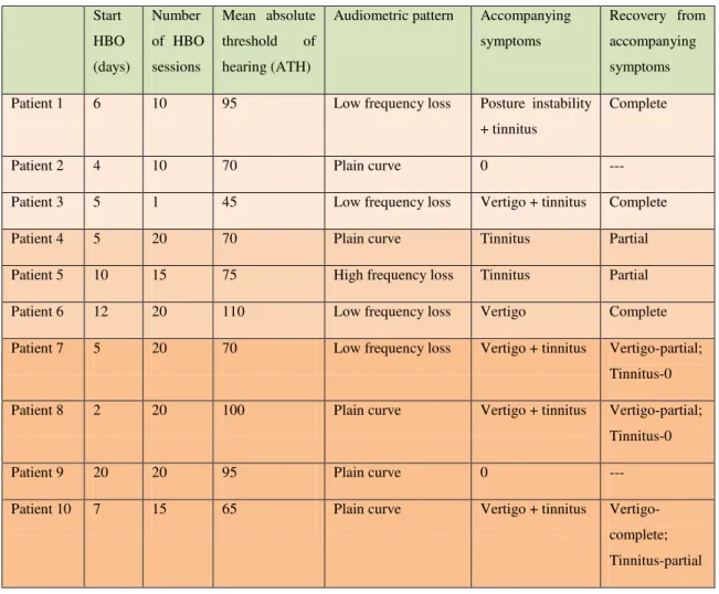
Concerning SSHL, subjects from the “no recovery” group were older (mean age of 44,3 y.o.) than those from the “complete” (38,7 y.o) and “partial” (33,3 y.o.) recovery groups. They also had a predominant plain audiometric curve (much more frequent than in the other groups) and high absolute thresholds of hearing (ATH > 80dB), suggestive of severe hearing loss – table 3.
Table 3 – SSHL characteristics in the study group Start
HBO
(days)
Number
of HBO
sessions
Mean absolute
threshold of
hearing (ATH)
Audiometric pattern Accompanying
symptoms
Recovery from
accompanying
symptoms
Patient 1 6 10 95 Low frequency loss Posture instability
+ tinnitus
Complete
Patient 2 4 10 70 Plain curve 0 ---
Patient 3 5 1 45 Low frequency loss Vertigo + tinnitus Complete
Patient 4 5 20 70 Plain curve Tinnitus Partial
Patient 5 10 15 75 High frequency loss Tinnitus Partial
Patient 6 12 20 110 Low frequency loss Vertigo Complete
Patient 7 5 20 70 Low frequency loss Vertigo + tinnitus Vertigo-partial;
Tinnitus-0
Patient 8 2 20 100 Plain curve Vertigo + tinnitus Vertigo-partial;
Tinnitus-0
Patient 9 20 20 95 Plain curve 0 ---
Patient 10 7 15 65 Plain curve Vertigo + tinnitus
Vertigo-complete;
DISCUSSION
First of all, it must be remembered that being a preliminary study, results cannot be interpreted rigorously or accurately, once the sample of subjects is too small to allow any inference. However, some observations may be pointed out:
Subjects from the control and study groups appeared to be similar and matched in terms of sex and age distribution.
Cardiovascular diseases appeared in both groups in a similar way, but patient 9 developed 2 rare auto-immune diseases in a short period of time after SSHL, which was unnoticed in the control group.
Negative prognostic factors were observed in the “no recovery” group (older age, severe hearing loss, plain audiometric pattern, accompanying vertigo) in accordance to published data.
That being said, discussion will focus problems and difficulties faced or anticipated during the preliminary test, which may influence the achievement of the study test.
The first difficulty to appear was the selection of subjects with valid phone numbers, especially among the earlier patients. Some of the times phone numbers were not written on the medical charts, in others they were incorrect or invalid.
Once the subjects were selected and the telephonic contacts begun, other obstacles arose. The most difficult to overcome was to get the subjects at the phone – either because they didn’t answer or because they weren’t at home. Another important obstacle was related to the absence of an eventual spouse/partner/family member – in cases of inexistence, patients were withdraw from the pretest; in cases of simple absence, further contact had to be established later. This difficulty was more or less easily overcome by contacting the selected subjects later during the day, from 6 to 9 pm, as described above.
Other anticipated problems were possible bias in this study. The selection bias may be a concern, since it is probable that earlier patients will be loss to follow-up due to lack of information in their charts, invalid or non-responding phone numbers, and death – of the patient himself or his spouse/partner. This last point is particularly true not only with earlier patients but also with older patients at the time of onset of SSHL.
Another important bias that was considered was confounding bias – this will hopefully be reduced through our defined control group (which is expected to be a good match for the study group) and stratified analysis of results. Accurate sample size calculation, based on the incidence of the disease and its recovery rates, on the alpha error and the study power, may also be an important point to reduce confounding bias.
Information bias is also of great concern in this study, especially because it is a retrospective study that is based on subjects’ memory (which may lead to recall bias). It is also based on incomplete, illegible medical files (written by different physicians, at different times of assessment) and telephonic questionnaires (that raise the possibility of wrong, incomplete or misleading answers from subjects who do not wish to fully expose their medical conditions). Strategies to reduce this bias may be to confront information given by patients and partners regarding each other medical conditions and time of onset (before or after SSHL), as well as reviewing with each subject all diseases specified in the questionnaire, even if they are globally denied at first.
less vigorous attempt to ascertain the partner’s diseases must be controlled and avoided by rigorous staff formation and questionnaire application.
Finally, a frequently anticipated problem is the total cost of the study. In this case, costs don’t represent a big concern, since this study is planned to be conducted by medical staff that ordinarily works or has contact to the selected patients and their respective charts at the UHMC. Extra costs, related to phone calls and papers, ink and envelopes needed to send the informed consent forms to the selected subjects don’t seem to constitute a major problem, since: a) the required information can be obtained by phone rather quickly as demonstrated in the preliminary study (at no costs with national home numbers and reduced costs for cell phone numbers), and b) all subjects live in Portugal, which significantly reduces mail prices.
CONCLUSION
SSHL is a relatively common pathology that carries substantial personal, medical and professional burden to patients’ lives. Besides its impact on quality of life, it is responsible for absenteeism and impaired professional opportunities (persons with earing disabilities are enabled to pursue military, aviation, aeronautics and musical careers, to name a few). Heavy medical costs are also a reality, mainly related to inpatient treatments (applied in some medical facilities before current guidelines) and HBO therapy (prices of full treatment – 10 to 15 sessions – are calculated to be around 760 to 1110 euros/patient). Adverse effects of some proposed therapies (including corticosteroids) also bring extra costs that are sometimes hard to handle.
HBO therapy, as a primary or adjunct treatment, appears to be effective, with fewer adverse effects than corticotherapy, but data is missing concerning real effectiveness and mechanisms of action. Likewise, SSHL is still a poorly understood entity with unclear etiopathology. Efforts must be made to clarify pathophysiology and identify causative agents, in order to ameliorate current treatments and prognosis.
As it is believed that SSHL is not a disease by itself but rather a symptom of an underlying disease, we believe that it is essential to take a step back and look closer to patients who had SSHL years ago. This epidemiologic study is therefore important, since it is intended to ascertain what medical conditions have arose since then and what clinical associations may be inferred.
REFERENCES
1. US National Institute for Deafness and Other Communication Disorders (NIDCD), National Institute of Health – Sudden Deafness [Em linha], actual. Mar.2003. [Consult. 20 Mar.2012]. Disponível em WWW: <URL: http://www.nidcd.nih.gov/health/hearing/pages/sudden.aspx>.
2. Byl, F.M. – Sudden hearing loss: eight years’ experience and suggested prognostic table. «Laryngoscope». 94 (1984) 647-661
3. Mattox, D.E.; Simmons, F.B. – Natural history of sudden sensorineural hearing loss. « Ann Otol Rhinol Laryngol». 86 (1977) 463-80
4. Hughes, G.B. [et al.] – Sudden sensorineural hearing loss. «Otolaryngol Clin North Am». 29 (1996) 393-405
5. Mathur, N.N.; Carr, M.M. – Inner ear, sudden hearing loss. Emedicine [Em linha]. 13Mar. (2012) [Consult. 21 Mar.2012]. Disponível em WWW: <URL: http://emedicine.medscape.com/article/856313-overview>.
6. De Klein, A. – Sudden complete or partial loss of function of the octavus-system in apparently normal persons. «Acta Otolaryngol». 32 (1944) 407-429
7. Mathieu, D. [et al] – Handbook on Hyperbaric Medicine. Lille: Springer, 2006. 8. Merchant, S.M.; Adams, J.C.; Nadol, J.B. – Pathology and Pathophysiology of
Idiopathic Sudden Sensorineural Hearing Loss. «Otology Neurotology». 26 (2005) 151-160
9. Schuknet, H.F.; Donovan, E.D. – The pathology of idiopathic sudden sensorineural hearing loss. «Arch Otorhinolaryngol». 243 (1986) 1-15
10. Takasaki, T. [et al.] – Serum antibodies to human herpesvirus 7, human herpesvirus 6 and cytomegalovirus in patients with idiopathic facial nerve palsy or sudden deafness. «Laryngol Otol». 112 (1998) 617-621
12. Kimura, R.S. – Animal models of inner ear vascular disturbances. «Am J Otolaryngol». 7 (1986) 130-139
13. Gussen, R. – Sudden hearing loss associated with cochlear membrane rupture. «Arch Otolaryngol». 107 (1981) 598-600
14. Yoo, T.J. [et al.] – Type II collagen-induced autoimmune inner ear lesions in guinea pigs. «Ann Otol Rhinol Laryngol Suppl» 113 (1984) 3-5
15. Boulassel, M.R. [et al.] – Identification of beta-actin as a candidate autoantigen in autoimmune inner ear disease. «Clin Otolaryngol Allied» 25 (2000). 535-541 16. Solares, C.A. [et al.] – Murine autoimmune hearing loss medited by CD4+T
cells specific for inner ear peptides. «J Clin Invest 2004» (113) 1210-1217 17. Nair, T.S. [et al.] – Identification and characterization of choline transporter-like
protein 2 (…) antibody-induced hearing loss. «J Neurosci» 24 (2004) 1772-1779
18. Nair, T.S. [et al.] – Monoclonal antibody induced hearing loss. «Hear Res» 83 (1995); 101-103
19. Stachler, R.J. [et al.] – Clinical Practice Guideline: sudden hearing loss. «Otolaryngol Head Neck Surg» [Em linha]. Vol.146:Nº 3suppl (2012) p.S1-S35. [Consult. 20 Mar. 2012]. Disponível em WWW: <URL: http://oto.sagepub.com/content/146/3_suppl/S1.full>
20. Newman, C.W.; Jacobson, G.P.; Spitzer, J.B. – Development of the tinnitus handicap inventory. «Arch Otolaryngol Head Neck Surg». 122:2 (1996) 143-148 21. Jacobson, G.P.; Newman, C.W. – The development of the dizziness handicap
inventory. «Arch Otolaryngol Head Neck Surg». 116:4 (1990) 424-427
22. Agarwal, L. [et al.] – Vasodilators and vasoactive substances for idiopathic sudden sensorineural hearing loss. «Cochrane Database Syst Rev». Issue 4 (2009)
24. Yue, W.L. [et al.] – Role of low-molecular-weight heparins in the treatment of sudden hearing loss. «Am J Otolaryngol». 24:5 (2003) 328-333
25. Joachims, Z. [et al.] – Dependence of noise-induced hearing loss upon perilymph magnesium concentration. «J Acoust Soc Am». 74 (1983); 104-108 26. Nageris, B.I. [et al.] – Magnesium for Sudden Hearing Loss. «Ann Otol Rhinol
Laryngol». 113 (2004) 672-675
27. Wilson, W.R.; Byl, F.M.; Laird, N. – The efficacy of steroids in the treatment of idiopathic sudden hearing loss. A double-blind clinical study. «Arch Otolaryngol». 106 (1980) 772–776
28. Conlin, A.E.; Parnes, L.S. – Treatment of sudden sensorineural hearing loss: I. A Systematic Review. «Arch Otolaryngol Head Neck Surg». 133 (2007) 573-581 29. Hamid, M.; Trune, D. – Issues, indications and controversis regarding
intratympanic steroid perfusion. «Curren Opin Otolaryngol Head Neck Surg». 16:5 (2008) 434-440
30. Plontke, S.K.; Salt, A.N. – Simulation of application strategies for local drug delivery to the inner ear. «J Otorhinolaryngol Relat Spec». 68:6 (2006) 386-392 31. Ahn, J.H. [et al.] – Therapeutic effectiveness over time of intratymapnic
dexamethasone as salvage treatment of sudden deafness. «Acta Oto-Laryngol». 128:2 (2008) 128-131
32. Xenelis, J. [et al.] – Intratympanic steroid treatment in idiopathic sudden sensorineural hearing loss: a control study. «Otolaryngol Head Neck Surg». 134:6 (2006) 940-945
34. Fujimura, T. [et al.] – Hyperbaric oxygen and steroid therapy for idiopathic sudden sensorineural hearing loss. «Eur Arch Otorhinolaryngol». 264 (2007) 861-866
35. Bennett, M. [et al.] – Hyperbaric oxygen for idiopathic sudden sensorineural hearing loss and tinnitus. « Cochrane Database Syst Rev». (2007) Issue I
36. Chang, N.C.; Ho, K.Y.; Kuo, W.R. – Audiometric patterns and prognosis in sudden sensorineural hearing loss in Southern Taiwan. «Otolaryngol Head Neck Surg». 133 (2005) 916-922
37. Albuquerque e Sousa, J.G. – Oxigenoterapia Hiperbárica: perspectiva histórica, efeitos fisiológicos e aplicações clínicas. «Rev Soc Portuguesa Med Interna». 14 (2007) 219-227
38. Jain, K.K. – Texbook of Hyperbaric Medicine. 3ª ed. Seattle: Hogrefe and Huber Publishers, 2004
39. Neumeister M. [et al] – Hyperbaric oxygen therapy. Emedicine [em linha]. 24 May. (2012) [Consult. 20 Jun.2012]. Disponível em WWW: < URL:http://www.emedicine.com/plastic/topic526.htm >
40. Mortensen, C. – Hyperbaric oxygen therapy. «Curr Anaesth Crit Care». 19 (2008) 333-337
42. Wilson, H.D.; Wilson, J.R.; Fuchs, P.N. – Hyperbaric oxygen treatment decreases inflammation and mechanical hypersensitivity in an animal model of inflammatory pain. «Brain Res». 1098:1 (2006) 126-128
43. Zhang Q. [et al] – Hyperbaric oxygen attenuates apoptosis and decreases inflammation in an ischemic wound model. «J Invest Dermatol». 128:8 (2008) 2102-12.
44. Niu K.C. [et al] – Hyperbaric oxygen causes both inflammation and anti-pyresis in rabbits. «Eur J Pharmacol». 606:1-3 (2009) 240-5.
45. European Committee for Hyperbaric Medicine – 7th European Consensus Conference on Hyperbaric Medicine [Em linha], actual. Mai. 2004. [Consult. 23
Jan.2010]. Disponível em WWW: <URL:
http://www.echm.org/documents/ECHM%207th%20Consensus%20Conference
%20Lille%202004.pdf>
Annex 1
Sudden Sensorineural Hearing Loss
1. Tinnitus Handicap Inventory
The THI is a 25-item questionnaire developed to assess the severity of the patients’ perceived tinnitus handicap, with items that are grouped into three subscales: functional, emotional and catastrophic responses. The functional subscale items reflect the effect of tinnitus on mental, social, occupational and physical functioning. The emotional subscale items probe the individual’s emotional reactions to the tinnitus and the catastrophic response items address whether tinnitus makes the respondent feel desperate, trapped, hopeless or out of control.
A “yes” response is given 4 points, a “sometimes” response is given 2 points and a “no” response is given 0 points. The questionnaire yields scores for each subscale and a total score that ranges from 0 and 100, with high scores indicating a greater handicap:
0–16 Slight or no handicap (Grade 1) Only heard in a quiet environment
18–36 Mild handicap (Grade 2)
Easily masked by environmental sounds and easily forgotten with activities 38–56 Moderate handicap (Grade 3)
Noticed in presence of background noise, although daily activities can still be performed
58–76 Severe handicap (Grade 4)
Almost always heard, leads to disturbed sleep patterns and can interfere with daily activities.
>78 Catastrophic handicap (Grade 5)
2. Dizziness Handicap Inventory
The DHI is a 25-item questionnaire developed to assess the severity of the patients’ perceived dizziness handicap, with items that are grouped into three subscales: functional, emotional and physical responses. The functional subscale items reflect the effect of dizziness on mental, social and occupational functioning. The emotional and physical subscale items probe the individual’s emotional and physical reactions to the dizziness, respectively.
A “yes” response is given 4 points, a “sometimes” response is given 2 points and a “no” response is given 0 points. The questionnaire yields scores for each subscale and a total score that ranges from 0 and 100, with high scores indicating a greater handicap:
0–14 Slight or no handicap 16–34 Mild handicap
Annex 2
Hyperbaric Oxygen
1. Multiplace hyperbaric chamber
The internal atmosphere is room air compressed up to 2,5 ATA. Patients inside the chamber bread compressed air, accruing a nitrogen load in their soft tissues similar to scuba divers. As such, they need to decompress to avoid decompression sickness – that is performed rigorously according to Navy tables of recompression. Hyperbaric oxygen is administered in periods of time via face mask; hood or endotracheal tube.
2. Gas laws
2.1. Dalton’s law
“The total pressure exerted by a gaseous mixture is equal to the sum of the pressures that would be exerted by the gases if they alone were present and occupied the total volume”.
Ptot = p1+ p2+ … + pn
where: p1,p2,…,pn represent the partial pressures of each component.
It allows calculating the partial pressure of each gas as follows:
“The partial pressure of a gas (p1) equals the product of total pressure of the gaseous mixture (Ptot) and the fraction of the gas (F1)”.
p1= Ptot x F1
where: F1 is defined as a part of 1; i.e. in air FO2 is 0,21..
2.2. Henry’s law
“The mass of a gas (C) that dissolves in a defined volume of liquid is directly proportional to the pressure of the gas (P) (provided the gas does not react with the solvent)”.
α x (p / C)= const, for T = const.
2.3. Fick’s law of diffusion
Rate of diffusion = (K x A xΔP) / D
3. Physiological effects of HBO
3.1. Increased fraction of O2 in circulating blood during HBO at different
levels of pressure in accordance with Dalton’s and Henry’s laws.
ATAs 1 1 2 3
FiO2 0,21 1 1 1
Alveolar oxygen pressure (PAO2, mmHg)
100 673 1433 2193
Hb(O2)4 (vol.%) 19,7 20,1 20,1 20,1
O2/Plasma (mL/100mL) 0,285 1,88 3,0 6
Hb(O2)4– hemoglobin’s oxygen saturation in circulating blood; O2/Plasma – dissolved fraction
of oxygen in the plasma.
3.2. Correction of tissue hypoxia under HBO, in accordance with Fick’s law.
ATA 1 3
Fi O2 0,21 1
Alveolar PO2 (mmHg) 100 2193
Hb(O2) 4 (vol.%) 19,7 20,1
Plasma O2 (vol.%) 0,285 6
Arterial PO2 (mmHg) 95-100 2052-2193
4. ECHM Recommendations (7th European Consensus Conference on Hyperbaric Medicine, Lille 2004)
The Jury issued its recommendations using a 3 grade scale according to the strength each recommendation had been evaluated:
- Type 1: Strongly Recommended. The implementation of the recommendation is considered of critical importance for final outcome of the patient/quality of practice/future specific knowledge.
- Type 2: Recommended. The implementation of the recommendation will positively affect final outcome of the patient/quality of practice/future specific knowledge.
- Type 3: Optional. The implementation of the recommendation is considered to be an option.
Such recommendations were supported by the following level of evidence (issue for clinical research):
- Level A: At least 2 concordant, large, double-blind, controlled randomized studies with no or little methodological bias (type 1 recommendation).
- Level B: Double-blind controlled, randomized studies but with methodological flaws; studies with only small samples, or one study only (type 2 recommendation).
- Level C: Consensus opinion of experts (type 3 recommendation).
- Level D: Only uncontrolled studies with no consensus opinion of experts.
- Level E: No evidence of beneficial action, or methodological or interpretation bias precluding any conclusion.
Annex 3
First questionnaire (study group)
Yes No
Identification
Age
Sex
Profession
Provenience
Past Medical History (date of occurence/diagnosis)
Smoking habits (cigarettes/day)
Ethanolic habits (g/day)
Drugs (cocaine, heroin, methadone)
Cardiovascular diseases
Arterial hypertension (HTN)
Dyslipidemia
Diabetes mellitus (DM)
Stroke / transient ischemic attack (TIA)
Acute coronary syndrome (ACS) / ischemic cardiomyopathy / coronary artery disease (CAD)
Dysrhythmia (atrial fibrillation – AF –, long QT syndrome)
Renal pathology
Obstructive sleep apnea syndrome (OSAS)
Autoimmune / rheumatologic diseases
Systemic lupus erythematosus (SLE) / rheumatoid arthritis (RA)
Ankylosing spondylitis (AS) / Sjogren
Polyarteritis nodosa / Wegener’s granulomatosis
Hashimoto’s thyroiditis / inflammatory bowel disease (IBD)
Multiple sclerosis (MS)
Neoplastic diseases
Vestibular schwanomma / other cerebellopontine angle tumors
Neurofibromatosis (type 2 – central nervous system)
Other brain tumors
Brain metastases / carcinomatous meningitis
Traumatic brain injury (TBI) / Barotrauma
Infections
Human Immunodeficiency Virus (HIV) / Acquired Immunodeficiency Syndrome (AIDS)
Parotiditis / measles / rubella
Cytomegalovirus (CMV) / Epstein-Barr Virus (EBV)
Hepatitis B Virus (HBV) / HSV (herpes simplex virus)
Tuberculosis (TB) / Syphilis (2ary/3ary)
Tropical infections (Chagas’ disease, Legionella, Lyme, Malaria)
Middle ear infections / labyrinthitis / vestibular neuronitis
Meningitis
Endocrine diseases
Hypo / Hyperthyroidism
DM
Adrenal pathology
Hyperviscosity
Sickle cell disease (SCD)
Antiphospholipids syndrome (APS)
Polycythemia vera (PV) / Essential thrombocytosis (ET)
Leukemia / lymphoma
Waldenstrom’s macroglobulinemia / Multiple myeloma (MM)
Medication
Aminoglycosides (gentamycin, neomycin)
Vancomycin
Tuberculostatics (streptomycin)
Antimalarials (primaquine, quinine)
Loop diuretics (furosemide)
Acetylsalicylic acid (ASA)
Phosphodiesterase type 5 inhibitors (PDE5-inhibitors: sildenafil, vardenafil, tadalafil)
Ribavarin / Pegylated interpheron alfa-2a (PEG-IFN)
SSHL
Date of occurrence / diagnosis
Laterality
Accompanying symptoms
Vertigo
Tinnitus
Precedents (30 days before SSHL)
Infectious diseases (upper or lower respiratory tract infections +/- acute otitis media, conjunctivitis, meningitis, exacerbated chronic otitis media, digestive tract or cutaneous infection; tuberculosis, rubella, mononucleosis, parotiditis, herpes, syphilis)
Brain ischemia (strong headache, syncope, stroke / TIA); asthma crisis; epilepsy crisis
Head trauma/TBI, intracranial hypertension (vomits, blurry vision)
Barotrauma / acoustic trauma (diving, snorkeling, explosion, aggression, sport / traffic accident)
Recent surgery under general anesthesia
Meds, drugs
New-onset of pathology
Audiometric exam
Severity
Further exams
Blood tests (abnormal results?)
Electrocardiogram (abnormal rhythm?)
Chest x-ray (pulmonary tuberculosis?)
Others (CT scan / MRI changes?)
Medical treatment
Corticosteroids
Antivirals
Vasodilatadors
Others
HBO therapy
Date of start
Date of completion
Number of sessions
Clinical evolution
Complete / Partial Recovery
Hearing loss?
Tinnitus?
Vertigo?
Annex 4
Second questionnaire (study and control groups)
Yes No
Identification
Age
Sex
Profession
Provenience
Past Medical History (date of occurence/diagnosis)
Smoking habits (cigarettes/day)
Ethanolic habits (g/day)
Drugs (cocaine, heroin, methadone)
Cardiovascular diseases
HTN, dyslipidemia, DM, stroke / TIA, ACS / ischemic cardiomyopathy / CAD, dysrhythmia (AF, long QT syndrome), renal pathology, OSAS
Autoimmune / rheumatologic diseases
SLE, RA, AS, Sjogren, Polyarteritis nodosa, Wegener’s granulomatosis, sarcoidosis, MS, Hashimoto’s thyroiditis, Behçet, DII
Neoplastic diseases (with or without brain metastases)
TBI / Barotrauma
Infections
HIV/AIDS, CMV, EBV, HBV, HSV; parotiditis, measles, rubella, CMV
TB, syphilis; tropical infections (Chagas’, Lyme’s, Malaria);
Menigitis; middle / internal ear infections
Endocrine diseases
Hypo/hypertiroidism, DM, adrenal pathology
Hyperviscosity
macroglobulinemia, MM
Other diseases
Hepatic disease (hepatic failure, cirrhosis, others)
Pulmonary disease (asthma, chronic obstructive pulmonary disease – COPD –, TB, pulmonary hypertension)
Meds
Chemotherapy, amynoglycosides, vancomycin, tuberculostatics, antimalarials, loop diuretics, ASA, PDE5 inhibitors, ribavarin / PEG-IFN
Follow-up (new onset of pathology)
Otologic diseases
Recurrent SSHL (if yes, report again to the 1st questionnaire – SSHL)
Meniere’s disease
Enlarged vestibular aqueduct syndrome/other congenital internal ear malformation
Cardiovascular diseases
Stroke / TIA (date of occurence, brain area damaged, number of episodes?)
Cerebrospinal fluid leak (cranial hypothension)
Autoimmune / rheumatologic diseases
Behçet, Sjogren, IBD
SLE, RA, Cogan’s syndrome, juvenile rheumatic arthritis Polyarteritis nodosa, Wegener’s granulomatosis, sarcoidis
MS, Hashimoto’s thyroiditis
Endocrine diseases
Hypothyroidism
Neoplastic diseases
Vestibular schwanomma / other cerebellopontine angle tumors
Brain metastases / carcinomatous meningitis
Neurofibromatosis (type 2 – central nervous system)
Leukemia / lymphoma, MM, Waldenstrom’s macroglobulinemia, PV, ET
Annex 5
Informed Consent Form
Sudden Sensorineural Hearing Loss – evaluation of co-morbidities and potential clinical associations
Sudden sensorineural hearing loss (SSHL) is characterized by a sudden, idiopathic loss of hearing of at least 30 decibels (dB) affecting at least 3 consecutive frequencies. It preferentially affects working adults in the 40ths, with no sex preponderance. Though its relatively low incidence (5-20/100.000 people/year), SSHL plays an important role in today’s medicine due to its huge impact on quality of life and its unknown pathophysiology, with subsequent controversial (and frequently ineffective) treatment.
Clinically, SSHL is characterized by sudden hearing loss, generally unilateral and frequently associated to aural fullness, tinnitus and vertigo. Spontaneous recovery is reported in as much as 60% of the patients, but it may take up to 6 months to occur. The remaining 40% of the patients experience variable responses to current treatments, with estimated rates of total and partial recovery rounding around 18 to 25% and 40 to 60%, respectively.
Possible causes of SSHL include infectious, circulatory, neoplastic, traumatic, metabolic, neurologic, immunologic, toxic and unidentified cochlear diseases. Nevertheless, its etiology remains unknown in the vast majority of the cases (more than 80%), giving rise to controversial choices of adequate treatment. Available therapies range from corticosteroids to antivirals, vasodilators, antiaggregants, anticoagulants, vitamins and hyperbaric oxygen, with different levels of (estimated) effectiveness and adverse effects.
Nonetheless, its real effectiveness remains unclear due to the lack of randomized controlled studies and the relatively high rate of spontaneous recovery.
Given the lack of data concerning SSHL’s etiology and mechanisms of disease, we intend to evaluate clinical evolution of patients with SSHL treated with hyperbaric oxygen in theUnderwater and Hyperbaric Medical Center at Lisbon from 2000 to 2005, during a minimum period of 5 years, in an attempt to identify eventual risk factors or clinical associations to SSHL.
If you belong to this group of patients, you and your spouse/partner/close family member will be asked to answer a clinical questionnaire performed telephonically. It will also be asked your permission to access and review your medical file from the Underwater and Hyperbaric Medical Center.
The participation in this study requires your consent. In order to help you to decide, this written information was elaborated and attached to the study’s protocol. Any questions you might have will be answered by the principal investigator, Dr. Sonia Pereira, physician at the Underwater and Hyperbaric Medical Center (contact number: +351 218840821).
Underwater and Hyperbaric Medical Center – Lisbon, January 2013
Sonia Pereira, MD Otolaryngology resident (Ordem dos Médicos nº 46181)
Annex 6
Informed Consent Form in Portuguese (Original)
Avaliação de co-morbilidades em doentes com surdez súbita e sua eventual relação
nosológica
A surdez súbita, caracterizada por uma perda súbita de audição superior ou
igual a 30 dB no mínimo de três frequências de audição, afecta preferencialmente
adultos na década de 40, independentemente do sexo. Apesar de não ser uma patologia
muito frequente (5-20/100.000 pessoas num ano), reveste-se de particular importância
pelo impacto significativo na qualidade de vida dos doentes e pela ausência da
identificação de causa na grande maioria dos casos (com consequências óbvias na
escolha do tratamento mais adequado).
As manifestações clínicas caracterizam-se por uma surdez de instalação súbita,
muitas vezes ao acordar, que se associa frequentemente a sensação de plenitude
auricular, acufenos e vertigem. A recuperação espontânea pode ir ter até aos 60%, mas
prolongar-se até 6 meses após o aparecimento da doença. Os 40% restantes dos
doentes obtêm respostas variáveis com os tratamentos actuais, com resultados a rondar
entre os 40 a 60% de recuperação parcial e os 18-25% de cura.
A escolha dos tratamentos mantém-se controversa, por desconhecimento
etiológico da doença, incluindo-se corticóides, anti-virais, vasodilatadores, expansores
de plasma, anti-agregantes e anti-coagulantes, vitaminas e sais minerais ou
oxigenoterapia hiperbárica, entre as modalidades de tratamento.
Apesar de não existir consenso absoluto entre especialistas, estudos recentes
apontam para um maior efeito dos corticóides e da oxigenoterapia hiperbárica, desde
que iniciados nos primeiros quinze dias de aparecimento da doença (coincidente com o
início da recuperação espontânea segundo a história natural da doença, o que dificulta
a avaliação da real eficácia da terapêutica instituída).
Atendendo à inexistência actual de dados esclarecedores da causa e dos
mecanismos desta doença, pretende-se avaliar a evolução dos doentes tratados por este
motivo no Centro de Medicina Subaquática e Hiperbárica entre o ano de 2000 e 2002
após um período mínimo de cinco anos, com identificação de eventuais factores de
Se pertencer a este grupo de doentes, será pedida a sua colaboração e a do seu
conjugue/parceiro(a) ou equiparado na resposta a um questionário clínico por via
telefónica. Será também pedida a sua autorização para a consulta do seu processo
médico existente no Centro de Medicina Subaquática e Hiperbárica.
A participação neste estudo necessita da sua autorização. Para o ajudar a
decidir, foi elaborada esta informação escrita, assim como foi anexado o protocolo do
estudo a efectuar, mas serão respondidas todas as eventuais questões ou dúvidas
persistentes pela médica investigadora, pertencente ao quadro clínico do Centro de
Medicina Subaquática e Hiperbárica (contacto telefónico: 218840821).
Centro de Medicina Subaquática e Hiperbárica,
Dra. Sonia Lopes Pereira
Interna de Otorrinolaringologia
Nº da Ordem dos Médicos: 46181