T
WO NEWSPECIES ANDAREVIEWOFTHEINSEMINATINGFRESHWATERFISHGENUS
M
ONOTOCHEIRODON(C
HARACIFORMES: C
HARACIDAE)
FROMP
ERUANDB
OLIVIAN
AÉRCIOA.M
ENEZES1S
TANLEYH. W
EITZMAN2I
RANIQ
UAGIO-G
RASSIOTTO3AbstrAct
Two new species of inseminating freshwater fishes of the genus Monotocheirodon, family Characidae, are described from Peru. Males and females of both new species have an external, visually obvious urogenital papilla that was not detected in the females in previous studies, with this longer in males, which use it as an inseminating organ. A third inseminating species from Bolivia, Monotocheirodon pearsoni, unstudied in any detail since its original description in 1924, is redescribed. This latter species lacks an inseminating organ. Monotocheirodon is re-described, its phylogenetic relationships are briefly discussed and it is suggested that it is possibly related to the stevardiin genera Ceratobranchia, Othonocheirodus, and Odontostoechus.
Key-Words: Inseminating characid fishes, new species and relationships of Monotocheirodon.
maintained Montocheirodon in the Cheirodontinae and although he considered the subfamily an “un-natural group,” he considered Monotocheirodon to be a cheirodontine “in the strict sense.” Further, Géry considered Othonocheirodus and Monotocheirodon to be “adaptations from some Odontostilbe-like species.” Malabarba (1998: 231) in a detailed phylogenetic revision of the Cheirodontinae removed Monotochei-rodon, Othonocheirodus and 54 other genera from that subfamily to incertae sedis in the Characidae. Mala-barba & Weitzman (2003: 73-88) in a phylogenetic study of inseminating and related non-inseminating characids placed Montocheirodon, Othonocheirodus, IntroductIon
Eigenmann and Pearson in Pearson (1924: 34) briefly described a new characid genus, Monotochei-rodon, with its single new species, M. pearsoni de-scribed by Eigenmann in the same paper (pp. 34-35). Eigenmann and Pearson stated that Montocheirodon was allied to “Cheirodon and Odontostilbe,” genera then assigned to the characid subfamily Cheirodon-tinae. Although Eigenmann did not specifically relate his new genus and species to Creagrutus Günther in his species description, he remarked that it had the “gen-eral appearance of Creagrutus.” Géry (1977: 546-547)
1. Museu de Zoologia, Universidade de São Paulo. Caixa Postal 42.494, 04218-970, São Paulo, SP, Brasil. E-mail: naercio@usp.br 2. Division of Fishes, Department of Zoology, National Museum of Natural History, MRC 0159, PO Box 37012,
Smithsonian Institution, Washington, D.C. 200013-7012, USA. E-mail: weitzmas@si.edu
Odontostoechus and many other characid genera, both inseminating and non-inseminating, in a new chara-cid subgroup they called Clade A. Weitzman et al. (2005) in a discussion of inseminating characids as-sociated with the inseminating characid subfamilies Stevardiinae and Glandulocaudinae found all three described species of Montocheirodon to be inseminat-ing and included them in a subgroup within Clade A consisting of genera with inseminating species. Mi-rande (2010) proposed that the members of Clade A characids along with a few additional characid genera were a monophyletic group which he named sub-family Stevardiinae. Their phylogenetic relationships were not resolved and as emphasized by Ferreira et al. (2011) were depicted as forming a large polytomy. This subfamily was also utilized as the representa-tive taxon of Clade A characids along with Marki-ana nigripinnis analyzed in a recent molecular study (Oliveira et al., 2011). The present contribution is designed to describe the two new species of Monoto-cheirodon, redefine this genus, and briefly discuss its relationships with the other members of the subfamily Stevardiinae.
MAterIAl And Methods
Count and measurement techniques are those de-scribed by Fink & Weitzman (1974: 1-2) and Menezes & Weitzman (2009: 296-297), except for the number of longitudinal scale rows below the lateral line which are counted from the pelvic-fin origin to the lateral line. In the descriptions, the range of meristic charac-ters is presented first, followed by the mean of the sam-ple and by counts of the holotypes and the lectotype in parentheses and total number of specimens counted. Measurements in all the tables, other than standard length (SL), are expressed as a percentage of SL ex-cept for subunits of the head that are presented as a percentage of head length. Total vertebral counts were taken from radiographs. These include the vertebrae of the Weberian apparatus as well as the complex cau-dal ossification, PU1 + U1 with the associated hypural bones and “half vertebrae” counted as one element. Meristic characters are presented in the description of the species. Tukey box plots were not used herein because no significant meristic differences were found among the species studied. Analyses for differences be-tween sexes using regressions were not performed due to the very limited number of male specimens of the three species. Basic descriptive statistics were prepared using BioEstat 5.0, in Ayres et al. (2007). A difference was considered significant when p ≤ 0.05.
All mature specimens of the species of Monoto-cheirodon were identified to sex by examination of their gonads. In most cases tissue samples for histol-ogy were taken only from particular organs. For ex-ample, in the case of the gonads, one entire gonad was removed from one side only, usually the right side.
For Transmission Electron Microscope (TEM) preparations the gonads were extracted from speci-mens previously fixed in a 10% formalin solution and preserved in 70% ethanol. Fragments of gonads were post-fixed for 48 h in solutions of 2% glutaraldehyde and 4% paraformaldehyde in 0.1 M Sorensen phos-phate buffer, pH 7.4. The material was post-fixed again for 2 h in the dark in 1% osmium tetroxide in the same buffer, stained in block with a aqueous solution of 5% uranyl acetate for 2 h, dehydrated in acetone, embedded in araldite, and sectioned and stained with a saturated solution of uranyl acetate in 50% ethanol and with lead citrate (Reynolds, 1963).
The following abbreviations are used for institutions: Academy of Natural Sciences, Philadelphia (ANSP); California Academy of Sciences, San Francisco (CAS); Museo de Historia Natural de la Universidad Mayor de San Marcos, Lima (MUSM); Museu de Ciências e Tecnologia, Pontifícia Universidade Católica do Rio Grande do Sul (MCP); University of Michigan, Mu-seum of Zoology, Ann Arbor (UMMZ); and National Museum of Natural History, Smithsonian Institution, Washington, D.C (USNM and NMNH).
Abbreviations in the text are: SL (standard length) and HL (head length).
results
Family characidae Agassiz, 1844
Monotocheirodon eigenmann & Pearson, 1924
Monotocheirodon Eigenmann & Pearson, 1924: 34 (type species: Monotocheirodon pearsoni, by monotypy).
characters, although useful for distinguishing Monoto-cheirodon are not unique to the genus.
1 – One enlarged scale on basal portion of each cau-dal-fin lobe (Figs. 1, 9, and 12).
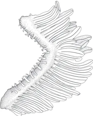
2 – Two rows (one external and one internal) of short and slender gill-rakers present on each branchial arch (Fig. 13).
3 – Single row of four distally compressed, pedun-culate, and multicuspid teeth present on the premaxilla (Figs. 2, 10, and 14).
4 – Ascending process of the premaxilla strongly bent ventrally (Figs. 2, 10, and 14).
5 – Posterior portion of maxilla strongly bent ven-trally (Figs. 2, 10, and 14).
6 – Anterior dentary teeth not notably larger than the remaining teeth on the bone. All dentary teeth gradually decreasing in size posteriorly (Figs. 2, 10, and 14)
7 – Two or three longitudinal scale rows from lateral line to pelvic-fin origin (Figs. 1, 9, and 12). 8 – Adipose fin absent (Figs. 1, 9, and 12).
9 – Anal fin short, with 8-12 branched rays (Figs. 1, 9, and 12).
10 – Hooks absent on pelvic and anal fins of males (Figs. 5 and 6).
Key to the species of Monotocheirodon
1. Horizontal diameter of eye 13.5-16.6% of HL (Fig. 3); anal-fin base length 9.3-12.7% of SL in females and juveniles (Table 1); adult sexually active males without externally visible urogeni-tal papilla (Fig. 1)...Monotocheirodon pearsoni Horizontal diameter of eye 18.0-22.6% of HL
(Fig. 3); anal-fin base length 13.2-16.3% of SL in females and juveniles (Tables 3 and 4); adult sexually active males with externally visible uro-genital papilla (Figs. 9 and 12) ...2 2. Premaxillary teeth with 5 cusps in adult males
and females (Fig. 10); dorsal-fin height 16.1-17.8% of SL in females and juveniles, 18-18.7 in males (Table 2); urogenital papilla of adult sexually active males short about twice length of anal-fin base (Fig. 9) ... ...Monotocheirodon drilos Premaxillary teeth with 7 cusps in adult males and
females (Fig. 14); dorsal-fin height 13.4-15.8% of SL in females and juveniles, 16.4-18.1 in males (Table 3); urogenital papilla of adult sexually ac-tive males elongate, about equal length of anal-fin base (Fig. 12)...Monotocheirodon kontos
Monotocheirodon pearsoni eigenmann, 1924 Figs. 1, 2, 5, 6, table 1
Monotocheirodon pearsoni Eigenmann, in Pearson, 1924: 34, pl. 11, fig. 1 (original description, type locality: Bolivia, Espia, Beni river basin). Mala-barba, 1998: 200 (structure of pseudotympa-num). Malabarba & Weitzman, 2000: 269-283 (insemination). Lima et al., 2003: 150 (maxi-mum length; distribution; remarks and refer-ences). Weitzman et al., 2005: 357 (listed in comparative specimens examined).
Specimens examined: All specimens from Bolivia.
Lectotype: CAS 59792 (SL 35.5 mm), Río Beni basin, confluence of Ríos La Paz and Miquilla where they form Río Bopi near Espia, 16°33’S, 67°51’W.
Paralectotypes: CAS 233970 (6, SL 19.5-29.1), UMMZ 66484 (4, SL 23.5-29.2), collected with lectotype; UMMZ 66485 (5, SL 23-27.8 mm), Río Iniqui (exact coordinates not found).
Diagnosis: Monocheirodon pearsoni can be easily dis-tinguished lacking the externally visible urogenital papilla present in males and females of M. drilos and M. kontos (see Figs. 1, 9, and 12). Aditionally it has a smaller horizontal eye diameter (13.5-16.6% of HL) than its congeners (18.3-22.6% in M. drilos and 18.0-21.6% in M. kontos).
Description: Morphometric data of lectotype and paralectotypes presented in Table 1. Stevardiine chara-cid reaching at least 35.5 mm SL. Body cylindrical in cross section; greatest depth situated between verticals through tip of pectoral fin and dorsal-fin origin. Dor-sal profile of head anterior to nape slightly convex to snout region dorsal to nostril. Snout bluntly convex with tip at about level of horizontal through mid-point of orbit. Lower jaw margin convex with jaw somewhat included below upper jaw. Ventral profile of head gen-tly convex, continuous with gengen-tly convex abdominal region extending to anal-fin origin. Body profile along anal-fin base approximately straight to slightly convex to posterior termination of anal fin. Ventral profile of caudal peduncle almost straight. Dorsal profile of body between nape and dorsal-fin origin gently convex. Base of dorsal fin slightly convex and somewhat inclined posteroventrally. Body profile between termination of base of dorsal fin and caudal-fin base slightly concave.
posterior ray not split to its base. Dorsal-fin height apparently sexually dimorphic (see discussion under sexual dimorphism). Adipose fin absent. Unbranched anal-fin rays ii or iii, usually ii; branched rays 8-10, 9.5, (10), n = 17, SD = 0.6; posterior ray split to its base and counted as one ray. No hooks on anal fin of males (Fig. 5). Pectoral-fin rays i, 8-10, 9, (9), n = 17, SD = 0.4. Tip of pectoral fin falling short of pelvic-fin origin. Pectoral-fin rays lacking hooks. Pelvic-fin rays i, 5, i, n = 17. Sexually active males without pelvic-fin hooks (Fig. 6). Pelvic-pelvic-fin length of sexually mature specimens apparently sexually dimorphic (see discus-sion under sexual dimorphism). Principal caudal-fin rays 10/9 in all specimens.
Scales cycloid: Lateral line complete; perforated scales 32-38, 35.1, (36), n = 14, SD = 1.5. Predorsal scales 13-17, 16, (13), n = 16, SD = 1.0. Scale rows between dorsal-fin origin and lateral line 4-5, 4.6, (5), n = 16, SD = 0.5. Scale rows from pelvic-fin origin to lateral line 2-3, 2.8, (3), n = 16, SD = 0.3. Scale rows around caudal peduncle 10 in all specimens, n = 16. Row of enlarged scales present along anal-fin base.
Premaxilla with single row of 4 multicuspid teeth (Fig. 2) in all specimens. All teeth compressed, pedunculate with wider distal parts spatulate with 6 or
7 cusps and two or three middle cusps largest. Small cusps sometimes appearing only as small rounded process. Maxillary teeth (Fig. 2) shaped like premax-illary teeth with larger anterior teeth bearing 6 or 7 cusps and smaller posterior teeth with 3 or 4 cusps. Total number of maxillary teeth 5-6, 5.7, (6), n = 16, SD = 0.5. Dentary teeth (Fig. 2) shaped like premax-illary and maxpremax-illary teeth, progressively decreasing in size posteriorly. Most dentary teeth with 5 cusps, with middle cusp usually largest. Total number of dentary teeth 7-9, 8.1, (8), n = 17, SD = 0.5.
Vertebrae 36-38, 37.1, n = 12, SD = 0.7. Dorsal limb gill rakers 8-10, 8.8, (8) n = 15, SD = 0.8; ventral limb gill rakers 11-15, 12.4, (12) n = 17, SD = 1.1. Branchiostegal rays 4 in one cleared and stained speci-men, 3 rays originating on anterior and one on pos-terior ceratohyal.
Color in alcohol: Pigmentation comparable in both sexes. Overall body color pale to yellowish-brown. Dark chromatophores widespread over all of body, more condensed dorsally and slightly darker in that re-gion than on ventral part of body. Dark roundish hu-meral blotch situated above anterior portion of pecto-ral fin and about two scales distant from posterodorsal part of opercle. Dark longitudinal stripe extending on tAble 1: Morphometrics of Monotocheirodon pearsoni. Standard length expressed in mm; measurements through headd length are per-centages of standard length; the last four entries are perper-centages of head length. Specimens are from CAS 59792 (lectotype), 233970 (paralectotypes); UMMZ 66484 (syntypes), 66485.
characters lectotype n Males n Females and juveniles
range mean sd range mean sd
Standard length 35.5 2 25.5-29.1 27.3 2.50 14 19.5-35.5 26.0 4.0
Depth at dorsal-fin origin 21.1 2 18.8-20.0 19.3 0.80 14 18.0-23.0 20.6 1.7
Snout to dorsal-fin origin 53.5 2 51.5-53.3 52.4 1.20 14 52.1-55.4 54.1 1.1
Snout to pectoral-fin origin 18.3 2 21.5-22.3 22.0 0.50 14 18.3-21.7 20.4 1.0
Snout to pelvic-fin origin 45.0 2 46.7-47.0 46.9 0.20 14 44.1-48.6 46.4 1.4
Snout to anal-fin origin 62.5 2 60.8-61.8 61.3 0.70 14 59.1-65.0 62.5 1.6
Caudal peduncle depth 10.1 2 09.0-10.0 09.5 0.60 14 08.2-11.0 09.6 1.0
Caudal peduncle length 22.0 2 19.6-21.6 20.1 0.70 14 19.1-22.7 20.8 1.0
Pectoral-fin length 18.8 2 18.8-19.0 18.8 0.05 14 17.4-19.8 18.7 0.7
Pelvic-fin length 14.3 2 13.7-20.6 17.1 4.87 14 10.8-14.7 13.0 1.2
Dorsal-fin base length 09.3 2 09.6-09.8 09.7 0.12 14 07.5-10.2 09.0 0.9
Dorsal-fin height 14.1 2 13.0-20.0 16.4 4.94 13 12.6-16.3 14.6 1.3
Anal-fin base length 12.6 2 10.0-12.0 11.0 1.57 14 09.3-12.7 10.7 1.0
Anal-fin lobe length 13.5 2 15.0-17.2 16.0 1.61 14 10.6-13.6 12.3 0.9
Eye to dorsal-fin origin 14.8 2 41.5-42.0 41.7 0.25 14 40.6-43.8 42.4 1.2
Dorsal-fin origin to caudal-fin base 44.2 2 44.6-45.0 44.8 0.30 14 42.8-46.4 44.4 1.1
Bony head length 19.7 2 21.1-22.3 21.7 0.82 14 19.7-22.2 21.2 0.6
Horizontal eye diameter 15.7 2 14.8-15.4 15.1 0.40 14 13.5-16.6 15.0 1.0
Snout length 17.1 2 15.4-16.6 16.0 0.90 14 13.3-17.3 15.5 1.2
Least interorbital width 25.7 2 24.6-26.0 25.2 0.92 14 21.6 -26.1 24.2 1.4
body from posterior border of blotch to base of me-dian caudal-fin rays. Stripe anteriorly narrower from posterior border of humeral blotch to point ventral to middle of dorsal-fin base and wider from this point to caudal-fin base. Terminal portion of stripe darker on caudal peduncle, forming inconspicuous dark spot.
Head darker dorsally and on anterior portion of snout; lighter and with scattered dark chromato-phores on region anterior to eye, infraorbitals, cen-tral portion of opercular area and lower jaw. Enalrged scales on base of each caudal-fin lobe with scattered dark chromatophores. All fins hyaline with scattered dark chromatophores.
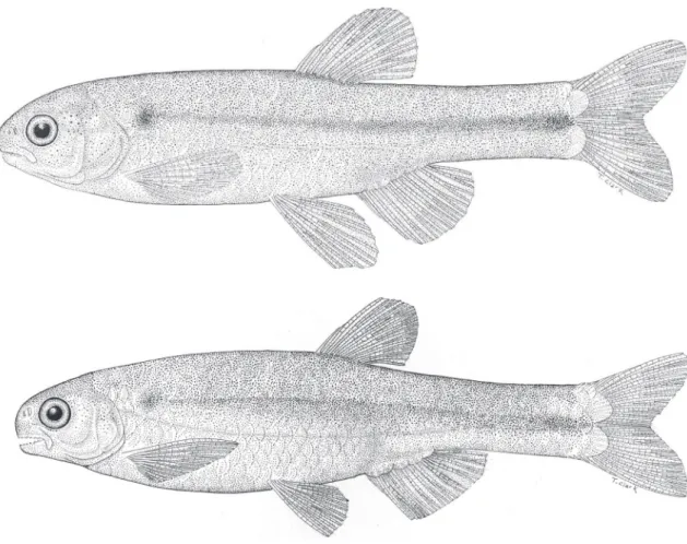
FIgure 1: Monotocheirodon pearsoni, CAS 59792, adult male above, 29.4 mm SL and adult female below, 35.5 mm SL.
FIgure 2: Monotocheirodon pearsoni, UMMZ 66485 (upper jaw), C&S, female, 26.3 mm SL, CAS 5792 (lower jaw), male, 29.4 mm SL, dentition, lateral view, left side, anterior at left.
Sexual dimorphism: Means corresponding to pelvic-fin length and dorsal-pelvic-fin height differ consider-ably between males and females (Table 1), but tests to evaluate if such differences would be statistically significant are meaningless since only two males are available.
Reproductive mode and gonad anatomy: Histologi-cal analysis revealed the presence of spermatozoa FIgure 4: Anal-fin base length as function of standard length for species of Monotocheirodon.
FIgure 5: Monotocheirodon pearsoni, CAS 59792, C&S, male, 29.4 mm SL; anal-fin rays, lateral view, left side.
FIgure 6: Monotocheirodon pearsoni, CAS 59792, C&S, male, 29.4 mm SL; pelvic-fin rays, ventral view, left side.
with spherical nuclei in ovaries from one mature female of Monotocheirodon pearsoni (Burns & Weitzman, 2006, fig. 1, Table 1). This indicates that the species can be classified as “aquasperm”, characteristic of externally fertilizing characids and is inseminating.
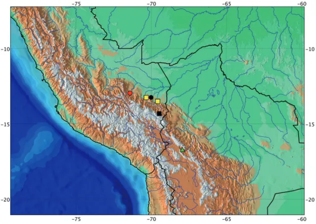
Distribution: Monotocheirodon pearsoni is known from headwaters of the Río Bopi, Río Beni basin and Río Iniqui (not exactly located), Bolivia, at about 5,000 m of elevation (Fig. 7).
Monotocheirodon drilos, new species Figs. 9‑11, table 2
Monotocheirodon sp. – Weitzman et al., 2005: 357, Burns & Weitzman, 2006: 529-530 (MUSM 11082, ANSP 143791, 143792).
Specimens examined: All specimens from Peru.
Holotype: MUSM 41541, male, SL 33.3 mm, Sandia, Zona Reservada Tambopata-Candamo, stream Ebe-bahuaeji (empties into Río Candamo), 13°14’56.4”S, 70°00’34.5”W, 31 March 1997, Fonchii Chang.
Paratypes: MUSM 11082, 2 (SL 28 and 32 mm), USNM 405296, 2 (31.8 and 34.8 mm) collected with holotype. ANSP 143790, 1 (SL 37.2 mm), Río Shintuya at Shintuya (exact coordinates not found). ANSP 143792, 6 (SL 300.8-37.8), border between Departamento of Cuzco and Departamento of Madre de Dios, Río Carbón (empties into Río Madre de Dios), 12°53’S, 71°20’W.
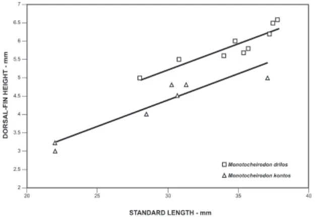
Diagnosis: Males and females of Monotocheirodon drilos have an externally visible urogenital papilla which is absent in M. pearsoni. The urogenital papilla FIgure 8: Dorsal-fin height as function of standard length for
Monotocheirodon drilos and M. kontos.
tAble 2: Morphometrics of Monotocheirodon drilos. Standard length expressed in mm; measurements through head length are percentages of standard legth; the last four entries are percentages of head length. Specimens are from MUSM 41541 (holotype), 11082 (paratypes); ANSP 143790, 143792 (paratypes). Values of p in bold indicates significant statistical differences.
characters holotype n Males sd n Females and juveniles sd p value
range mean range mean
Standard length 33.3 3 31.8-33.3 32,3 0.81 9 28.0-37.8 34.5 3.2
Depth at dorsal-fin origin 22.5 3 21.8-23.0 22.4 0.54 9 21.4-23.5 22.2 0.7 0.4055
Snout to dorsal-fin origin 52.0 3 51.0-52.0 51.5 0.51 9 50.6-53.7 52.4 1.1 0.2294
Snout to pectoral-fin origin 19.5 3 19.5-20.0 19.7 0.24 9 17.2-20.4 19.4 1.0 0.9263
Snout to pelvic-fin origin 44.1 3 44.1-45.3 44.7 0.58 9 43.1-47.6 45.7 1.3 0.1655
Snout to anal-fin origin 57.0 3 57.0-58.7 58.1 0.91 9 57.0-61.4 59.6 2.0 0.1655
Caudal peduncle depth 10.8 3 10.8-11.0 10.9 0.99 9 00.9-10.7 10.1 0.6 0.0126
Caudal peduncle length 21.0 3 21.0-21.8 21.5 0.45 9 20.6-22.8 21.4 0.6 0.6439
Pectoral-fin length 19.5 3 19.5-20.3 19.7 0.46 9 17.0-19.6 18.1 0.8 0.0335
Pelvic-fin length 16.5 3 16.5-16.8 16.7 0.18 8 12.4-13.0 12.6 0.2 0.0100
Dorsal-fin base length 09.0 3 08.8-09.3 09.1 0.28 9 08.2-10.0 09.2 0.5 0.6439
Dorsal-fin height 18.0 3 18.0-18.7 18.3 0.37 9 16.1-17.8 17.0 0.7 0.0126
Anal-fin base length 15.0 3 13.8-15.0 14.3 0.62 9 13.2-15.3 14.0 0.7 0.6439
Anal-fin lobe length 14.4 3 14.4-14.7 14.6 0.14 9 13.3-15.6 14.5 0.7 0.7815
Eye to dorsal-fin origin 39.6 3 39.6-40.8 40.4 0.65 9 40.2-42.4 41.5 0.7 0.0790
Dorsal-fin origin to caudal-fin base 49.5 3 49.5-51.5 50.9 1.16 9 47.0-50.0 48.2 1.0 0.0208
Bony head length 20.1 3 19.8-20.3 20.1 0.25 9 19.5-21.4 20.3 0.5 0.6439
Horizontal eye diameter 21.0 3 19.0-21.0 20.0 0.90 9 18.3-22.6 20.3 1.3 0.7115
Snout length 16.4 3 15.8-17.0 16.4 0.52 9 16.1-19.7 18.0 1.1 0.0522
Least interorbital width 26.8 3 26.8-28.5 27.7 0.85 9 26.6-29.0 27.4 0.7 0.5175
of sexually active males of M. drilos is shorter (half length of anal-fin base versus about equal to length of anal fin-base in M. kontos). Females and juveniles of
M. drilos and M. kontos can be differentiated in the height of the dorsal fin (16.1-17.8% SL in M. dri-los versus 13.4-15.8% SL in M. kontos). The number of premaxillary tooth cusps (5 in M. drilos vs 7 in M. kontos) is also useful to distinguish adult males and females of both species (Figs. 10 and 14).
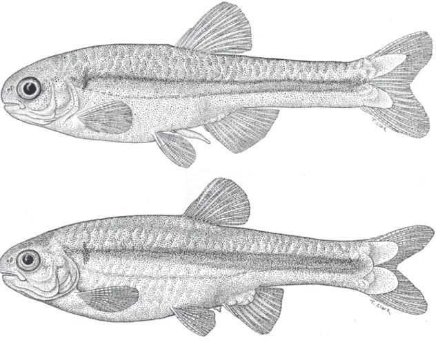
Description: Morphometric data of holotype and para-types presented in Table 2. Stevardiine characid reach-ing at least 37.8 mm SL. Body cylindrical in cross sec-tion; greatest body depth between verticals through middle and tip of pectoral fin. Dorsal profile of head anterior to nape strongly convex to snout region in males, less so in females. Snout bluntly convex; tip of snout at about horizontal through mid-point of orbit. Lower jaw convex in profile and somewhat included below upper jaw. Ventral profile of head gently con-vex, and continuous with strongly convex abdominal region as far as anal-fin origin. Body profile along anal-fin base approximately straight to slightly convex to posterior termination of anal fin. Ventral profile FIgure 9: Monotocheirodon drilos, MUSM 11082, adult male above, 33.6 mm SL and adult female, below, 33.0 mm SL.
of caudal peduncle slightly convex. Dorsal profile of body between nape and dorsal-fin origin gently con-vex. Base of dorsal fin slightly convex and somewhat inclined posteroventrally. Body profile between poste-rior terminus of dorsal fin and caudal-fin base slightly convex in males and almost straight in females.
Unbranched dorsal-fin rays 2 in all specimens, branched rays 7-8, 7.1, (7) n = 12, SD = 0.4); poste-rior ray not split to its base. Dorsal-fin height appar-ently sexually dimorphic (see discussion under sexual dimorphism). Adipose fin absent. Unbranched anal-fin rays ii in all specimens; branched rays 9-12, 10, (9), n = 12, SD = 0.9; posterior ray split to its base and counted as one ray. No hooks on anal fin of males. Pectoral-fin rays i, 9-10, 9.1, (9), n = 12, SD = 0.6. Pectoral fin longer in mature males, with tip almost reaching pelvic-fin origin; shorter in immatures and females, with tip distant from pelvic-fin origin. Pecto-ral-fin rays without hooks. Pelvic fin with one anterior and one posterior unbranched ray, and branched rays 4-5, 4.7, (7) n = 12, SD = 0.4. Sexually active males lacking pelvic-fin hooks. Pelvic-fin length of sexually mature specimens sexually dimorphic (see discussion under sexual dimorphism). Principal caudal-fin rays 10/9 in all specimens.
Scales cycloid: Lateral line complete, perforated scales 33-39, 36.8, (33), n = 12, SD = 1.9. Predoral scales 13-15, 14.5, (14), n = 12, SD = 0.7. Scale rows be-tween dorsal-fin origin and lateral line 4-5, 4.5, (5), n = 12, SD = 0.5. Scale rows from pelvic-fin origin to lateral line 3, n = 12. Scale rows around caudal pe-duncle 10 in all specimens, n = 12. Row of enlarged scales present along anal-fin base.
Premaxilla with single row of 4 multicuspid teeth (Fig. 10) in all specimens. All teeth compressed, pedunculate with distal parts spatulate with 5 cusps; three middle cusps largest, and marginal cusps re-duced. Maxillary teeth (Fig. 10) identical in form to premaxillary teeth, also with 5 cusps, but with three middle cusps slightly smaller than those of premax-illary teeth. Total number of maxpremax-illary teeth 6-9, 7.4, (6), n = 10, SD = 1.2. Dentary teeth (Fig. 10) identical to premaxillary and maxillary teeth, with 3 large middle cusps and reduced marginal cusps. To-tal number of dentary teeth 8-11, 9.3, (9), n = 12, SD = 0.4.
Vertebrae 37-38, 37.7, n = 8, SD = 0.4. Dor-sal limb gill rakers 9-10, 9.3, (9), n = 11, SD = 0.4; ventral limb gill rakers 11-15, 13.6, (12), n = 11, SD = 0.4. Branchiostegal rays 4 in one cleared and stained specimen; 3 rays originating on anterior and one on posterior ceratohyal.
Color in alcohol: Background body color pale to yel-lowish brown, darker dorsally due to presence of dark chromatophores largely concentrated towards poste-rior border of scales. Dark chromatophores fewer on posterior border of scales of midlateral and ventral parts of body. Dark longitudinal dark stripe extends from posterodorsal part of opercle to caudal-fin base. Stripe anteriorly inconspicuous and slightly arched dorsally from upper part of opercle to point below dorsal-fin origin; bordered ventrally by lateral line. Stripe more conspicuous and wider from point above anal-fin origin to caudal-fin base. Dark vertically elongate humeral blotch, located about two scales posterior of posterodorsal portion of opercle, and ex-tending one scale ventral of lateral line.
Head darker on upper part of snout and area dorsal to eye with scattered dark chromatophores on ventral portion of infraorbital bones and opercular region. Urogenital papilla and all fins with scattered dark chromatophores. Large scales on basal portion of each caudal-fin lobe with dark chromatophores most-ly concentrated on their basal and median portions.
Sexual dimorphism: The p value in Table 2 suggests that the caudal peduncle depth, pectoral-fin length, pelvic-fin length, dorsal-fin height and the distance from dorsal-fin origin to caudal-fin base are sexually dimorphic. Regression data to test the differences more accurately were not used due to the limited number available mature males.
Reproductive mode and gonad anatomy: Males of Mo-notocheirodon drilos (MUSM 11082, ANSP 143791 and 143792) identified as Monotocheirodon sp. were used by Burns & Weitzman (2006) for histological sections of the urogenital papilla, which was charac-terized as a large intromittent organ used for insemi-nation of the females. The sperm was found to have elongate nuclei 1.8-2.1 µm in length, usually char-acteristic of inseminating and internally fertilizing fishes.
strongly asymmetric (Fig. 11, A-B) and 0.6 µm in length (SD ± 0.1 µm). A single flagellum emerges from the midpiece (Fig. 1 I-L).
Etymology: The name drilos is Greek masculine mean-ing penis. The word is used here in reference to the prominent male inseminating organ. A noun in apposition.
Distribution: This species is known from headwaters of Ríos Tambopata and Madre de Dios, Río Madre de Dios basin, Peru (Fig. 7).
Monotocheirodon kontos, new species Figs. 12‑15, table 3
Monotocheirodon sp. – Burns & Weitzman, 2006: 529-530 (MUSM 6756 and 11250). Monotocheirodon personi [sic] – Ferreira et al. 2011
(misidentification; MUSM 11416, listed in comparative material).
Specimens examined: All specimens from Peru.
Holotype: MUSM 41542, male, SL 33.5 mm, Río Inambari, Sandia, Muspaypampa, 14°14’41”S, 69°25’51”W, 6 July 1994, Fonchii Chang.
Paratypes: MUSM 6756 (4, SL 22-37.1 mm) col-lected with holotype. MUSM 11644 (2, SL 30.7 and 31.3 mm), Ouno, Sandia, Zona Reservada Tam-bopata-Candamo, cuenca Ebebahuaeji, 13°24’ 52”S, 70°00’48”W. MUSM 11250 (4, SL 24.5-31.3 mm), USNM 405297 (2, SL 26.8 and 28.5 mm), Río Malinowski (empties into Rio Madre de Dios), Zona Reservada Tambopata-Candamo, 13°08’00”S, 70°17’00”W.
Diagnosis: M. kontos has a urogenital papilla in sexual-ly active males and females which is absent in M. pear-soni. The urogenital papilla in M. kontos is about equal length of anal-fin base versus half length of anal-fin base in M. drilos. Females and juveniles of M. kon-tos and M. drilos can be distinguished in the height
of dorsal fin (13.4-15.8% of SL in the former versus 16.1-17.8 in the latter). The number of premaxillary tooth cusps (7 in M. kontos versus 5 in M. drilos) dif-ferentiate adult males and females of both species.
Description: Morphometrics of holotype and paratype presented in Table 3. Stevardiin characid reaching at least 37.1 mm SL. Body cylindrical in cross sec-tion; greatest body depth situated between verticals through tip of pectoral fin and dorsal-fin origin. Dor-sal profile of head anterior to nape strongly convex in males, slightly convex in females to snout region dorsal to nostril. Snout bluntly convex; tip of snout situated along horizontal through approximate mid-point of orbit. Lower jaw convex in ventral profile and somewhat included below upper jaw. Ventral profile of head gently convex, continuous with gently convex in males and strongly convex abdominal region in fe-males extending to anal-fin origin. Body profile along anal-fin base approximately straight to slightly convex to posterior termination of anal fin. Ventral profile of caudal peduncle almost straight in males, slightly con-vex in females. Dorsal profile of body between nape and dorsal-fin origin gently convex. Base of dorsal fin straight and somewhat inclined posteroventrally. Body profile between basal of last dorsal-fin ray and
caudal-fin rays almost straight in males, slightly con-cave posterodorsally in females
Unbranched dorsal-fin rays 2 in all specimens, branched rays 7-8, 7.7, (8), n = 13, SD = 0.5; poste-rior ray not split to its base. Dorsal-fin height sexually dimorphic (see discussion under Sexual dimorphism). Adipose fin absent. Unbranched anal-fin rays ii or iii, most usually ii; branched rays 10-11, 10.1, (10), n = 13, SD = 0.4; posterior ray split to its base and counted as one ray. No hooks present on anal fin of mature males. Pectoral-fin rays i, 8-9, 8.5, (8) n = 13, SD = 0.5. Tip of pectoral fin falling short of pelvic-fin origin. Pectoral-fin rays lacking hooks. Pelvic-fin rays i, 5, i, n = 13. Sexually active males lacking pelvic-fin hooks. Pelvic-pelvic-fin length of sexually mature males sexually dimorphic (see under Sexual dimorphism). Principal caudal-fin rays 10/9 in all specimens.
Scales cycloid: Lateral line complete, perforated scales 36-38, 37.1, (37) n = 12, SD = 0.6. Predorsal scales 13-15, 14.2, (15), n = 12, SD = 0.7. Scale rows be-tween dorsal-fin origin and lateral line 4-5, 4.6, (4), n = 12, SD = 0.5. Scale rows from pelvic-fin origin to lateral line 2-3, 2.7, (2), n = 12, SD = 0.4. Scale rows around caudal peduncle 10 in all specimens, n = 12. Row of enlarged scales along anal-fin base.
tAble 3: Morphometrics of Monotocheirodon kontos. Standard length expressed in mm; measurements through head length are percent-ages of standard length; the last four entries are percentpercent-ages of head length. Specimens are from MUSM 41542 (holotype), 6756, 11250, 11644 (paratypes); USNM 405297 (paratypes). Values of p in bold indicates significant statistical differences
characters holotype n Males sd n Females and juveniles sd p value
range mean range mean
Standard length 33.5 5 24.0-33.5 27.2 3.7 8 22.0-37.1 29.1 5.0
Depth at dorsal-fin origin 20.0 5 20.0 -21.8 20.8 0.6 8 19.6-24.0 21.6 1.6 0.6084
Snout to dorsal-fin origin 52.5 5 52.2-53.4 52.8 0.5 8 51.2-54.5 52.6 1.0 0.6606
Snout to pectoral-fin origin 20.9 5 18.3-21.0 19.6 1.0 8 18.1- 20.4 19.1 0.9 0.3798
Snout to pelvic-fin origin 45.6 5 44.7-46.5 45.5 0.7 8 43.1-47.1 45.1 1.3 0.4642
Snout to anal-fin origin 58.0 5 57.5-61.2 59.0 1.5 8 56.8-61.7 59.1 1.7 0.7697
Caudal peduncle depth 11.3 5 10.4-11.3 10.8 0.3 8 09.6-11.3 10.4 0.7 0.2416
Caudal peduncle length 21.5 5 19.4-21.5 20.3 1.5 8 19.0-23.0 20.5 1.5 0.7697
Pectoral-fin length 19.4 5 19.4-21.2 20.1 0.8 8 18.2-20.3 19.5 0.8 0.3798
Pelvic-fin length 15.5 5 14.5-15.5 15.0 0.8 8 10.8-13.2 12.2 0.8 0.0034
Dorsal-fin base length 07.4 5 07.4-09.0 08.3 0.5 8 08.0-08.4 08.1 0.1 0.1432
Dorsal-fin height 16.4 5 16.4-18.1 17.3 0.7 8 13.4-15.8 14.6 0.8 0.0034
Anal-fin base length 14.0 5 14.0-15.2 14.5 0.4 8 13.4-16.3 15.2 0.8 0.1073
Anal-fin lobe length 12.8 5 12.8-14.7 13.8 0.6 8 12.7-13.5 13.0 0.3 0.0192
Eye to dorsal-fin origin 41.0 5 38.7-40.9 39.7 0.7 8 37.3-40.4 39.3 1.1 0.9417
Dorsal-fin origin to caudal-fin base 47.7 5 46.6-48.3 47.7 0.6 8 45.3-49.8 47.1 1.7 0.3055
Bony head length 21.2 5 20.4-23.0 21.1 1.0 8 19.8-22.7 20.8 1.0 0.6606
Horizontal eye diameter 21.1 5 18.3-21.1 19.6 1.0 8 18.0-21.6 19.7 1.2 0.8833
Snout length 18.3 5 16.3-18.3 17.6 0.7 8 15.8-18.3 17.0 0.8 0.3055
Least interorbital width 28.1 5 26.5-28.1 27.3 0.7 8 26.0-28.2 27.1 0.8 0.9273
Premaxilla with single row of 4 multicuspid teeth (Fig. 14) in all 13 specimens. All teeth com-pressed and pedunculate with distal parts spatulate with 7 cusps in adult males and females; 3 middle cusps largest, and marginal cusps smaller. Maxil-lary teeth (Fig. 14) identical in form to premaxil-lary teeth; most teeth with 6-7 cusps, 5-8, 7.1, (7), n = 13, SD = 1. Dentary dentition identical in form to that on premaxilla and maxilla; with 3 middle cusps slightly larger than marginal cusps. Total number of dentary teeth 7-11, 8.6, 9 (13), n = 13, SD = 1.2.
Vertebrae 37-39, 37.8, (38), n = 14, SD = 0.5. Dorsal limb gill rakers 8-9, 8.4, (8), n = 13, SD = 0.5; ventral limb gill rakers 12-14, 12,7, (13), n = 13, SD = 0.7. Branchiostegal rays 4 in one cleared and stained specimen; 3 rays originating on anterior and one on posterior ceratohyal.
Color in alcohol: Identical to that of M. drilos, except that the head is dark overall with the central portions of maxilla, infraorbitals, preopercle, dorsal and ventral parts of opercle, subopercle and branchiostegal rays and basal portion of pectoral fin lighter with scattered dark chromatophores. Longitudinal dark stripe on body identical to that of M. pearsoni, but dark chro-matophores are more densely concentrated along its dorsal and posterior portions.
Sexual dimorphism: The p values in Table 3 indicate that pelvic-fin length, dorsal-fin height and anal-fin lobe length are sexually dimorphic, but testing these differences through regression analysis is inappropri-ate in light of using the limited number of available males and females.
Reproductive mode and gonad anatomy: Males and fe-males of this species (MUSM 6756 and 11250), iden-tified as Monotocheirodon sp. were also used by Burns & Weitzman (2006) for histological analysis of the urogenital papilla and the ovary of mature females. The results revealed that the intromittent organ of Monoto-cheirodon kontos, which is larger than that of M. drilos is also used to inseminate females and that the more elon-gate nuclei of the spermatic cells are 4.1 µm in length. As in Monotocheirodon drilos, the sperm nucle-us contains highly condensed granular chromatin, but it is more elongate toward the flagellar axis be-ing approximately 3.95 µm in length (SD ± 0.4µm) (Fig. 15, A-F). The flagellum originates along the first quarter of the nuclear length (Fig. 15, A-B). In cross section the flagellum shows an irregular outline with depressions (Fig. 15, C-F) and in the centrio-lar complex the centrioles are oblique to one another (Fig. 15 B-inset). Other distinctive sperm nucleus fea-tures of M. kontos are: the mitochondria are elongate, display a longitudinal position relative to the flagel-lar axis (Fig. 15 B) and are mainly accumulated in the depressions on the nuclear outline (Fig. 15 E-F); the vesicular system is formed by a large number of small interconnected vesicles positioned very close to one another giving the system an alveolar appearance (Fig. 15, F-L); due to the superposition of the mem-brane, the points where the vesicles are connected are FIgure 13: Monotocheirodon kontos, MUSM 11250, female,
37.1 mm SL; gill rakers on first gill arch, lateral view, left side.
seen as electron dense dots (Fig. 15 J); the vesicles are intermingled with the mitochondria and are mainly external to them; this vesicular system and also the mitochondria are found in the midpiece at the base of the nucleus (Figs. 15, H-L). The midpiece, identi-cal to that of M. drilos (Fig. 15 B) is about 0.7 µm in length (SD ± 0.1 µm), and as in that species a single flagellum also emerges from the midpiece (Fig. 15 G).
Etymology: The name kontos is Greek masculine mean-ing a long pole. The word is used here in reference to the prominent male inseminating organ. A noun in apposition.
Distribution: Monotocheirodon kontos was collected in tributaries of the Río Madre de Dios basin, Peru (Fig. 7) between 350 and 3,200 m of altitude. It is sympatric with M. drilos in the Río Ebebahuaeji basin.
dIscussIon
After its inclusion with inseminating clade A characids (Weitzman et al., 2005) and subsequently the subfamily Stevardiinae (Mirande, 2010; Oliveira et al., 2011) the first attempt to resolve the relation-ships of Monotocheirodon with the other stevardiin genera was by Ferreira et al. (2011). This involved 153 characters, including features of morphology, repro-duction and sperm ultrastructure. Monotocheirodon kontos (the M. personi of those authors) was included as representative of the genus. Character analysis in-dicated that Monotocheirodon is closely related to and forms with the genus Otonocheirodus a separate clade within the Stevardiinae.
The two new species described herein share with M. pearsoni the presence of a single row of pedun-culate, distally compressed multicuspid teeth on the premaxilla, the absence of an adipose fin, and the anal fin short with only 8-12 branched rays (characters 3, 8, and 9 of the diagnosis of the genus). These fea-tures are putatively considered non-exclusive synapo-morphies that support the monophyletic condition of the genus. In the remaining stevardiines there are two rows of thick nearly rounded usually tricuspidate teeth, the adipose fin is usually present and the anal fin longer with more tham 12 rays.
Preliminary examination of Ceratobranchia cf. delotaenia (MZUSP 89678), Bryconacidnus el-lisi (MUSM 11628), Rhinopetitia cf. myersi (MZUSP 36813), Rhinopetitia sp. (MZUSP 97176), Otonochei-rodus sp. (MEPN 2787) and Odontostoechus lethostig-mus (MCP 10774) cleared and stained for an ongoing
study of relationships, indicated that these genera share with Monotocheirodon at least characters 2, 6, and 7, described above in the diagnosis of that genus. These preliminary findings suggest that these five gen-era are more closely related among themselves than to any other stevardiin genus and probably represent a separate clade within the subfamily; however, a more comprehensive analysis of characters is required to confirm this hypothesis.
Oliveira et al. (2012) state that gill glands were found via histological preparations in mature insemi-nating males of Monotocheirodon species. No histo-logical sections were conducted in this study to detect the presence of such structures.
The discovery that females of Monotocheirodon drilos and M. kontos also have a urogenital papilla, al-beit smaller than that of the males, is intriguing. As demonstrated by Burns & Weitzman (2006) the male intromittent organs receive the sperm ducts from each testis and have a special circularly oriented skeletal muscle probably to avoid reflux of sperm during the process of its introduction into the oviduct. The func-tion of the female urogenital papilla is unknown, but it might be used as a storage organ either for fertilized or unfertilized ovules prior to eggs release.
It is interesting that the absence of a male intro-mittent organ is correlated with the nearly spherical shape of the sperm nucleus in the externally fertiliz-ing (aquasperm) of Monotocheirodon pearsoni whereas a small intromittent organ with ovoid sperm nuclei in M. drilos and a larger intromittent organ with more elongate nuclei in M. kontos occur in two internally fer-tilizing species. Other differences in sperm ultrastruc-ture between the last two species as discussed above provide further evidence that they are different species.
resuMo
Palavras-Chave: Peixes caracídeos inseminadores, novas espécies e relações de Monotocheirodon.
AcKnowledgMents
The authors thank Tamara Clark for prepar-ing Figs. 1, 2, 5, 6, 9, 10, 12, 13 and 14 with sup-port from the Herbert R. and Evelyn Axelrod Chair in Systematic Ichthyology in the Division of Fishes, NMNH, which also provided funds for a trip of NAM to NMNH. The Conselho Nacional de Desenvolvi-mento Científico e Tecnológico (CNPq) supported research work of NAM in MZUSP and NMNH through a research fellowship. Lisa Palmer (NMNH) prepared radiographs and provided general curatorial assistance. Luiz Malabarba and Richard Vari supplied valuable input regarding discussions of inseminating characid fishes. Richard Vari provided assistance and working space to the senior author during his visit to USNM to study material of Monotocheirodon, read the manuscript, added useful suggestions and helped to improve the English version. Hernán Ortega (MUSM) and Jeffrey Clayton (NMNH) greatly con-tributed to this study by providing museum catalog numbers, loan of specimens, locality information and specimen data. Eduardo G. Baena (MZUSP) com-pleted some drawings and provided computer assis-tance. José Birindelli, André Luiz Netto-Ferreira and Manoela Maria F. Marinho (MZUSP) helped with computer programs. Two anonymous reviewers pro-vided valuable comments on the manuscript. We are gratefully indebted to all.
reFerences
Ayres, M.; Ayres Jr., M.; Ayres, D.L. & dos Santos, A. de A.S. 2007. BioEstat. Aplicações estatísticas nas áreas de ciências biomédicas. Belém, Pará, xvii + 359 p.
Burns, J.R. & Weitzman, S.H. 2006. Intromittent organ in the genus Monotocheirodon (Characiformes: Characidae). Copeia, 2006(3):529-534.
Ferreira, K.M.; Menezes, N.A. & Quagio-Grassiotto, I. 2011. A new genus and two new species of Stevardiinae (Characiformes: Characidae) with a hypothesis on their relationships based on morphological and histological data. Neotropical Ichthyology, 9(2):281-298.
Fink, W.L. & Weitzman, S.H. 1974. The so-called cheirodontin fishes of Central America with a description of two new species (Pisces: Characidae). Smithsonian Contributions to Zoology, 172:1-46.
Géry, J. 1977. Characoids of the World. Neptune City NJ, T.F.H. Publications. 672 p.
Lima, F.C.T.; Malabarba, L.R.; Buckup, P.A.; da Silva, J.F.P.; Vari, R.P.; Herald, A.; Benine, R.; Oyakawa, O.T.; Pavanelli, C.S.; Menezes, N.A.; Lucena, C.A.S.; Malabarba,
M.C.S.L.; Lucena, Z.M.S.; Reis, R.E.; Langeani, F.; Casatii, L.; Bertaco, V.A.; Moreira, C. & Lucinda, P.H.F. 2003. Genera Incertae Sedis in Characidae. Pp. 106-169. In: Reis, R.E.; Kullander, S.O. & Ferraris, Jr., C.J. (Eds.). Check List of the Freshwater Fishes of South and Central America. Porto Alegre, Edipucrs. 729 p.
Malabarba, L.R. & Weitzman, S.H. 2000. A new genus and species of inseminating fish (Teleostei: Cheirodontinae: Compsurini) from South America with uniquely derived caudal-fin dermal papillae. Proceedings of the Biological Society of Washington, 1139(1):269-283.
Malabarba, L.R. 1998. Monophyly of the Cheirodontinae, characters and major clades (Ostariophysi: Characidae). In: Malabarba, L.R.; Reis, R.E.; Vari, R.P.; Lucena, Z.M. & Lucena, C.A. (Eds.). Phylogeny and Classification Neotropical Fishes. Porto Alegre, Edipucrs. p. 193-233.
Malabarba, L.R. & Weitzman, S.H. 2003. Description of a new genus with six new species from Southern Brazil, Uruguay and Argentina, with a discussion of a putative characid clade (Teleostei: Characiformes: Characidae). Comunicações do Museu de Ciências e Tecnologia, PUCRS, Série Zoologia, 16(1):67-151.
Menezes, N.A. & Weitzman, S.H. 1990. Two new species of Mimagoniates (Teleostei: Characidae: Glandulocaudinae), their phylogeny and biogeography and a key to the glandulocaudin fishes of Brazil and Paraguay. Proceedings of the Biological Society of Washington, 103(2):380-426.
Menezes, N.A. & Weitzman, S.H. 2009. Systematics of the neotropical fish subfamily Glandulocaudinae (Teleostei: Characiformes: Characidae). Neotropical Ichthyology, 7(3):295-370.
Mirande, J.M. 2010. Phylogeny of the famlily Characidae (Teleostei: Characiformes): from characters to taxonomy. Neotropical Ichthyology, 8(3):385-568.
Oliveira, C.; Avelino, G.S.; Abe, K.T.; Mariguela, T.C.; Benine, R.C.; Orti, G.; Vari, R.P. & Castro, R.M.C. 2011. Phylogenetic relationships within the speciose family Characidae (Teleostei: Ostariophysi: Characiformes) based on multilocus analysis and extensive ingroup sampling. BMC Evolutionary Biology, 11:1-25.
Oliveira, C.L.C. de; Malabarba, L.R. & Burns, J.R. 2012. Comparative morphology of gill glands in externally fertilizing and inseminating species of cheirodontine fishes (Actinipterygii: Cheirodontinae), with implications on the phylogeny of the family Characidae. Neotropical Ichthyology, 10(12):349-360.
Pearson, N.E. 1924. The fishes of the eastern slope of the Andes, I: The fishes of the Rio Beni Basin, Bolivia, collected by the Mulford expedition. Indiana Universities Studies, 11(64):1-8. Reynolds, E.S. 1963. The use of lead citrate at high pH as an
electron opaque stain in electron microscopy. Journal of Cell Biology, 17:208-212.
Weitzman, S.H.; Menezes, N.A.; Evers, H-G. & Burns, J.R. 2005. Putative relationships among inseminating and externally fertilizing characids, with a description of new genus and species of Brazilian inseminating fish bearing an anal-fin gland in males (Characiformes: Characidae.). Neotropical Ichthyology, 3(3):329-360.