GASTROINTESTINAL DISORDERS
Laura Lúcia Cogo1,2∗∗∗∗; Cristina Leise BastosMonteiro2
; Marilis DallarmiMiguel3; Obdulio Gomes Miguel3; Miriam Machado Cunico3; Marcelo Lima Ribeiro4; Eloá Ramalho de Camargo4; Gislene Maria Botão Kussen1; Keite da Silva
Nogueira1,3; Libera Maria Dalla Costa1,2,3
1
Hospital de Clínicas da Universidade Federal do Paraná, Curitiba, PR, Brasil; 2Programa de Pós Graduação em Processos
Biotecnológicos da Universidade Federal do Paraná, Curitiba, PR, Brasil; 3Programa de Pós Graduação em Ciências
Farmacêuticas da Universidade Federal do Paraná, Curitiba, PR, Brasil; 4Laboratório de Microbiologia e Biologia Molecular da
Unidade Integrada de Farmacologia e Gastroenterologia da Universidade São Francisco, Bragança Paulista, SP, Brasil.
Submitted: April 07, 2009; Returned to authors for corrections: May 19, 2009; Approved: October 06, 2009.
ABSTRACT
The antibacterial activity of plant extracts obtained from Bixa orellana L., Chamomilla recutita L., Ilex
paraguariensis A. St.-Hil., Malva sylvestris L., Plantago major L. and Rheum rhaponticum L. has been
evaluated against two reference strains and eleven clinical isolates of Helicobacter pylori. All the plant
species chosen are used in popular Brazilian cuisine and folk medicine in the treatment of gastrointestinal
disorders. Initial screening was made by the disk diffusion test and then minimum inhibitory concentration
was determined by the agar dilution method. The results presented in this work demonstrated that among
the plant preparations analyzed, B. orellana L., C. recutita L., I. paraguariensis A. St.-Hil. and M.
sylvestris L. were capable of inhibiting the in vitro growth of H. pylori.
Key words: Helicobacter pylori, antibacterial activity, plant extracts.
INTRODUCTION
Helicobacter pylori is a Gram-negative spiral-shaped
bacterium that was first isolated by Barry Marshall and J.
Robin Warren. Since its discovery in 1983, the microrganism
has been associated with the etiopathogenesis of several
diseases of the digestive system, such as gastritis, peptic ulcer
disease and gastric cancer (11). Conventional treatment for
eradication therapy of these infections is mainly based on the
use of multiple drugs, such as clarithromycin, amoxicillin,
furazolidone, tetracycline and metronidazole with bismuth or a
proton pump inhibitor (15).
Although the conventional treatment for eradication
____________
therapy of H. pylori allows obtaining high cure rates,
eradication failure rate remains of 5-20 %. This fact may be
partially explained by non-compliance in some patients who do
not follow the treatment properly and by the development of
resistance to antibiotics used (10). Therefore, there is a
growing need to search new therapeutic agents that can
hopefully eradicate this significant human pathogen and
medicinal plants are a useful source of novel drugs. Several
natural products have demonstrated antibacterial activity
against H. pylori (18) and for centuries a wide variety of plants
and substances derived from plants have been used to treat
gastrointestinal disorders (2).
Many plants used in Brazil to treat these infections do not
present any scientific evidence of efficacy. It is interesting to
determine whether their traditional uses are supported by
pharmacological effects or merely based on folklore. Within
this context, extracts obtained from Bixa orellana L. (annatto),
Chamomilla recutita L. (chamomile), Ilex paraguariensis A.
St.-Hil.(roasted and green yerba maté), Malva sylvestris L.
(mallow), Plantago major L. (plantain) and Rheum
rhaponticum L. (rhubarb) - all of which are used in popular
Brazilian cuisine and folk medicine in the treatment of
gastrointestinal disorders - were investigated for their anti-H.
pylori activity.
MATERIALS AND METHODS
General
Roots, rhizomes or aerial parts (leaves, stems, seeds,
inflorescence) of the plants Bixa orellana L., Chamomila
recutita L., Ilex paraguariensis A. St.-Hil., and Plantago major
L. were collected in Paraná state, Southern region of Brazil
(cities of Morretes, Lapa, Piraquara, and Curitiba respectively)
and identified by Dr. Gerdt Hatschbach from Museu Botânico
Municipal da Prefeitura de Curitiba, Paraná (MBM), where the
vouchers have been deposited. The plants Malva sylvestris L.
and Rheum rhaponticum L. were obtained commercially
(Flores & Ervas, Piracicaba, SP, Brazil); the voucher
specimens, including identification and classification of plant
materials, had been preserved by the company.
The parts of each plant examined and voucher numbers are
shown in Table 1.
Extraction of materials
A total of 50g of each plant species was exhaustively
extracted with aqueous 96% ethanol (v/v) by maceration at
room temperature. The extracts were obtained after filtration
and concentration of the material under reduced pressure until
the final volume of 50 ml.
Stock solutions of the extracts were made with sterile
distilled water at concentration of 100 mg/ml which were used
in the disk diffusion test. Another was made at the same
concentration, now with dimethylsulphoxide (DMSO), to
perform the determination of the minimum inhibitory
concentration. Final concentration of DMSO in the culture
medium did not exceed 1% (12).
Bacterial strains
A total of eleven clinical isolates of H. pylori obtained
from the gastric mucosa of patients submitted to upper
endoscopy and subsequently diagnosed with gastritis, peptic
ulcer disease or gastric cancer were used in the present study.
Clinical isolates were coded with the numbers of access BP-84,
BP-667, BP-660, BH-27, BP-446, BP-650, BP-118, BP-713,
BP-132, BP-652 and F-39 in order to preserve the identity of
the patients from whom they were obtained and were
previously approved by the Ethics Committee with the issuing
of protocol number 982.021/2005-01.
Reference strains H. pylori 26695 (23) and J99 (1), that
had their genomes completely sequenced, were tested as
control. All the strains were previously evaluated against
clarithromycin, amoxicillin, furazolidone, tetracycline and
metronidazole, which are antibiotics commonly used in
conventional therapy.
Preparation of bacterial suspensions
An inoculum of each strain used in susceptibility tests was
prepared by transferring fresh colonies of the microrganisms in
tubes containing sterile physiological saline solution and
adjusting the turbidity to the 2.0 McFarland standard (7).This
turbidity produces a suspension that corresponds to
approximately 6.0 x 10⁸ CFU/mL of H. pylori.
Disk diffusion test
In the initial phase, the disk diffusion test was used as
screening to analyze the susceptibility of reference strains H.
pylori 26695 and J99 against to different plant extracts. The
bacterial suspensions were spread-plated onto Columbia Agar
plates (Oxoid, Basingstoke, UK) supplemented with 10%
defibrinated sheep blood (Newprov, Curitiba, Brazil). Filter
extract (50µl of stock solutions) were placed onto the surface
of the inoculated agar. The plates were incubated at 37ºC under
microaerophilic conditions and observed after 3 to 5 days. The
tests were performed in triplicate and the antimicrobial activity
was expressed in terms of the mean diameter of the inhibition
zone around the disks impregnated with the plant extracts
tested, as presented in Table 1.
Determination of the minimum inhibitory concentration All the extracts that had produced an inhibition zone
greater than 6 mm in the disk diffusion test were separated to
determinate the MIC by the agar dilution method. In addition to
reference strains, 11 clinical H. pylori isolates were subjected
to this test.
The stock solutions made with DMSO were further
serially diluted in distilled sterile water and 1 mL of each
dilution was incorporated into 19 mL of molten Columbia agar
(Oxoid, Basingstoke, UK) containing 10% defibrinated sheep
blood (Newprov, Curitiba, Brazil) to be then transferred
separately into Petri dishes. The final concentrations of the
extracts in the culture medium ranged from 5.0 to 0.625
mg/mL.
Bacterial suspensions were prepared as described above,
and 1 µL of each suspension was spotted with a multipoint
inoculator onto the surface of the agar plates containing
consecutive dilutions of plant extracts. After that, plates were
incubated at 37ºC in a microaerophilic atmosphere for 72 hours
and MIC, which is defined as the lowest concentration of an
extract that inhibits the visible growth of a microrganism, was
determined. For clinical isolates, MIC50 and MIC90 were
determined and defined as the concentrations that inhibited,
respectively, 50 and 90% of the strains evaluated. All tests
were conducted in triplicate, in addition to growth controls
with and without DMSO.
RESULTS AND DISCUSSION
According to the data reported in Table 1, of all the plant
extracts submitted to the screening test, B. orellana L., C.
recutita L., I. paraguariensis A. St.-Hil. (green and roasted
Yerba Maté varieties) and M. sylvestris L. produced inhibition
zone diameters by the disk diffusion test. However, there is a
disadvantage to this method in that it yields only qualitative
results. The absence of objective quantification inherent in the
method makes it impossible to compare the degree of
antimicrobial activity of the extracts against the H. pylori
strains investigated (3). For that reason, in the next stage of the
study, MIC values were determined by the agar dilution
method. The results obtained are shown in Table 2.
The agar dilution test confirmed an anti-H. pylori activity
of all the plant extracts evaluated, with C. recutita L. and I.
paraguariensis A. St.-Hil. (green Yerba Maté variety) showing
to be more potent (MIC50: <0.625 mg/ml) than B. orellana L.
(MIC50: 1.25 mg/ml), I. paraguariensis A. St.-Hil. (roasted
Yerba Maté variety) (MIC50: 1.25 mg/ml) and M. sylvestris L
(MIC50: >5.0 mg/ml). The MIC90 values demonstrated that I.
paraguariensis A. St.-Hil. was able to inhibit a higher number
of clinical isolates when compared with other extracts,
although the green Yerba Maté variety (MIC90: 5.0 mg/ml) was
slightly less active than the roasted variety (MIC90: 2.5 mg/ml).
Previous investigations have demonstrated that I.
paraguariensis A. St.-Hil., widely consumed as part of the
usual diet in Brazil in the form of tea (roasted yerba maté) and
chimarrão (green yerba maté), presents several secondary
metabolic products that have antimicrobial activity, including
phenolic compounds, triterpenes and flavonoids (21). As for C.
recutita L., this plant has anti-inflammatory and calming
properties and is also used to treat gastric colic, and several
forms of gastritis, stomatitis, laryngitis and pharyngitis (17).
Flavonoids - particularly apeginine - and essential oils are
among the main constituents of the plant extract (13).
Research conducted by Stamatis et al. (22) confirmed the
anti-H. pylori activity of C. recutita L. extract. Although, the
plant part used to produce the extract in their work was not
specified, which may directly influence the development of
results (5).
B. orellana L. and M. sylvestris L. were other plant
Table 1. Analysis of anti-Helicobacter pylori activity of plant extracts by disk diffusion test.
Mean of inhibition zone ∗
∗ ∗ ∗ (mm)
Species (voucher numbers) Family Plant part used
H. pylori J99
H. pylori 26695 Bixa orellana L.
(MBM 212752)
Bixaceae Seed 7 10
Chamomilla recutita L. (MBM 189637)
Asteraceae Inflorescence 10 11
Ilex paraguariensis A. St.-Hil. (MBM 113738)
Aquifoliaceae green leaves 9 10
Ilex paraguariensis A. St.-Hil. (MBM 113738)
Aquifoliaceae roasted leaves 9 9
Malva sylvestris L. (Flores & Ervas)
Malvaceae inflorescence and leaves 10 8
Plantago major L. (MBM 243458)
Plantaginaceae above-ground parts < 6 < 6
Rheum rhaponticum L. (Flores & Ervas)
Polygonaceae Root < 6 < 6
*Final concentration of each extract = 5 mg/disk
Table 2. MIC (mg/mL) values of plant extracts against clinical isolates and reference strains of Helicobacter pylori.
Plant extracts
H. pylori strains
B . o re ll a n a C . re cu ti ta I. p a ra g u a ri en si s (g re en y er b a m a te ) I. p a ra g u a ri en si s (r o a st ed y er b a m a te ) M . sy lv es tr is
H. pylori 26695 < 0.625 < 0.625 < 0.625 < 0.625 < 0.625
H. pylori J99 < 0.625 < 0.625 < 0.625 2.5 1.25
BP-84 >5.0 >5.0 5.0 1.25 >5.0
BP-667 >5.0 >5.0 5.0 5.0 >5.0
BP-660 >5.0 >5.0 5.0 1.25 >5.0
BH-27 1.25 < 0.625 < 0.625 < 0.625 >5.0
BP-446 1.25 < 0.625 < 0.625 2.5 >5.0
BP-650 >5.0 >5.0 5.0 < 0.625 >5.0
BP-118 < 0.625 < 0.625 < 0.625 < 0.625 0.625
BP-713 >5.0 < 0.625 2.5 5.0 2.5
BP-132 >5.0 < 0.625 < 0.625 2.5 5.0
BP-652 < 0.625 < 0.625 < 0.625 2.5 2.5
F-39 1.25 < 0.625 < 0.625 < 0.625 >5.0
MIC50 1.25 <0.625 <0.625 1.25 >5.0
widely used in Brazilian home cooking - is known to contain
an essential oil rich in all-E-geranylgeraniol, oxygenated
monoterpenes and sesquiterpenes (8). The second one is
composed of mucilage, tannins, essential oils and flavonoids
(4) reasons why it is used as anti-inflammatory and support in
the treatment of different types of infections (14).
Moreover, it is important to note that the most active
substances found in the plants screened in these experiments
have recognized properties in gastrointestinal digestive
diseases and presented stable activity at acid pH (9).
Increasing antimicrobial resistance is a serious global problem
that is present in this important human pathogen (6). Mendonça
et al. reported the susceptibility profile involving Brazilian H.
pylori strains. Resistance rates were observed as to
metronidazole, amoxicillin and clarithromycin of 42%, 29%
and 7% respectively; values of furazolidone (4%) and
tetracycline (7%) were also presented (16).
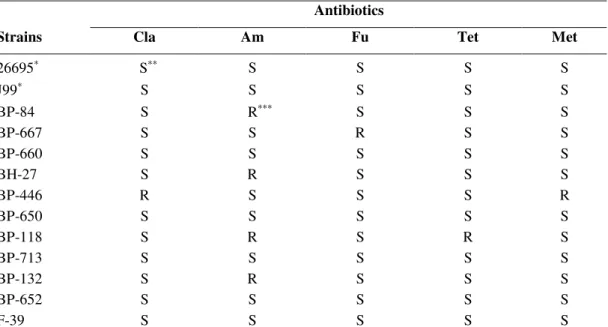
In this study, for each H. pylori strain evaluated for the
antimicrobial activity of plant extracts, susceptibility to
antibiotics used in conventional therapy, was also characterized
as shown in Table 3. These strains presented different
susceptibility profiles and, in some cases, resistance to one or
more antibiotics. Interestingly, the resistant strains evaluated
against the different extracts, demonstrated a similar profile
when compared to sensitive ones (Table 2).
Table 3. Susceptibility test of Helicobacter pylori reference strains and clinical isolates.
Antibiotics
Strains Cla Am Fu Tet Met
26695∗ S∗∗ S S S S
J99∗ S S S S S
BP-84 S R∗∗∗ S S S
BP-667 S S R S S
BP-660 S S S S S
BH-27 S R S S S
BP-446 R S S S R
BP-650 S S S S S
BP-118 S R S R S
BP-713 S S S S S
BP-132 S R S S S
BP-652 S S S S S
F-39 S S S S S
Cla - Clarithromycin, Am - Amoxicillin, Fu - Furazolidone, Tet - Tetracycline, Met – Metronidazole ∗Reference strains, ∗∗ Susceptibility, ∗∗∗Resistance.
In summary, a variety of plant species is capable of
synthesizing many substances which show antibacterial
activity. These properties have been described to extracts of
many plants found in Brazilian flora (19,20). However, as
regards the plant extracts included in this work, there are no
previous studies that evaluate the proposed feature, except for
C. recutita L. (22). Results demonstrate that the extracts
obtained from plants B. orellana L., C. recutita L., I.
paraguariensis A. St.-Hil. and M. sylvestris L. were capable of
inhibiting the in vitro growth of H. pylori and could form a
promising basis for further investigation in the discovery of
ACKNOWLEDGEMENTS
The authors would like to thank Dr. Gerdt Guenther
Hatschbach from Museu Botânico Municipal da Prefeitura de
Curitiba, for the botanical identification and NEBaC (Núcleo
de Estudos em Bacteriologia Clínica de Curitiba), for the
financial support.
REFERENCES
1. Alm, R.A.; Ling, L.S.; Moir, D.T.; King, B.L.; Brown, E.D.; Doig, P.C.; Smith, D.R.; Noonan, B.; Guild, B.C.; deJonge, B.L.; Carmel, G.; Tummino, P.J.; Caruso, A.; Uria-Nickelsen, M.; Mills, D.M.; Ives, C.; Gibson, R.; Merberg, D.; Mills, S.D.; Jiang, Q.; Taylor, D.E.; Vovis, G.F.; Trust, T.J. (1999). Genomic-sequence comparison of two unrelated isolates of the human gastric pathogen Helicobacter pylori. Nature, 397(6715):176-80.
2. Borrelli, F.; Izzo, A.A. (2000). The plant kingdom as a source of anti-ulcer remedies. Phytother. Res., 14, 581-591.
3. Bresolin, T.M.B.; Cechinel Filho, V. (2003). Ciências Farmacêuticas - Contribuição ao Desenvolvimento de Novos Fármacos e Medicamentos. Editora Univali, Itajaí, Santa Catarina, Brasil.
4. Buffon, M.C.M.; Lima, M.L.C.; Galarda, I.; Cogo, L. (2001). Avaliação da eficácia dos extratos de Malva sylvestris, Calêndula officinalis, Plantago major e Curcuma zedoarea no controle do crescimento das bactérias da placa dentária. Estudo “in vitro”. Visão Acadêmica, 2 (1), 31-38.
5. Cechinel Filho, V.; Yunes, R.A. (1998). Estratégias para a obtenção de compostos farmacologicamente ativos a partir de plantas medicinais. Conceitos sobre modificação estrutural para otimização da atividade. Quím. Nova, 21 (1), 99-105.
6. Chatterjee, A.; Yasmin, T.; Bagchi, D.; Stohs, S. (2004). Inhibition of Helicobacter pylori in vitro by various berry extracts, with enhanced susceptibility to clarithromycin. Mol. Cell. Biochem, 265, 19-26. 7. Clinical and Laboratory Standards Institute (2006). Performance
standards for antimicrobial susceptibility testing. Sixteenth informational supplement M100-S16. Clinical and Laboratory Standards Institute, Wayne, PA.
8. Coelho, A.M.S.P.; Silva, G.A.; Vieira, O.M.C.; Chavasco, J.K. (2003). Atividade antimicrobiana de Bixa orellana L. (Urucum). Revista Lecta, 21 (1/2), 47-54.
9. Friedman, M.; Jürgens, H.S. (2000). Effect of pH on the stability of plant phenolic compounds. J. Agr. Food Chem., 48 (6): 2101-2110.
10. Gadhi, C.A.; Benharref, A.; Jana, M.; Lozniewski, A. (2001). Anti-Helicobacter pylori activity of Aristolochia paucinervis Pomel extracts. J. Ethnopharmacol., 75 (2-3), 203-205.
11. Kusters, J.G.; Van Vliet, A.H.; Kuipers, E.J. (2006). Pathogenesis of Helicobacter pylori infection. Clin. Microbiol.,Rev.19 (3), 449-490.
12. Li, Y.; Xu, C.; Zhang, Q.; Liu, J.Y.; Tan, R.X. (2005). In vitro anti-Helicobacter pylori action of 30 Chinese herbal medicines used to treat ulcer diseases. J. Ethnopharmacol., 98 (3), 329-33.
13. Mapeli, N.C.; Viera, M.C.; Heredia, Z.N.A.; Siqueira, J.M. (2005). Produção de biomassa e de óleo essencial dos capítulos florais da camomila em função de nitrogênio e fósforo. Hortic. Bras., 23 (1), 32-37. 14. Martinazzo, A.P.; Martins, T. (2004). Plantas medicinais utilizadas pela
população de Cascavel/PR. Arq. Ciênc. Saúde Unipar, 8 (1), 3-6. 15. Megraud, F.; Lehours, P. (2007). Helicobacter pylori detection and
antimicrobial susceptibility testing. Clin. Microbiol. Rev., 20 (2), 280-322.
16. Mendonça, S.; Ecclissato, C.; Sartori, M.S.; Godoy, A.P.; Guerzoni, R.A.; Degger, M.; Pedrazzoli Jr, J. (2000). Prevalence of Helicobacter pylori resistance to metronidazole, clarithromycin, amoxicillin, tetracycline, and furazolidone in Brazil. Helicobacter, 5(2):79-83 17. Morais, T.C.; Vieira, M.C.; Heredia, Z.N.A.; Teixeira, I.R.; Ramos,
M.B.M. (2006). Produção de biomassa e teor de óleos essenciais da camomila (Chamomilla recutita (L.) Rauschert) em função das adubações com fósforo e nitrogênio. Rev. Bras. Pl. Med., 8 (4), 120-125. 18. Nostro, A.; Cellini, L.; Di Bartolomeo, S.; Di Campli, E.; Grande, R.;
Cannatelli, M.A.; Marzio, L.; Alonzo, V. (2005). Antibacterial effect of plant extracts against Helicobacter pylori. Phytother. Res., 19 (3), 198-202.
19. Oliveira, D.F.; Pereira, A.C.; Figueiredo, H.C.P; Carvalho, D.A.; Silva, G.; Nunes, A.S.; Alves, D.S.; Carvalho, H.W.P. (2007). Antibacterial activity of plant extracts from Brazilian southeast region. Fitoterapia, 78, 142-145.
20. Sartoratto, A.; Machado, A.L.M.; Delarmelina, C.; Figueira, G.M.; Duarte, M.C.; Rehder, V.L.G. (2004). Composition and antimicrobial activity of essential oils from aromatic plants used in Brazil. Braz. J.Microbiol., 31, 247-256.
21. Schubert, A.; Zanin, F.F.; Pereira, D.F.; Athayde, M.L. (2006). Variação anual de metilxantinas totais em amostras de Ilex paraguariensis A. St. - Hil. (erva-mate) em Ijui e Santa Maria, Estado do Rio Grande do Sul. Quím. Nova, 29 (6), 1233-1236.
22. Stamatis, G.; Kyriazopoulos, P.; Golegou, S.; Basayiannis, A.; Skaltsas, S.; Skaltsa, H. (2003). In vitro anti-Helicobacter pylori activity of Greek herbal medicines. J. Ethnopharmacol., 88, 175-179.