Plant arginyltransferases (ATEs)
Tatiana Domitrovic
1, Anna K. Fausto
1, Tatiane da F. Silva
2, Elisson Romanel
2and Maite F. S. Vaslin
11
Laboratório de Virologia Molecular Vegetal, Departamento de Virologia IMPPG,
Universidade Federal do Rio de Janeiro, Rio de Janeiro, RJ, Brazil.
2
Departamento de Biotecnologia, Escola de Engenharia de Lorena, Universidade de São Paulo,
Lorena, SP, Brazil.
Abstract
Regulation of protein stability and/or degradation of misfolded and damaged proteins are essential cellular pro-cesses. A part of this regulation is mediated by the so-called N-end rule proteolytic pathway, which, in concert with the ubiquitin proteasome system (UPS), drives protein degradation depending on the N-terminal amino acid se-quence. One important enzyme involved in this process is arginyl-t-RNA transferase, known as ATE. This enzyme acts post-translationally by introducing an arginine residue at the N-terminus of specific protein targets to signal deg-radation via the UPS. However, the function of ATEs has only recently begun to be revealed. Nonetheless, the few studies to date investigating ATE activity in plants points to the great importance of the ATE/N-end rule pathway in regulating plant signaling. Plant development, seed germination, leaf morphology and responses to gas signaling in plants are among the processes affected by the ATE/N-end rule pathway. In this review, we present some of the known biological functions of plant ATE proteins, highlighting the need for more in-depth studies on this intriguing pathway.
Keywords:ATE, N-end rule, plant, argyniltransferase, protein degradation.
Received: April 01, 2016; Accepted: August 16, 2016.
Introduction
Protein post-translational modifications are responsi-ble for biological regulation of many important physiologi-cal processes and are the focus of intense research. However, the cellular functions associated with certain pro-tein modifications described many years ago are only re-cently being revealed. This is the case for the incorporation of Arg residues into proteins by a mechanism that is de-pendent on tRNA but indede-pendent of conventional transla-tion. Discovered in 1963 by Kaji et al. (1963a,b), the modification known as arginylation occurs in some bacte-ria and all eukaryotic organisms and is mediated by the en-zyme arginyltransferase (ATE).ATE genes are found in organisms ranging from yeasts to humans. The enzyme is encoded by a single gene in lower eukaryotes and by multi-ple isoforms in higher eukaryotes (Kwonet al., 1999; Huet al., 2005; Rai and Kashina, 2005). In this short review we summarize some of the knowledge about plant ATEs, high-lighting the biological process in which they modulate pro-tein stability. Although Manahan and App (1973) first
identified a plant arginyltransferase, approximately 30 years elapsed before further details about the cellular pro-cesses regulated by this modification emerged.
Knockouts of yeast and Caenorhabditis elegans ATE1genes give rise to viable organisms with no obvious phenotypes, whereas ATE1 deletion in Drosophila melanogasterandMus musculusresults in embryonic le-thality (Kwon et al., 2002; Crowe and Candido, 2004; Mutsuddi et al., 2004). Lee et al. (2012) showed that
ATE1-knockout mouse embryos present cardiovascular and angiogenesis defects. These observations suggest that arginylation has acquired new functions and more special-ized and essential roles in higher eukaryotes.
In plants, however, ATE is not required for viability (Yoshidaet al., 2002; Gracietet al., 2009; Holmanet al., 2009). Regardless, as presented in the following sections,
ArabidopsisATE isoform mutants have revealed important phenotypes associated with seed germination and plant de-velopment, suggesting that this post-translational modifi-cation is involved in diverse regulatory functions in eukaryotes.
Chemistry of arginylation and the N-end rule
pathway of protein degradation
Arginyltransferases from eukaryotes and the related family of L/F transferases from bacteria are the only known class of enzymes that can transfer amino acid residues from aminoacyl-tRNA to proteins in a way that is independent of ribosomal protein synthesis (Kaji et al., 1963a,b, 1968; Bulinski,2009). Studies with partially purified ATE prepa-rations, and more recently with recombinant mouse ATE isoforms, have revealed that ATE does not require any fac-tors other than the target protein and charged tRNA to cata-lyze arginine transfer (Wanget al., 2011). Proteins with N-terminally exposed Glu or Asp residues are typical ATE targets. The reaction is thought to result in a peptide bond between the alpha amino group of the N-terminal residue of a protein and the carboxy group of the added Arg that be-comes the first residue (Kaji, 1968). However, recent stud-ies have demonstrated that arginylation is not restricted to the N-terminus of proteins but can also occur at internal Asp and Glu side chains, adding a new layer of complexity to the reaction (Wang et al., 2011, 2014; Wong et al., 2007). It was also demonstrated that ATE catalyzes self-arginylation and that different mouse ATE isoforms exhibit different kinetic properties and substrate specificity, which suggests that the differential expression of ATE variants is important for target selectivity (Wanget al., 2011).
The functional implications of arginylation accompa-nied the observation that proteins with an N-terminally ex-posed Arg or arginylated proteins are unstable in the cytosol of cells from different organisms (Bachmairet al., 1986; Gondaet al., 1989). Furthermore, it was shown that cellular degradation of proteins with an acidic N-terminus is dependent on arginylation and on the ubiquitin proteasome system (UPS) (Elias and Ciechanover, 1990). Therefore, arginylation is now considered an important as-pect of the so-called N-end rule pathway of protein degra-dation.
The N-end rule pathway relates to a series of orderly reactions that control protein degradation depending on the nature of the N-terminal residue (Bachmairet al., 1986). The presence of the classic destabilizing amino acids Arg, His and Lys at the N-terminus of a protein is a degradation signal, or degron, that is recognized by a specific type of E3 ubiquitin ligases, the N- recognins, called proteolysis pro-tein or PRT in plants (Potuschaket al., 1998; Staryet al., 2003) and ubiquitin system recognition component or UBR in animals (Sriram et al., 2011). In association with the destabilizing residues, the target protein must have an opti-mally positioned downstream Lys residue as a site for polyubiquitination and an appropriate secondary and/or ter-tiary structure (Tasakiet al., 2009, Varshavsky, 2011; Kim
et al., 2013). E3 ubiquitin ligases direct degron-containing proteins toward degradation via the 26S proteasome (Tasakiet al., 2012).
The basic amino acid residues Arg, His, and Lys as well as hydrophobic Phe, Trp, Tyr, Leu, and Ile residues are called primary destabilizing amino acids because their presence at the N-terminus of a protein is a determinant for proteolysis. Asp, Glu or oxidized Cys are ATE substrates, and the protein may become a substrate for E3 ligases fol-lowing arginylation. Therefore, these amino acids are known as secondary destabilizing residues. Gln, Asn and Cys are considered tertiary destabilizing amino acids; after deamidation of Gln and Asn by the action of Nt-amidase enzymes NTAN1 and NTAQ1 or Cys oxidation, these resi-dues are converted to secondary destabilizing amino acids and consequently substrates for ATE1 (Graciet and Wellmer, 2010). It is important to note that in all cases, these amino acids can act as destabilizers only if they are exposed at the N-terminus. To serve as a degradation sub-strate, the protein must loose the first Met by the action of a Met-aminopeptidase (MAP) (Bradshawet al., 1998). This reaction can occur during translation if the second amino acid is non-bulky, or by selective cleavage by cellular endoproteases.
It is interesting to note that N-recognins have diversi-fied in different ways in plants and animals. Yeast cells en-code a single protein, UBR1, which recognizes basic and hydrophobic N-terminal residues (Sriramet al., 2011). In contrast, two types of structurally unrelated PRTs are known in plants, PRT1 and PRT6, which recognize hydro-phobic and basic residues, respectively. Animals encode different UBR isoforms, but similar to yeast N-recognins, these proteins can bind to both types of primary destabilizing residues (Sriramet al., 2011).
Plant ATEs and their evolutionary relationship
with other ATEs
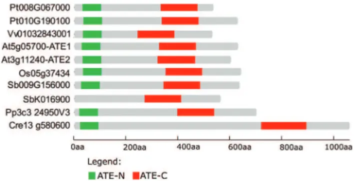
ATE protein sequences contain two Pfam domains named ATE-N (PF04376) and ATE-C (PF04377), which are located at N- and C-termini, respectively (Figure 1). Comparative alignment and identity analysis of ATE pro-teins from human, mouse, fruit fly, yeast andArabidopsis
have revealed high conservation in domain regions but variability in inter-domain sequence and size (Figure 1A, B). Interestingly, the observed identity of more than 60% between the C-terminal ATE domains of mammals and fruit flies may reflect conserved functions or interaction partners in these taxa (Figure 1B). It is also remarkable that the plant ATE C-domain shares 50% identity with the same domain fromC. elegans to human, demonstrating strong protein conservation among multicellular eukaryotes.
Plant evolutionary analysis has identified ATE
orthologous genes from the green alga Chlamydomonas reinhardtiito angiosperms (Figure 2). In general, only one
ATEgene is detected in a given plant species, with the two conserved ATE domains located at the N- and C-termini (Figures 1, 2). Some species, such asArabidopsis,Populus
andSorghum, have experienced gene duplication. How-ever,Sorghumappears to have generated a paralog gene that contains only the ATE-C domain and is likely not a functional arginyltransferase.
Recently, it was demonstrated that ATE protein abun-dance is spatially and temporally regulated by hormones and light and is highly abundant in meristematic cells from the mossPhyscomitrella patens(Schuesseleet al., 2016). This work also showed that arginylation is necessary for moss gametophyte development, which is not observed in flowering plants. These findings support the conservation of N-end rule pathway components in land plants, even though the regulated processes may have diverged during evolution.
Plant ATE biological functions
Arabidopsis encodes two closely relatedATEgenes,
AtATE1(At5g05700) andAtATE2(At11240), which have at least partially redundant functions.ATE1was first func-tionally investigated by Yoshidaet al.(2002) in the mutant
delayed leaf senescence1(dls1), which exhibits extremely slow age-dependent, dark-induced leaf senescence. Such a phenotype was linked to a disruptedATE1indls1. Years later, Holmanet al.(2009) showed that the N-recognin E3 ligase PRT6 andAtATE1andAtATE2are involved in seed germination controlled by abscisic acid. A double mutant forAtATE1andAtATE2(ate1.ate2) displays lost sensitivity to this hormone and consequently uncontrolled seed germi-nation and establishment. At the end of the same year, Gracietet al.(2009) showed that apical dominance, stem elongation and regulation of leaf morphology were also linked to the N-end rule pathway, as demonstrated by ana-lyzing AtATE1and/or AtATE2T-DNA insertion mutants (Graciet et al., 2009; Graciet and Wellmer, 2010). The
arginylation branch of the N-end rule pathway is also re-sponsible for repressing expression of the meristem-promoting brevipedicellus (BP) gene during leaf develop-ment, acting in a redundant way with the asymmetric leaves 1 (AS1) transcription factor complex, a known negative regulator ofBP expression (Gracietet al., 2009). The R-transferase target in this mechanism, however, has not yet been identified. Another important discovery by Gracietet al.(2009) was that the N-recognin PRT6 is the E3 ligase N-recognin of plants and acts downstream of ATE.
One of the most studied and understood signaling pathways in plants controlled by arginylation is that involv-ing the ethylene responsive transcription factor VII (ERFVII). It was observed thatArabidopsis ate1/ate2 or
prt6mutants could not degrade ERFVII, and as a conse-quence showed increased expression of hypoxia–respon-sive genes involved in fermentation and sugar consumption even under oxygen-rich conditions (Gibbs et al., 2011; Licausi et al., 2011). The mechanism responsible for ERFVII regulation was suggested by previous studies on mammalian ATE1, which revealed that these enzymes can recognize oxidized N-terminal Cys residues as substrates for arginylation. The occurrence of this was later demon-strated in plants, with oxidized Cys also acting as a tertiary destabilizing N-terminal residue (Graciet et al., 2010). Gibbset al.(2011) demonstratedin vitrothat all members ofArabidopsisgroup VII ERFs are N-end rule substrates that function as sensors of molecular oxygen via oxidation of the tertiary destabilizing cysteine residue.Arabidopsis
has five ERFVII proteins: hypoxia-responsive ERF 1 and 2 (HRE1 and HRE2) and three more proteins related to apetala 2.2: RAP2.2, RAP.2.3, RAP2.12 (Nakano et al., 2006). Alignment of the five Arabidopsis VII ERFs re-vealed that all share the same five first N-terminal residues (Gibbset al., 2011, 2015), with the Nt-MCGGAII/L do-main being highly conserved in flowering plants (Licausiet al., 2011). Moreover, all five ERFVII proteins from
Arabidopsisaccumulate under conditions of low oxygen and are destabilized by the presence of oxygen. Oxidation of the N-terminal Cys residue is catalyzed by plant cysteine oxidases (PCOs) under specific conditions in which the Nt-Met residue is removed by MAP activity. Therefore, arginylation is directly involved in the mechanism that en-ables this transcription factor family to function as a sensor of hypoxia in plants (Gibbs et al., 2011; Licausi et al., 2011).
Another important biological process mediated by Cys arginylation was demonstrated years later. Gibbset al.
Figure 1- ATE protein sequences across distinct kingdoms. A) Alignment of ATE full-length protein sequences fromHomo sapiens(NP_001001976.1),
RGS a substrate for ATE and the N-end rule pathway (Huet al., 2005; Jabaet al., 2013). Gibbset al.(2014) found that the same mechanism regulates ERFVII stability in plants. In the presence of oxygen and/or NO, ERFVII degradation leads to hypocotyl growth inhibition, seed germination, photomorphogenesis, stomatal closure, and hypoxia-responsive transcriptional repression. Under hypoxia, how-ever, accumulation of group VII ERFVs promotes hypocotyl growth, seed dormancy, photomorphogenesis repression, stomatal opening, and anaerobic-responsive gene expression (Gibbset al., 2015). Together, these stud-ies suggest that group VII ERFs function as a central hub for the perception of oxygen and NO and that the N-end rule pathway is an important integrator of gas signaling in plants (Gibbset al., 2014). The regulatory role can be even broader, as ERFVII arginylation can also respond to other signals such as ethylene (Gibbset al., 2015).
By studying water submergence and starvation stress responses in N-end rule pathway mutants, Riber et al.
(2015) observed that these mutant plants were generally more tolerant to starvation conditions, such as prolonged darkness or water submergence. Interestingly, increased tolerance was a direct consequence of the impaired N-end rule pathway rather than up-regulation of the expression of genes involved with sugar consumption and fermentation (Riberet al., 2015). This work showed the potential of reg-ulating the ATE branch of the N-end rule pathway to gener-ate stress-resistant plant variants.
The identification of ATE arginylation targets is an intriguing area of research and can reveal new biological processes regulated by the N-end rule pathway. Based on sequence analysis only, a large number of proteins appear to be potential substrates for ATE-mediated desta-bilization. However, the experimental identification and characterization of ATE substrate proteins remain
techni-cally challenging. The best characterized targets proteins are mammalian RGS proteins, especially RGS4, RGS5 and RGS16, and plant group VII ERFs (Huet al., 2005; Gibbs
et al., 2011; Licausiet al., 2011; Bailey-Serreset al., 2012; Lee,et al., 2012; Gibbset al., 2014). As mentioned above, both RGS and ERFVII substrates have a Cys as the second Nt residue which, after oxidation, acts as a tertiary desta-bilizing residue dependent on ATE arginylation (Gibbset al., 2011; Licausiet al., 2011; Bailey-Serreset al., 2012; Lee et al., 2012). During oxygen-limiting conditions, ERFVII and RGS accumulate, leading to transcription of hypoxia-response genes in plants (Gibbs, et al., 2011; Licausi et al., 2011; Gibbs et al., 2014) and decreased cardiomyocyte proliferation by RGS accumulation and downregulation of G protein signaling in mammals (Leeet al., 2012).
The proteins’ resistance to Pseudomonas syringae
1-interacting protein 4 (RIN4), ethylene response factor 72 (EBP) and vernalization 2 (VRN2) have been identified as putative ATE substrates by transcriptome analysis of
ate1.ate2 mutants. However, their modification by ATE has not been shown at the molecular level (Gibbset al., 2011).
Mass spectrometry approaches are being developed to screen for arginylated proteins and to reveal new cellular processes controlled by ATE modification. To identify plant ATE substrates, Majovsky et al. (2014) analyzed plant proteomes fromA. thaliana ate1,ate2,prt1andprt6
knockout mutants, and new putative substrates of ATE were identified among Nt Met-Cys proteins. The relative abundance of methylesterase 10 (MES10), nucleoside diphosphate kinase family protein (NDPK1), and two asparagine synthetases (ASNs) was augmented inate1/ate2
mutants. The MES10 protein hydrolyzes methyl salicylate to salicylic acid, NDPK1 plays a role in the response to re-active oxygen species (ROS) stress, and ASNs are compo-nents of the L-asparagine biosynthesis pathway (Peeterset al., 2000; Fukamatsuet al., 2003; Yanget al., 2008). Other putative substrates found by Majovskyet al.(2014) that do not have a Nt-Met-Cys were glyceraldehyde 3-phosphate dehydrogenase subunits GAPA, GAPA2 and GAPB, the granulin repeat cysteine protease family protein (RD21), glucoside (GGD), thioglucoside glucohydrolases (TGG1 and TGG2), two glycine-rich proteins (GRPs), cold circa-dian rhythm (CCR1 and CCR2), RNA-binding 1 and 2 and nicotinamide adenine dinucleotide-dependent malic en-zyme (NAD-MED2). The functional implication of these modifications awaits experimental validation.
Other interesting roles for mammalian ATEs are emerging and could point to new functions in plants. Re-cently, Cha-Molstadet al.(2015a) showed that in human cells, ATE1 mediates N-terminal arginylation of binding immunoglobulin protein (Bip) and possibly other endo-plasmic reticulum (ER)-residing chaperones. Under certain stress conditions, such as the presence of dsDNA in the
cy-Figure 2- Domain location ofATEprotein sequences among plant spe-cies. ATE coding sequences from distinct plant species, such as the eudicotsPopulus trichocarpa,Arabidopsis thalianaandVitis vinifera, monocotsSorghum bicolorandOryza sativa, mossPhyscomitrella patens
toplasm or proteasome inhibition, Bip was directed to the cytosol, where it was arginylated by ATE. Further experi-ments showed that Nt-Arg residues function as a determi-nant of autophagic delivery to autophagosomes by acting as an activating ligand of p62, an important component of the autophagy pathway that interacts with degradation targets and drives autophagosome formation by recruiting other components of the pathway (Yamano and Youle, 2013; Cha-Molstadet al., 2015a,b). Intriguingly, ATE was also reported to act in mammals directing the degradation of caspase- and calpain-generated fragments of cohesin (Rai and Kashina, 2005; Piatkovet al., 2012, 2014). Nonethe-less, the involvement of plant ATEs in the autophagy path-way and in the degradation of caspase-like and/or calpain-generated fragments in plants remains elusive. Combining these putative ATE mechanisms with those already studied in plants, we propose a model of biological processes regu-lated by ATE arginylation in plants (Figure 3).
Concluding remarks
Although important efforts have been made towards an understanding of the role of ATE/N-end rule functions in plants, many open questions remain, for example, the functions of different isoforms of ATE in plants, how ATE activity is regulated, and the identification of interaction partners. It is possible that plant ATEs can also arginylate
internal amino acids of proteins and that this type of modifi-cation has other signaling functions.
The discovery of the physiological substrates of R-arginylation is essential for understanding N-end rule functions. In addition to its biological importance, elucidat-ing some of the processes regulated by the N-end rule path-way in plants is relevant for biotechnological purposes, as this pathway has a direct impact on developmental pro-cesses and tolerance to starvation and/or abiotic stress.
References
Bachmair A, Finley D and Varshanvsky A (1986)In vivohalf-life of protein is a function of its amino-terminal residue. Sci-ence 234:179-186.
Bailey-Serres J, Fukao T, Gibbs DJ, Holdsworth MJ, Lee SC, Licausi F, Perata P, Voesenek LA and van Dongen JT (2012) Making sense of low oxygen sensing. Trends Plant Sci 17:129-138.
Bradshaw RA, Brickey W and Walker KW (1998) N-terminal processing: The methionine aminopeptidase and N alpha-acetyl transferase families. Trends Biochem Sci 23:263-267. Bulinski JC (2009) Tubulin posttranslational modifications: A
Pushmi-Pullyu at work? Dev Cell 16:773-774.
Cha-Molstad H, Sung KS, Hwang J, Kim KA, Yu JE, Yoo YD, Jang JM, Han DH, Molstad M, Kim JG, et al. (2015a) Amino-terminal arginylation targets endoplasmic reticulum chaperone BiP for autophagy through p62 binding. Nat Cell Biol 17:917-929.
Cha-Molstad H, Kwon YT and Kim BY (2015b) Amino-terminal arginylation as a degradation signal for selective autophagy. BMB Reports 48:487-488.
Crowe E and Candido EP (2004) Characterization ofC. elegans
RING finger protein 1, a binding partner of ubiquitin-conjugating enzyme 1. Dev Biol 265:446-459.
Elias S and Ciechanover A (1990) Post-translational addition of an arginine moiety to acidic NH2 termini of proteins is re-quired for their recognition by ubiquitin-protein ligase. J Biol Chem 265:15511-15517.
Fukamatsu Y, Yabe N and Hasunuma K (2003) Arabidopsis NDK1 is a component of ROS signaling by interacting with three catalases. Plant Cell Physiol 44:982-989.
Gibbs DJ, Lee SC, Isa NM, Gramuglia S, Fukao T, Bassel GW, Correia CS, Corbineau F, Theodoulou FL, Bailey-Serres J,
et al.(2011) Homeostatic response to hypoxia is regulated by the N-end rule pathway in plants. Nature 479:415-418. Gibbs DJ, Md Isa N, Movahedi M, Lozano-Juste J, Mendiondo
GM, Berckhan S, Marín-de la Rosa N, Vicente Conde J, Sousa Correia C, Pearce SP,et al.(2014). Nitric oxide sens-ing in plants is mediated by proteolytic control of group VII ERF transcriptional factors. Mol Cell 53:369-379.
Gibbs DJ, Conde JV, Berckhan S, Prasad G, Mendiondo GM and Holdsworth MJ (2015) Group VII Ethylene Response Fac-tors coordinate oxygen and nitric oxide signal transduction and stress responses in plants. Plant Physiol 169:23-31. Gonda DK, Bachmair A, Wunning I, Tobias JW, Lane WS and
Varshavsky A (1989) Universality and structure of the N-end rule. J Biol Chem 264:16700-16712.
Graciet E, Hu RG, Piatkov K, Rhee JH, Schwarz EM and Varshavsky A (2006) Aminoacyl-transfersases and the N-end rule pathway of prokaryotic/eukaryotic specificity in a human pathogen. Proc Natl Acad Sci U S A 103:3078-3083. Graciet E, Walter F, Maoileidigh DO, Pollmann S, Meyerowitz
EM, Varshavsky A and Wellmer F (2009) The N-end rule pathway controls multiple functions during Arabidopsis shoot and leaf development. Proc Natl Acad Sci U S A 106:13618-13623.
Graciet E and Wellmer F (2010) The plant N-end rule pathway: Structure and functions. Trends Plant Sci 15:447-453. Graciet E, Mesiti F and Wellmer F (2010) Structure and
evolu-tionary conservation of the plant N-end rule pathway. Plant J 61:741-751.
Holman TJ, Jones PD, Russell L, Medhurst A, Tomas S, Talloji P, Marquez J, Schmuths H, Tung SA, Taylor I,et al.(2009) The N-end rule pathway promotes seed germination and es-tablishment through removal of ABA sensitivity in Arabidopsis. Proc Natl Acad Sci U S A 106:4549-4554. Hu RG, Sheng J, Qi X, Xu Z, Takahashi TT and Varshavsky A
(2005) The N-end rule pathway as a nitric oxide sensor con-trolling the levels of multiple regulators. Nature 437:981-986.
Jaba IM, Zhuang ZW, Li N, Jiang Y, Martin KA, Sinusas AJ, Papademetris X, Simons M, Sessa WC, Young LH,et al.
(2013) NO triggers RGS4 degradation to coordinate angio-genesis and cardiomyocyte growth. J Clin Invest 123:1718-1731.
Kaji H (1968) Further studies on the soluble amino acid incorpo-rating system from rat liver. Biochemistry 7:3844-3850.
Kaji A, Kaji H and Novelli GD (1963a) A soluble amino acid in-corporating system. Biochem Biophys Res Commun 10:406-409.
Kaji H, Novelli GD and Kaji A (1963b) A soluble amino acid-incorporating system from rat liver. Biochim Biophys. Acta 76:474-477.
Kim ST, Tasaki T, Zakrzewska A, Yoo YD, Sa Sung K, Kim SH, Cha-Molstad H, Hwang J, Kim KA, Kim BY,et al.(2013) The N-end rule proteolytic system in autophagy. Autophagy 9:1100-1103.
Kwon YT, Kashina AS and Varshavsky A (1999) Alternative splicing results in differential expression, activity, and local-ization of the two forms of arginyl-tRNA-protein trans-ferase, a component of the N-end rule pathway. Mol Cell Biol 19:182-193.
Kwon YT, Kashina AS, Davydov IV, Hu RG, An JY, Seo JW, Du F and Varshavsky A (2002) An essential role of N-terminal arginylation in cardiovascular development. Science 297:96-99.
Lee MJ, Kim DE, Zakrzewska A, Yoo YD, Kim SH, Kim ST, Seo JW, Lee YS, Dorn 2nd GW, Oh U,et al.(2012) Character-ization of arginylation branch of N-end rule pathway in G-protein-mediated proliferation and signaling of cardio-myocytes. J Biol Chem 287:24043-24052.
Licausi F, Kosmacz M, Weits DA, Giuntoli B, Giorgi FM, Voesenek LA, Perata P and van Dongen JT (2011) Oxygen sensing in plants is mediated by an N-end rule pathway for protein destabilization. Nature 479:419-422.
Majovsky P, Naumann C, Lee CW, Lassowskat I, Trujillo M, Dissmeyer N and Hoehenwarter W (2014) Targeted proteomics analysis of protein degradation in plant signaling on an LTQ-orbitrap mass spectrometer. J Proteome Res 13:4246-4258.
Manahan CO and App AA (1973) An arginyl-transfer ribonucleic acid protein transferase from cereal embryos. Plant Physiol 52:13-16.
Mutsuddi M, Marshall CM, Benzow KA, Koob MD and Reba YI (2004) The spinocerebellar ataxia 8 noncoding RNA causes neurodegeneration and associates with staufen in Drosophila. Curr Biol 14:302-308.
Nakano T, Suzuki K, Fujimura T and Shinshi H (2006) Ge-nome-wide analysis of the ERF gene family in Arabidopsis and rice. Plant Physiol 140:411-432.
Peeters NM, Chapron A, Giritch A, Grandjean O, Lancelin D, Lhomme T, Vivrel A and Small I (2000) Duplication and quadruplication of Arabidopsis thaliana cysteinyl- and asparaginyl-tRNA synthetase genes of organellar origin. J Mol Evol 50:413-423.
Piatkov KI, Colnaghi L, Békés M, Varshavsky A and Huang TT (2012) The auto-generated fragment of the Usp1 deubiquitylase is a physiological substrate of the N-end rule pathway. Mol Cell 48:926-933.
Piatkov KI, Oh JH, Liu Y and Varshavsky A (2014) Calpain-generated natural protein fragments as short-lived substrates of the N-end rule pathway. Proc Natl Acad Sci U S A 111:E817-E826.
Rai R and Kashina A (2005) Identification of mammalian arginyltransferases that modify a specific subset of protein substrates. Proc Natl Acad Sci U S A 102:10123-10128.
Riber W, Müller JT, Visser EJW, Sasidharan R, Voesenek LACJ and Mustroph A (2015) The greening after extended darkness1 is an N-end rule pathway mutant with high toler-ance to submergence and starvation. Plant Physiol 167:1616-1629.
Schuessele C, Hoernstein SN, Mueller SJ, Rodriguez-Franco M, Lorenz T, Lang D, Igloi GL and Reski R (2016) Spatio-temporal patterning of arginyl-tRNA protein transferase (ATE) contributes to gametophytic development in a moss. New Phytol 209:1014-1027.
Sriram SM, Kim BY and Kwon YT (2011) The N-end rule path-way: Emerging functions and molecular principles of sub-strate recognition. Nat Rev Mol Cell Biol 12:735-747.
Stary S, Yin XJ, Potuschak T, Schlögelhofer P, Nizhynska V and Bachmair A (2003) PRT1 of Arabidopsis is a ubiquitin pro-tein ligase of the plant N-end rule pathway with specificity for aromatic amino-terminal residues. Plant Physiol 133:1360-1366.
Tasaki T, Zakrzewska A, Dudgeon DD, Jiang Y, Lazo JS and Kwon YT (2009) The substrate recognition domains of the N-end rule pathway. J Biol Chem 284:1884-1895.
Tasaki T, Sriram S, Park K and Kwon YT (2012) The N-end rule pathway. Annu Rev Biochem 81:261-289.
Varshavsky A (2011) The N-end rule pathway and regulation by proteolysis. Protein Sci 20:1298-1345.
Wang J, Han X, Saha S, Xu T, Rai R, Zhang F, Wolf YI, Wolfson A, Yates 3rd JR and Kashina A (2011) Arginyltransferase is an ATP-independent self-regulating enzyme that forms dis-tinct functional complexes in vivo. Chem Biol 28:121-130. Wang J, Han X, Wong CC, Cheng H, Aslanian A, Xu T, Leavis P,
Roder H, Hedstrom L, Yates 3rd JR, et al. (2014) Arginyltransferase ATE1 catalyzes midchain arginylation of proteins at side chain carboxylatesin vivo. Chem Biol 20:331-337.
Wong CCL, Xu T, Rai R, Bailey AO, Yates JR, Wolf YI, Zebroski H and Kashina A (2007) Global analysis of posttranslational protein arginylation. PLoS Biol 5:e258.
Xia Z, Webster A, Du F, Piatkov K, Ghislain M and Varshavsky A (2008) Substrate-binding sites of URB1, the ubiquitin ligase of the N-end rule pathway. J Biol Chem 283:24011-24028. Yamano K and Youle RJ (2013) PINK1 is degraded through the
N-end rule pathway. Autophagy 9:1758-1769.
Yang Y, Xu R, Ma CJ, Vlot AC, Klessig DF and Pichersky E (2008) Inactive methyl indole-3-acetic acid ester can be hy-drolyzed and activated by several esterases belonging to the AtMES esterase family of Arabidopsis. Plant Physiol 147:1034-1045.
Yoshida S, Ito M, Callis J, Nishida I and Watanabe A (2002) A de-layed leaf senescence mutant is defective in arginyl-tRNA:protein arginyltransferase, a component of the N-end rule pathway in Arabidopsis. Plant J. 32:129-137.
Associate Editor: Marcia Pinheiro Margis