Analysis of Polycomb Target
Chromatin
José Manuel Menino Ventura Antão
Analysis of Polycomb Target
Chromatin
José Manuel Menino Ventura Antão
Dissertation presented to obtain the Ph.D degree in Molecular Biology Instituto de Tecnologia Química e Biológica | Universidade Nova de Lisboa
the following entities
Table of Contents
Page
List of Figures and Tables……..………2
Acknowledgments………..………..3
Abstract……….………..5
Sumário………..……….7
Chapter 1: Introduction……….………..9
Chapter 2:
Zeste interacts with Polyhomeotic and contributes to the recruitment of PRC1 to PRE sequences………...20
Chapter 3:
The Protein Landscape at Drosophila Telomere- Associated Sequence Repeats………55
Chapter 4: Discussion……….…..99
References……….…106
List of Figures and Tables
Page
Figure 2.1: Zeste interacts specifically with Polyhomeotic………..…35
Figure 2.2: Zeste increases the efficiency of PCC-mediated inhibitionof Swi/Snf remodeling………39
Figure 2.3: Functional interaction between Zeste, Ph and PSC………..…41
Figure 2.4: Influence of various PRE-binding proteins on the activity of PSC……….…43
Figure 2.5: Chromatin-IP analysis of Zeste-HA and Ph occupancy at target loci……….46
Figure 2.6: Model for the contribution of Zeste and other PRE-binding proteins to the activities of PRC1……….…52
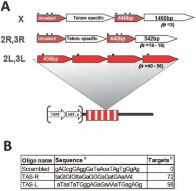
Figure 3.1: Structure of the TAS repeats and PICh capture probes………...…70
Figure 3.2: TAS sequence unit repeats and probe hybridization sites………...71
Figure 3.3: The optimized PICh protocol……….…73
Figure 3.4: TAS repeats chromatin purification……….…….……75
Figure 3.5: Candidate TAS repeat factor enrichment levels by Western blot……….…78
Figure 3.6: ChIP analysis of candidate TAS protein binding at TAS repeats……….………84
Figure 3.7: brm2 effects on Df(2L)M26 suppression of TPE……….…89
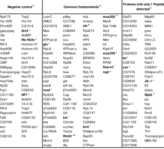
Table 3.1: Proteins removed from the final list of candidates………...…77
Table 3.2: TAS-L proteins, ranked by confidence score………...79
Table 3.3: TAS-R proteins, ranked by confidence score………...80
Table 3.4: Genes eliminated in dominant Su(TPE) deficiencies………..86
Acknowledgments
The work reported on the following pages did not result only from the many hours at the bench, but also from the entire context in which they happened. I was extremely fortunate to land in an environment where people were knowledgeable, enthusiastic, kind and supportive. I thank Jonathan Dennis for having been an example in conduct and
enthusiasm, Hua-Ying Fan for having been a teacher and a lot of fun, Jérôme Déjardin for all of the above and below. I thank all other
labmates I was fortunate to interact with in the Kingston lab. You are too many to name, but you should know you all have been important. And a special word for my Graduate School brothers Aaron and Dan: I have outlived you.
I thank the staff of the Department of Molecular Biology at MGH for making everything run smoothly and for your dedication. Special thanks to Panfilo Federico, Neil Henderson, Melanie Ransom, Pauline Lim and Janet McLeod.
I thank Bob Kingston. Bob is as much a good scientist as he is a good manager. And he is as decent a human being as I can conceive of an advisor. Today, a whole 9 years since I first met him in person, I can say that my tenure under his watch would have been substantially shorter had I listened to what he said more often. I did not do it on purpose, but in the end I just came to appreciate him more.
I don’t thank friends for being friends, but I need to reflect their
importance on my path. Boston is a wonderfully diverse environment and I was fortunate to meet many extraordinary people. Many are forever inseparable from my experience of growing-up. Some are friends for life. Some are family. Thank you Elena, Eyleen, Colo, Rita, Miguel, Sachiko, Ben, Anita, KJ, Caroline, Jesse, Kika, Tiago, Rodrigo, Sara, J-Lo, Maria, Alex, Rui, Inbal, Cátia, Gilberto, Susana, Dinis, Cláudia, André, Eduardo, Linn, Luís, Anna, Nuno, Xana, Bernardo, Gisele, Luciana, Nelly,
I thank Instituto Gulbenkian de Ciência, Fundação para a Ciência e Tecnologia and Programa Gulbenkian de Doutoramento em Biomedicina for the investment in my education and the trust deposited on me. I thank Manuela Cordeiro and Ana Maria Portocarrero for the help with bureaucracies. I am especially indebted to my local advisor Lars Jansen, thesis committee members Alekos Athanasiadis and Vasco Barreto and to the members of the jury for my dissertation: Luísa Figueiredo, João Ferreira, Hélder Maiato and Jorge Carneiro.
And I feel very fortunate to have had the opportunity to collaborate with Jim Mason. Jim is an extremely knowledgeable and thorough scientist and his generous contribution to my research was invaluable.
Agradeço ao meu país e aos cidadãos que, através dos seus impostos, contribuiram para a minha educação. A oportunidade que me deram foi extraordinária. O peso da responsabilidade de devolver esse
investimento é considerável, mas vou dar o meu melhor.
Agradeço acima de tudo à minha família, pelo apoio incondicional ao longo destes anos. Apesar de tanto estudo, é-me difícil compreender a origem do vosso orgulho. Por isso, acredito que seja amor. Obrigado mãe, pai, Teresa, João, Madalena, Paulo e tios Isabel, Vieira, Quim e todos os outros Meninos e Ferreiras.
Abstract
In metazoans, chromatin-mediated gene silencing phenomena fall into
three main categories: Heterochromatin-mediated gene silencing,
Telomeric gene silencing and Polycomb Group (PcG)-mediated
silencing. The function of the Drosophila PcG system is best known at
the homeotic gene clusters, where it keeps the transcriptional state of
developmental regulators repressed, contributing to the maintenance of
cell identity during embryo development. This system is also present at
the TAS repeat sequences, which are responsible for Telomeric
silencing in Drosophila, although the mechanisms may differ.
Here I report on the discoveries from different approaches to study the
function of PcG-regulated chromatin at two genomic locations: the bxd
Polycomb Response Element, from the Ubx locus, and the TAS repeat
sequences. I show that Zeste, one of the PRC1 PcG complex subunits,
interacts specifically with the Polyhomeotic protein, likely as a dimer.
Using an in vitro system with purified factors I show that the
PRC1-mediated chromatin compaction over a bxd sequence is enhanced by
Zeste in a dimerization- and DNA binding- dependent way and that other
factors known to interact with the PcG machinery also display an
enhancement effect. These results are consistent with a targeting
function and suggestive of a resilient mechanism for the targeting of
PRC1 function.
To study the less-characterized TAS sequences, I developed a
biochemical purification procedure to identify proteins associated with
the TAS loci. With this protocol, over 70 new candidates were identified
and 5 were validated for direct physical association with TAS sequences.
Position Effect and three other factors were identified as potential
modifiers. These findings have suggested a predominantly modifier
dose-independent silencing mechanism at Drosophila telomeres and will
allow a more directed study of the silencing mechanism mediated by
Sumário
Em organismos metazoários existem três categorias principais de
silenciamento de genes pela cromatina: silenciamento através da
Heterocromatina, silenciamento Telomérico e silenciamento através de
proteínas da família Polycomb (PcG). A função mais estudada das
proteínas PcG em Drosophila é exercida nos complexos de genes
homeóticos, onde mantêm a repressão destes reguladores do
desenvolvimento, contribuindo para manter a identidade celular durante
o desenvolvimento embrionário. Este sistema também está presente
nas sequências repetitivas TAS, que são responsáveis pelo
silenciamento Telomérico em Drosophila, apesar de os mecanismos
poderem ser diferentes.
Apresento aqui as descobertas feitas através de abordagens
experimentais distintas ao estudo da cromatina regulada por proteínas
PcG em duas regiões do genoma: o Elemento de Resposta a Polycomb
bxd, do locus Ubx, e as sequências repetitivas TAS. Mostro que a
proteína Zeste, um dos componentes do complexo PRC1 da família
PcG, interage especificamente com o componente Polyhomeotic,
provavelmente como um dímero. Através de experiências in vitro com
componentes purificados, mostro que a actividade de compactação da
cromatina associada à sequência do elemento bxd pelo complexo PRC1
é potenciada pela proteína Zeste, com o envolvimento dos domínios
proteicos de dimerização e de ligação ao ADN. Outras proteínas de que
se conhece a associação à actividade das proteínas PcG apresentam
um efeito de potenciação semelhante ao de Zeste. Estes resultados são
interpretáveis à luz de uma função de Zeste e dos outros factores no
direccionamento do complexo PRC1 para os alvos apropriados e
Para estudar as menos caracterizadas sequências TAS, desenvolvi um
método bioquímico de purificação para identificar proteínas associadas
aos loci TAS. Através deste protocolo, foram identificados mais de 70
candidatos e a associação de 5 deles com as sequências TAS foi
confirmada por outros métodos. O complexo Brahma foi identificado
como um modificador dominante do Efeito Posicional Telomérico de
silenciamento e outros três factores foram também identificados como
potenciais modificadores. Estes resultados sugerem um mecanismo de
silenciamento Telomérico em Drosophila predominantemente
independente da dose dos modificadores e vão permitir um estudo mais
direccionado do mecanismo de silenciamento mediado pelas
Chapter 1
Chromatin Structure in Eukaryotic Cells
The confinement of the eukaryotic genome within nuclear boundaries
poses very significant physical challenges. On the one hand, the
structure needs to solve the packaging problem, by condensing the
genetic material roughly 10,000-fold from its linear length to fit within the
nuclear space, while managing the large amount of negative charge of
the DNA molecule. On the other hand, packaging needs to be done in a
plastic enough manner, to allow disassembly and re-assembly with cell
cycle progression and during genome regulation.
The levels of organization of the genome are likely several, and a
common rule probably does not exist for every genomic region. It is
consensual that the basic unit of organization of the genome is the
nucleosome, formed by the wrapping of roughly 150 base pairs of DNA
around a histone octamer, composed of two copies of each H2A, H2B,
H3 and H4 histones (1). These basic proteins counter the negative
charge of DNA, stabilizing the electrostatic potential inside the nucleus.
At the same time, they provide a 5-fold stacking of the DNA molecule
length, which is an important, if insufficient level of shrinking to solve the
folding problem. Levels of organization above the nucleosome are
contentious. The organization of a “beads-on-a-string” linear
multi-nucleosome fragment into the next predicted hierarchical level, the 30nm
fiber, has been proposed to be done by a 2-start zig-zag model (2,3) or a
1-start solenoid model (4,5), depending on the in vitro reconstitution
conditions, the method used to determine the structure (X-ray
crystallography, biochemical analysis or electron microscopy) or the
presence of the linker histone H1.
The 30nm fiber is proposed to organize into a 100nm chromonema and
Criticism of these models has always been latent (7), due to the lack of
in situ evidence and the un-physiological conditions of the in vitro data.
Recent in situ cryo-EM studies of mitotic chromosomes have found no
evidence for the existence of the 30nm fiber, or of any higher-order of
organization beyond an 11nm structure, and have proposed an irregular
fractal-like structure for chromatin in the nucleus (8,9), consistent with
the recent analysis of chromatin interaction maps from human cells (10)
and Drosophila embryos (11). Such “molten fractal” models are
proposed to grant a higher fluidity to, and efficiency in the regulation of,
the chromatin structure, which are in line with a contemporary view of
chromatin as a dynamic structure, regulating access to genomic DNA
and being regulated by a plethora of partner non-histone proteins, as
opposed to the classical view of a more static frame of structural
elements organizing the genome (12).
The association of genomic DNA with histone and non-histone proteins,
and their higher-order organization into more complex chromatin
structures and nuclear sub-compartments, create a very intricate
substrate for genome regulation. Transcription factors and other
DNA-binding proteins which recognize specific motifs in the DNA sequence
display a different ability to access their substrate whether it is in a
nucleosomal context or un-protected by the absence of histones (13).
This access is regulated by various chromatin remodeling activities. On
one extreme, these can involve the eviction of the whole histone
octamer, or a part of it from the nucleosome (14), leaving the DNA
accessible, or substituting one of the multiple histone variants with
diverse properties (15) for the canonical histone form within the octamer.
Subtler forms of regulation involve the breaking of contacts between
histones and DNA, leading to the sliding, looping, or otherwise exposing
activities are carried out by a variety of protein complexes, at the
expense of ATP hydrolysis (16).
Moreover, DNA-binding proteins often act in the context of multi-protein
complexes, or transiently associate with other proteins which, by virtue
of their affinity for specific histone NH2-terminal tail covalent
modifications at certain aminoacid residues, confer an extra level of
specificity for target loci. The combination of DNA target presence,
affinity and accessibility, histone tail covalent modifications, and the
presence and amount of DNA-binding protein and interacting factors for
each locus in the genome determines, to a great extent, the processes
of genome regulation and cell identity.
Chromatin Silencing Mechanisms
Stable silencing of genes is a desirable property of genome regulation,
be it for developmental reasons or to defend the cell from deleterious
transcripts, such as transposons. It generally involves the creation of a
compacted chromatin structure. The first mechanistic models of
chromatin-mediated gene silencing came from yeast. In Saccharomyces
cerevisiae, although no cytological features of heterochromatin are
visible, specialized genomic structures at the silent mating loci (17), the
Ribosomal DNA (18,19) and the telomeres (20), are found in a
transcriptionally silent chromatin state. This repressive environment is
stable and depends on histones, SIR proteins and on the relative
position of the repressive loci (21). Transgenes inserted in the vicinity of
these regions are subjected to the repressive chromatin environment
and can be silenced to different extents. In higher eukaryotes, the
to the accumulation of non-coding sequences in the genome, particularly
at the pericentromeric region. Translocation of a white gene into the
vicinity of pericentric heterochromatin in Drosophila causes variegating
expression of the white gene in the eye, due to stochastic silencing by
the juxtaposed heterochromatin in primordial cells and clonal growth into
the adult eye (22). This effect is mediated by the local chromatin
structure (23) and factors that influence this structure control the extent
of silencing. Mutants that cause an increase in the repression of white on
this background are called Enhancers of Position Effect Variegation
[E(PEV)], whereas mutants which alleviate silencing of white are called
Suppressors of PEV [Su(PEV)]. Collectively, they can be referred to as
Modifiers of PEV [Mod(PEV)]. In most cases studied, these mutants are
dominant, i.e. mutation of one of the two copies in the diploid genome is
enough to cause a phenotype (24). The dose-dependence of this
phenomenon has led some to propose a mass-action model for the
silencing of chromatin by specialized complexes (25).
Unlike in yeast, the chromatin silencing mechanisms at the telomeric
regions are not equivalent to other constitutive heterochromatic regions
in Drosophila. Modifiers of PEV don’t show an effect on Telomere
Position Effect (TPE), the variegating phenotype of white-bearing
P-element insertions at the telomeric regions (26). Furthermore, the
silencing effect is conveyed by the Telomere-Associated Sequence
(TAS) repeat loci, rather than the telomeres themselves, and has been
shown to be regulated by the Polycomb-Group (PcG) of proteins (27),
which are associated with developmental regulation in higher eukaryotes
and are mostly found at nuclear regions cytologically classified as
Drosophila
Development and PcG Protein functions
A classic model for the study of cell identity phenomena is the
developing embryo of Drosophila melanogaster. Within the first few
hours of embryo development, patterns of homeotic master regulatory
gene expression are formed along the anterior-posterior axis, leading to
the establishment of the body segments which will result in the
appropriate organ formation in the adult (30). These master regulatory
genes are located in two genomic clusters on the Chromosome 3R: the
Antennapedia Complex (ANT-C) and the Bithorax Complex (BX-C)
[hereafter collectively called Homeotic Complex (HOM-C)]. Genes in
these clusters are regulated through cis-acting sequences, relatively
distant from gene promoters, and by trans-acting transcription factors
which, depending on the position in the developing body plan, bind to
regulatory regions in combinations that activate or repress these master
regulatory identity genes. Such trans-acting transcription factors are
classified as Gap, Pair-Rule and Segment-Polarity proteins (30) and they
act sequentially to determine the appropriate pattern of homeotic gene
expression. They are very short-lived, though, their transcripts decaying
sharply after the first four hours of development. Concomitantly with their
decline, another two families of factors – the Trithorax-Group (TrxG) and
the PcG – replace them at the HOM-C and stably maintain the active or
repressed states, respectively, of transcription previously established.
The Drosophila Brahma complex is an example of a TrxG complex and it
is the homolog of the human ATP-dependent chromatin remodeling
complex Swi/Snf, which is one of the most ubiquitous chromatin
remodeling enzymes. PcG proteins exist in the context of multi-protein
complexes as well and their activities are varied. The Polycomb
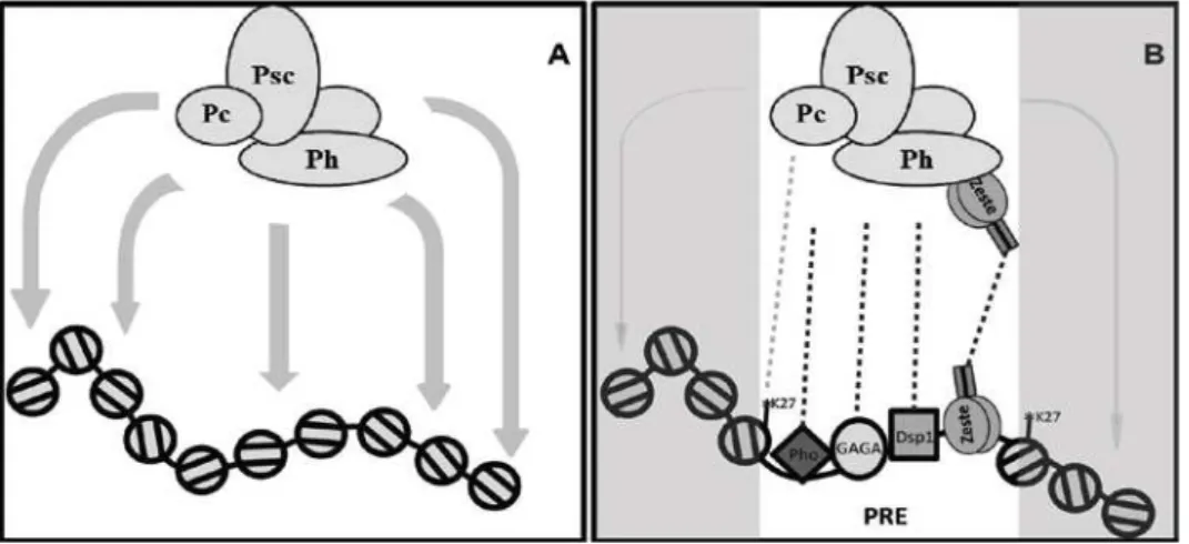
Repressive Complex 1 (PRC1) binds histone H3 methylated at residue
due to its in vitro compaction activity (31). dRING-Associated Factors
(dRAF) complex mono-ubiquitylates histone H2A at residue K119 and
de-methylates histone H3 at residue K36 (32). PR-DUB de-ubiquitylates
histone H2A, counteracting the activity of dRAF (33). PRC2
tri-methylates histone H3 at residue K27 (34-36), and the Pcl-PRC2 variant
carries the same activity, but more efficiently (37). Pho Repressive
Complex (PhoRC) binds a specific sequence within PcG target sites and
interacts with di-methylated histone H3 and histone H4 at residues K9
and K20, respectively (38). All these complexes are found enriched at
Polycomb Response Elements (PREs), which are cis-regulatory regions
found at the HOM-C and elsewhere in the genome. Mutations in ANT-C,
BX-C, Gap, Pair-Rule, Segment Polarity, TrxG, PcG or PREs all can
lead to homeotic transformation, due to the loss of the appropriate cell
identity. This suggests a highly inter-dependent regulation of the
segmentation mechanism.
PRE chromatin has been characterized by a list of structural features:
the reduced accessibility of the underlying DNA to an exogenous
methylase, in a PcG-dependent manner, suggestive of a condensed
structure (39), the sensitivity to nucleases DNase I and MNase (40-42),
the depletion in histones (42-44), and the low-density on a CsCl gradient
(45). Besides these, there is a characteristic organization of DNA motifs
recognized by a group of factors known or thought to recruit PcG
proteins (46). The combination of these properties, along with a
particular topology and genomic organization (47,48), is the basis of
some of the functional readouts that identify these regions as PREs.
Namely, the strong physical association with PcG proteins (49), the
homing of P-elements containing a PRE sequence (50,51), or the
Pairing-Sensitive Silencing (PSS) of an associated mini-white transgene
shown to also require the proteins Pleiohomeotic (Pho), GAGA (53),
Zeste (54) and other unidentified factors (55).
Some of this information has been used to predict the occurrence of
PREs on the human homeotic (HOX) clusters, leading to the
identification of a few elements that exhibit the hallmarks of Drosophila
PREs (56); on the other hand, the DNA motifs recognized by proteins
associated with PREs (GAGA, Zeste, Pho) were used to build algorithms
for the in silico identification of clusters of these motifs, thus expanding
the catalog of potential PREs in Drosophila (57,58). Both cases typify the
strengths and weaknesses of predictive tools as, on the one hand, they
allowed the discovery of the first human PREs and of new Drosophila
PREs but, on the other hand, they probably missed most human PREs,
while identifying a substantial number of probable false positive
Drosophila PREs, as several studies have not found any PcG proteins
bound at many of the in silico predicted PREs (59-61).
Long-range communication in chromatin
One of the most interesting features of the PcG machinery is the
involvement with long-range communication between chromatin
locations in cis and in trans. The PSS phenomenon is just one case
where this feature is detected, but other communication events, such as
chromatin fiber looping (62), PRE clustering (11,48) and transvection,
depend on PcG function and, often, they also depend on PRE-binding
proteins.
Transvection has been extensively studied in Drosophila, but evidence
from other organisms is very limited. This can be, in part, due to the long
the genome of Drosophila is organized in a very peculiar way: the
homologous chromosomes are extensively paired in interphase nuclei,
allowing a physical proximity between the alleles throughout most of the
genome (63). Given this organization, the distance between regulatory
regions and promoters in the homologous chromosomes may be
sufficiently short to allow cross-talk. This is the original concept that led
to the coinage of the term “trans-vection”, in opposition to “cis-vection”,
when regulation involves the promoters and regulatory regions in one of
the chromosomes alone (64). This pairing phenomenon is likely made
possible by the action of general chromatin structural proteins, such as
Topoisomerases (63) and Condensins (65). But the close association of
homologs by the action of general chromatin factors is not enough,
though necessary, for transvection to occur. Many cases have been
reported where the wild type function of the zeste gene product is
needed. These are the cases of decapentaplegic (66), apterous (67),
yellow (68), or eyes absent (69). Several cases, though, such as
vestigial (70), or men (71) don’t require wild-type zeste function. Due to
the close association of homologous chromosomes in Drosophila nuclei,
it has been suggested that transvection has evolved from cis-regulatory
phenomena. It is thus interesting to find cases where zeste function is
also required for enhancer-promoter communication in cis (72).
At the BX-C, the dependence on zeste for transvection seems to be the
predominant case at the Ubx locus (73,74), whereas transvection at
Abd-A and Abd-B loci was shown to be independent of zeste in all cases
studied. Interestingly, transvection at the iab-5, -6, and -7 within these
locations is also independent of local chromosome pairing and indeed
very difficult to disrupt (75,76), although this is not the case for
observations make it plausible that transvection effects operate in
organisms without extensive homolog pairing.
The study of Chromatin Processes
Experimental approaches to study all of these genome regulation
parameters have evolved in parallel for the past century, but technical
developments in the last two decades have made progress in their
understanding extremely fast. Genome-wide binding motifs for
DNA-binding proteins have become possible to identify with the development
in genome sequencing technology. Likewise, the combination of
microarray technology, and later of high-throughput sequencing, with
Chromatin Immunoprecipitation (ChIP) allowed the identification of all
genome sites where a particular protein is found in a specific cell or
tissue type (78), and how differentiation, signaling, stress or mutations
alter that protein localization. The computational analysis of these data
has also allowed the discovery and refinement of DNA sequence motifs
recognized by DNA/chromatin-binding factors. Finally, the
high-throughput sequencing analysis now available has combined with the
Micrococcal Nuclease (MNase) mapping technology, to study the
nucleosomal genome occupancy (79), while recent Chromosome
Conformation Capture (3C) derivatives, such as Hi-C, started to offer
insights into the higher-order chromatin structures and the principles of
genome organization (10).
All these innovations and breakthrough discoveries notwithstanding,
there isn’t at the moment enough information to predict all the factors
which associate with a defined genomic locus at a given time or
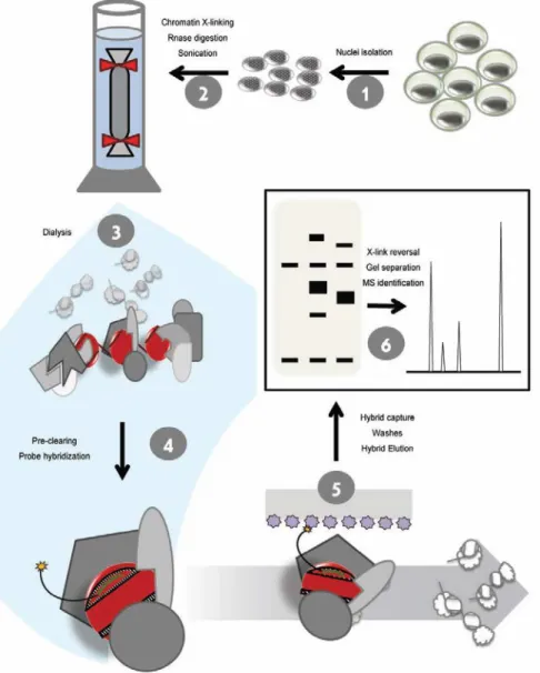
Chromatin segments (PICh) technology allowed the unbiased
identification of chromatin-associated proteins at least for one locus (80),
paving the way to expand the application of this tool to other loci. Of
special interest in this regard are regulatory regions involved in
developmental processes. The pattern of association of structural and
regulatory proteins with such loci has the potential to provide deep
insights into the mechanisms of development, cell identity and disease.
Here I present and discuss the results of experiments on two projects. In
Chapter 2, I show the biochemical analysis of the association of the
PRE-binding factor Zeste with the PRC1 complex and the functional
interaction of Zeste and other PRE-binding proteins with the chromatin
compacting activity of PRC1. Activity assays and in vivo targeting
experiments support a model of robust concentration of PRC1 activity at
target loci, through multiple layers of interaction.
In Chapter 3, I describe the technical optimization of the PICh technique
and its application to the isolation of proteins associated with TAS
sequences from Drosophila. This work has extended considerably the
number of proteins known to associate with TAS sequences and
provided an extensive list of further candidates. I discuss the implications
of these findings for the mechanisms of TPE and other
Chapter 2
Zeste interacts with Polyhomeotic and
contributes to the recruitment of PRC1 to PRE
Abstract
Control of homeotic gene expression during early Drosophila embryo
development involves an intricate web of interactions between
transcription factors, Polycomb- and Trithorax-Group proteins and
regulatory regions. The association of Polycomb-group proteins with the
appropriate targets at the appropriate times is assured by a set of
DNA-binding factors which recognize specific sequences at target regions.
Here we show that one of these factors, Zeste, physically associates
with the PRC1 subunit Polyhomeotic, and that this interaction depends
on the dimerization domain of Zeste. We further show that both the
dimerization domain and the DNA-binding domain are required for
stimulating the PRC1 chromatin compacting activity in vitro and that this
apparent stimulation is probably due to a targeting effect. Other
Polycomb targeting factors show a similar in vitro behavior.
Overexpression of Zeste in cell cultures increased the occupancy of
Polyhomeotic at legitimate targets, but did not create ectopic
Polyhomeotic sites, suggesting the existence of a resilient mechanism
Introduction
The crucial function of the PcG machinery during the early Drosophila
embryo development is the maintenance of the silencing of target genes,
such as the master regulators of the Antennapedia and Bithorax
complexes, through the creation of a repressive chromatin structure.
Due to its in vitro chromatin compaction activity, PRC1 is thus
considered the main engine of PcG chromatin-mediated gene repression
in Drosophila.
The PRC1 complex is formed by the association of four PcG proteins –
Posterior Sex Combs (PSC), Polyhomeotic (Ph), Polycomb (Pc) and Sex
Combs Extra (SCE, or dRING) – with several TFIID general transcription
factors and accessory proteins (82). A recombinant complex assembled
with the four core components of PRC1 [Polycomb Core Complex
(PCC)] maintains all of the in vitro activities of the whole
multi-Megadalton PRC1 complex, and indeed the PSC subunit alone
conserves the compaction activity to a large extent (83-85).
One of the accessory PRC1 proteins is Zeste, has been described as a
sequence-specific transcriptional activator of the homeotic gene Ubx
(86) and as a PRE-binding recruiter of the TrxG Brahma complex (87),
with which it cooperates in the gene activating remodeling activity at
promoter sequences (88). One of the most intriguing roles of Zeste,
however, is its function on transvection (89). The most studied cases of
transvection are at the Ubx locus, where Zeste is necessary for the
activation of the Ubx promoter by an enhancer in trans, and the zeste1
-white interaction, in which the neomorphic zeste1 allele is responsible for silencing paired copies of white, in a PcG-dependent fashion. These
silencing. Its stable association as a PRC1 subunit, thus, suggested a
central role in the targeting of the PcG silencing activity.
zeste-deleted adult flies without visible homeotic defects can be
obtained, though (90). This observation suggested that the protein is part
of a redundant mechanism, as an added layer of safety when other
mutations are present at the Bithorax Complex, in which cases it can
compensate through transvection. The notion of redundancy is
reinforced by the fact that multiple sequence-specific DNA-binding
factors associate with PRE sequences, and are thought to contribute to
the recruitment of PcG proteins. Besides Zeste, GAGA (54), Dsp1 (91),
Grainyhead (92), Pho (93), Pho-like (94), Pipsqueak (95) and Spps (96)
have a recruitment function at PREs. Furthermore, Sex Comb on Midleg
(SCM) (97), Polycomb-like (98) and the methylation mark at Lysine 27 of
the histone H3 tail (99) were all proposed to be involved in the
recruitment of PcG proteins.
Nevertheless, Zeste appears to have a significant role in the
maintenance of Ubx in a silenced state (100). And importantly, like
GAGA, Zeste was shown to significantly increase the in vitro compaction
activity of PCC (101), suggesting an important function in PcG
regulation.
Here we have dissected further the physical association between Zeste
and PRC1 and analyzed their functional interaction in the compaction of
a chromatin template. We have found that the dimerization domain of
Zeste is required for the physical association with Ph and that Zeste and
other PRE-binding factors are able to stimulate the compaction activity of
PSC when target sequences are present. The failure of Zeste
PcG targeting mechanism, likely dependent on multiple interaction
surfaces.
Materials and Methods
Cloning
Primer sequences can be found in the Annex Table X. The pFastBac
baculovirus expression constructs for dRING-Flag, Flag-PSC, Flag-Ph,
Flag-Pc, dRING, PSC, Pc (83), Zeste and
pVL1392-HA-Zeste-Flag (88), pMT-Zeste tagged expression vector (102), GST-PhSAM
(103) and His-PhSAM (104) were described. The pFB-Zeste∆DBD plasmid
was constructed by amplifying the Zeste coding sequence downstream
of the DNA-binding domain (starting at aminoacid 127) with primers
NdeDBD X Zeste-CT and cloning into the NdeI X EcoRI sites of the
pFB-Zeste plasmid, maintaining the in-frame fusion with the N-terminal HA
tag. The pFB-Zeste∆AD plasmid was built by amplifying the regions
N-terminal and C-N-terminal to the Activation domain with primers Zeste-NT
X BssHII-N2 and Zeste-CT X BssHII-C, respectively, which introduced
BssHII sites before and after the Activation domain. These were cloned
with the Topo-TA cloning kit (Invitrogen) into the pCRII plasmid.
Assembly into pFastBac was done in two steps, cloning the N-terminal
fragment into the BamHI X EcoRI sites and then cloning the C-terminal
fragment into the BssHII X EcoRI sites. The pFB-Zeste∆CT plasmid was
built by cloning the Zeste sequence upstream of the BlpI site (aminoacid
418) with the BlpCT X Zeste-NT primers into the BamHI X EcoRI sites
on the pFastBac plasmid. The pFB-Zeste∆Pro plasmid was constructed by
first introducing a PstI site to cleave at the sequence corresponding to
ZPst429F and ZPst429R, using the Quickchange mutagenesis kit
(Stratagene). The Leucine-Zipper region was then amplified with primers
ZPstLeu and Zeste-CT and this fragment was cloned into the first
plasmid, replacing the PstI X EcoRI fragment. For pFB-Zeste∆LZ, the
Zeste sequence upstream of the Leucine-Zipper domain (aminoacid
500) with the ProCT X Zeste-NT primers into the BamHI X EcoRI sites
on the pFastBac plasmid. The pFB-Zeste1 plasmid was constructed by
site-directed mutagenesis, using primers ZK425M2F and ZK425M2R
and the Quickchange mutagenesis kit (Stratagene). For the
pFB-Zeste∆DBD–Flag, pFB-Zeste∆Pro–Flag and pFB-Zeste∆AD-Flag plasmids,
the Zeste-CTFlag primer was used instead of the Zeste-CT primer. For
pFB-Zeste∆LZ-Flag, primer ProFlag2 substituted for ProCT. For
pFB-Zeste∆CT -Flag, the BlpFlag2 primer substituted the BlpCT primer.
Dsp1-Flag, Psq-Dsp1-Flag, Pho-Flag and GAGA-Flag were cloned into pFastBac by
amplifying the ORFs out of cDNA vectors obtained from the Drosophila
Genomics Resource Center (DGRC), using the following primer pairs
and cloning sites on pFastBac: Dsp1-NT X Dsp1Flag-CT into the EcoRI
X XbaI site, Psq-NT X PsqFlag-CT into the SpeI X XhoI site, Pho-NT X
PhoFlag-CT into the EcoRI X XbaI site, GAGA-NT X GAGAFlag-CT into
the SpeI X XbaI site. The 5S-G5bxd array construct was built by first
creating SalII and EagI restriction sites flanking the E4 central region of
the 5S-G5E4 sequence, by site-directed mutagenesis with the
Quickchange kit (Stratagene) and primer pairs G5E4mutSalIA X
G5E4mutSalIB and G5E4mutEagIA X G5E4mutEagIB, respectively. The
PB fragment of the bxd PRE (105) was amplified from genomic DNA
with primers bxdSal and bxdEag and cloned into the SalII X EagI sites of
the mutagenized p5S-G5E4 vector, replacing the E4 fragment, to create
the p5S-G5bxd plasmid. The GST-ZesteLZ construct was made by
ZesteLZBam and ZesteLZEco and cloning into the BamHI X EcoRI sites
of the pGEX-4T-1 vector.
Protein Expression and Purification
The baculovirus system was used to express proteins in Sf9 insect cells.
pFastBac plasmids were transformed into DH10bac cells to produce the
expression bacmid by recombination. Bacmids were isolated and
transfected into Sf9 cells following the instructions of the Bac-to-Bac
expression system (Life Technologies). For the expression of HA-Zeste
and HA-Zeste-Flag, the baculovirus production was done using the
Baculogold system (BD Biosciences), due to the difference in the donor
vector backbone (pVL1392). Baculovirus amplification was done
consecutively in 2cm, 10cm and 25cm tissue culture plates, by infecting
70% confluent cultures with 1% volume of baculovirus from the previous
amplification step. Amplification was carried out at 27ºC for 4 days, or
until signs of infection were obvious (swelling of the cells and
detachment from the plate). For protein production, individual or
combinations of different baculoviruses at a Multiplicity of Infection of
between 1 and 10 pfu/cell were added to suspension cultures of Sf9
cells in the early to middle exponential growth phase (~5X10⁵ cells/ml).
Cultures were grown at 27ºC, in a shaking incubator at 100rpm for 40 hr.
Nuclear protein extracts were prepared based on a previously described
protocol (107): cells were washed with 1X PBS (137mM NaCl; 2.7mM
KCl; 10mM Na2HPO4; 2mM KH2PO4; pH 7.4), then with Hypotonic buffer
(10mM HEPES, pH 7.9; 1.5mM MgCl2; 10mM KCl; 0.2mM PMSF; 1mM
DTT) and resuspended to 3 packed cell volumes (pcv) with Hypotonic
buffer. Cells were swollen on ice for 10 min. and lysed with 10 strokes of
a tight-fitting homogenizer pestle. Nuclei were collected by centrifugation
buffer (20mM HEPES, pH 7.9; 25% Glycerol; 1.5mM MgCl2; 20mM KCl;
0.2mM EDTA; 0.2mM PMSF; 1mM DTT). An equal volume of High-Salt
buffer (same composition as Low-Salt buffer, but with 1.2M KCl) was
added dropwise, while vortexing, and the nuclear protein was extracted
by incubating in a rotating platform for 45 min. Insoluble materials were
removed by centrifugation and the cleared extracts were used for protein
purification. All procedures were carried out at 4ºC or on ice.
A 400µl volume of M2 agarose beads slurry (Sigma), equilibrated in BC
buffer (20 mM HEPES, pH 7.9, 0.2 mM EDTA, 10% glycerol) with 0.3M
KCl (BC300), was added to the nuclear extract per liter of originally
infected Sf9 culture. The mixture was incubated on a rotating platform for
4 hr., then poured onto an empty Econo-pac column (Bio-Rad) and the
flow-through was discarded. Beads were washed sequentially with 20
volumes of BC buffer containing 0.3, 0.6, 1.2, 2.0, 1.2, 0.6 and 0.3M of
KCl, with a 20 min. incubation at the 2.0M step, to allow a more stringent
wash. The protein was eluted from the beads by replacing the buffer with
a 0.8mg/ml solution of the Flag peptide (DYKDDDDK) in BC300,
incubating for consecutive elution steps of 30 min. and collecting
eluates. All solutions contained 0.05% NP-40, 10µM ZnCl2, 1mM DTT,
0.2mM PMSF and 1X cOmplete EDTA-free protease inhibitor cocktail
(Roche). Protein concentration was determined by Bradford assay.
The hSwi/Snf complex used in REA assays was purified from nuclear
extracts of the Ini1-11 Hela cell line (108), using the same protocols as
for Sf9 cells, but with a maximum of 0.6M KCl in the BC buffer washes.
GST-pulldown
pET-PhSAM (His-PhSAM construct) was transformed into BL21(DE3)
IPTG and incubated for 3 hr. at 30ºC. Cells were resuspended with Lysis
buffer [50 mM Tris-HCl, pH 8.0; 0.2M NaCl; 10 mM β-mercapto-ethanol
(BME); 10 mM Imidazole, pH 7.5], lysed with five 10 sec. pulses of
sonication, using a Misonix sonicator and the extracts were cleared by a
15 min. centrifugation at 20,000g. Extract was applied to a Ni-NTA
agarose resin (Qiagen) equilibrated with Lysis buffer and incubated on a
rotating platform for 30 min. at 4ºC. The extract with resin was poured
onto an Econo-column (Bio-Rad), washed with 20 resin volumes of Lysis
buffer, 20 volumes of Lysis buffer with 50mM Imidazole, and the protein
was eluted with 5 volumes of Lysis buffer with 0.5M Imidazole. Imidazole
was removed from the eluate by dialyzing twice (4 hr., then overnight)
against 100 volumes of Assay buffer (10 mM Tris-HCl, pH 8.0; 50 mM
NaCl; 2 mM DTT).
pGEX-ZesteLZ and pGEX-PhSAM were transformed and induced similarly
to pET-PhSAM. Bacterial pellets were resuspended with 1X PBS
containing 0.2mM PMSF, lysed by sonication and cleared by
centrifugation as above. 0.05% NP-40 was added to the extracts before
adding to Glutathione Sepharose 4 Fast Flow beads (Pharmacia)
equilibrated in 1X PBS. Extracts were incubated 1 hr., rotating with the
beads at 4ºC. Beads were collected by a 1 min. centrifugation at 1000g
and washed 5 times with 20 bead volumes of 1X PBS, 5 min. each,
collecting the beads between washes by centrifugation as above. 40µl of
beads with bound fusion protein were added into each of 4 tubes. PBS
or purified 10mg His-PhSAM, Ph or Zeste were added to the beads and
incubated 30 min. at room temperature. Beads were collected and
washed 5 times as above. Protein was eluted by adding 40µl of 2X
reducing sample buffer (Thermo Scientific) and incubating 5 min. at
95ºC. Samples were separated by SDS-PAGE and stained with Imperial
Western Blot
For Zeste interaction analysis, 0.1% Input chromatin was separated
along with 0.01% of the purified protein by SDS-PAGE and transferred
onto a PVDF membrane. Membrane was blocked for 1 hr. with 5% milk
in PBST (1X PBS; 0.1% Tween-20), and incubated overnight with
1:10,000 dilutions of rabbit anti-HA tag (Abcam) or mouse M2 anti-Flag
(Sigma) antibodies in PBST-3% milk at 4oC. For Zeste protein levels in
wild-type and transfected cells, the protein from 10,000 cells was
separated by SDS-PAGE, transferred and blocked as above, and probed
with the same anti-HA antibody or with rabbit anti-Zeste (1:1000).
Membranes were washed 3 times, 15 min. each with PBST, and then
incubated 1 hr. with a 1:10,000 dilution in PBST of HRP-conjugated
secondary anti-rabbit (GE), or anti-mouse (GE) antibodies. Membranes
were washed 3 times, 15 min. each with PBST, incubated 5 minutes with
SuperSignal West Pico Chemiluminescent Substrate (Thermo Scientific),
exposed and developed to Kodak Biomax film (PerkinElmer). Films were
digitalized and images processed using Adobe Photoshop (Adobe
Systems).
Nucleosomal Template Assembly
The 5S-G5bxd DNA template was excised from 2mg of the pG5bxd
plasmid by digestion with 700U Asp718 and 480U ClaI (Roche), 180U
DraIII and 450U DdeI (NEB) at 37ºC overnight. Digested DNA was
purified by phenol:chloroform separation and precipitated with ethanol.
DNA was re-suspended with TEN1000 (10mM Tris-HCl pH 8.0; 1mM
EDTA, pH 8.0; 1M NaCl) and the 2.5Kb 5S-G5bxd fragment was
separated from the digested backbone by sequential PEG-6000
precipitation: 50% (w:v in 0.5M NaCl solution) PEG-6000 was added to
centrifuged 10 min., at room temperature, 10,000g. The supernatant was
recovered into a new tube and PEG-6000 solution was added to
increase the concentration to 4%, repeating the previous procedure.
Further PEG-6000 concentration steps of 5% and 6% were produced
and the pellets of each centrifugation were re-suspended in TE buffer
(10mM Tris-HCl, pH 8.0; 1mM EDTA, pH 8.0) and analyzed on an
agarose gel, to determine the presence of the 2.5Kb fragment and
contaminant bands. Typically, >90% of the fragment was on the 4% and
5% PEG pellets, but complete separation from the smaller fragments
was only achieved after 3 to 4 re-iterations of the precipitation protocol.
80µg of the purified 5S-G5bxd fragment were end-labeled with 32
P-αdATP through a Klenow (NEB) fill-in reaction at the Asp718 site. The
reaction was carried out for 50 min. at room temperature, followed by a
clean-up spin through a Microspin G-25 column (Amersham-GE) to
remove unincorporated nucleotides. Nucleosomal arrays were
assembled with histone octamers purified from Hela cells (109) by
gradient salt dialysis (110). Briefly, 10mg of labeled template DNA were
added to the assembly reaction (20mM Tris-HCl, pH 7.7; 1mM EDTA,
pH 8.0; 10mM DTT; 0.1mg/ml BSA; 0.5mM Benzamidine-HCl; 2M NaCl;
in a 100µl reaction volume). To each assembly reaction, Hela histone
octamer solution was added at varying weight ratios (typically 1:0.9, 1:1,
1:1.1, but wider ratios need to be tested with new histone octamer
batches, due to the frequently imprecise protein quantification).
Assembly reactions were placed inside pre-boiled 6-8KDa MWCO
dialysis membranes (Spectrum Labs) and into 200ml of High Salt
assembly buffer (20mM Tris-HCl, pH 7.7; 2M NaCl; 1mM EDTA, pH 8.0;
10mM DTT; 0.5mM Benzamidine-HCl). The gradient dialysis was set-up
by establishing a constant flow rate, with a peristaltic Rabbit Pump
pH 7.7; 0.25M NaCl; 1mM EDTA, pH 8.0; 10mM DTT; 0.5mM
Benzamidine-HCl) into the High-Salt assembly reaction, and from here
to waste. The dialysis proceeded for 48 hr. (roughly until 1000ml of the
Low-Salt assembly buffer was used), after which time the dialysis bags
were transferred into 1000ml of TE-low (10mM Tris-HCl, pH 7.7; 0.25mM
EDTA, pH 8.0; 1mM DTT) and dialyzed for an extra 12 hr.
Restriction Enzyme Accessibility Assay
Pre-incubation with PcG proteins was carried out at 30ºC for 60 min. in
8.8µl reaction mixture [2ng (1.6nM) nucleosomal array; 4.5mM MgCl2;
2.25mM ATP; 45mM KCl; 9mM HEPES, pH 7.9; 0.09mM EDTA, pH 8.0;
9% Glycerol]. REA assay reactions were carried out at 30ºC for 60 min.
in 10µl reaction mixture (4 mM MgCl2; 2mM ATP; 52mM KCl; 12%
glycerol; 10 mM HEPES, pH 7.9; 0.1mM EDTA, pH 8.0; 1mM DTT) in
the presence of 5U XbaI (NEB) and 100ng hSwi/Snf. Reactions without
PcG proteins were supplemented with 2µM Bovine Serum Albumin
(BSA). Reactions were stopped by the addition of 3µl of Stop buffer
[6.7mg/ml Proteinase K (Roche); 67mM EDTA, pH 8.0; 33mM Tris-HCl,
pH 7.7; 0.67% SDS; 17% Glycerol; Bromophenol Blue and Xylene
Cyanol) and incubation at 55ºC for 40 min. Samples were separated on
a 1% Agarose gel in 1X TAE (40mM Tris-HCl, pH 7.5; 20mM Acetic
Acid; 1mM EDTA, pH 8.0), and the gel was dried in a gel drier (Bio-Rad)
before exposing to a PhosphorImager screen and detecting in a
Typhoon scanner (Molecular Dynamics - GE). Bands were quantified
using the ImageQuant software (GE) and the inhibition of hSwi/Snf
remodeling was determined using the equation:
(% uncut with PcG protein and hSwiSnf−% uncut with hSwiSnf) (% uncut without hSwiSnf−% uncut with hSwiSnf) X100
Pho, Dsp1, Psq, GAGA) in the PcG pre-incubation, the ratio of the levels
of hSwi/Snf inhibition with and without such proteins was determined at
each concentration point.
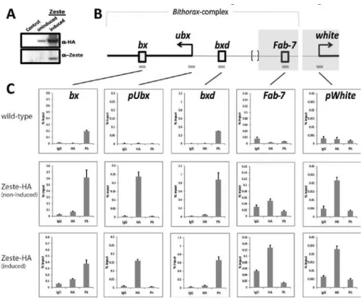
Chromatin Immuno-precipitation
Sg4 cells were grown in CCM3 medium (Hyclone), supplemented with
Pen/Strep (Gibco) at 27ºC, in T-flasks. Zeste-Flag-HA tagged expression
vector [pMK33-CFH-BD; (102)] was co-transfected with pCoBlast
(Invitrogen) using the FuGENE HD Transfection Reagent (Qiagen),
following the manufacturer’s protocol. 25µg/ml Blasticidin (Invivogen)
was added to the culture medium 2 days after transfection. 24 hr. before
crosslinking, transgene expression was induced by the addition of 1mM
CuSO4 and ChIP was performed. ChIP experiments were performed
essentially as described (111), with minor changes: All operations,
unless otherwise noted, at 4oC. Cells were cross-linked at a density of
5X10⁶/ml for 10 min. with 1.8% Formaldehyde (Fisher), at room
temperature, stopped with Glycine to 0.125M, washed with 1X PBS,
re-suspended and incubated 10 min. in ChIP Wash buffer A (10mM
HEPES, pH 7.6; 10mM EDTA, pH 8.0; 0.5mM EGTA, pH 8.0; 0.25%
Triton X-100), and repeated with ChIP Wash Buffer B (10mM HEPES,
pH 7.6; 0.1M NaCl; 1mM EDTA, pH 8.0; 0.5mM EGTA, pH 8.0). Nuclei
were isolated by a brief incubation with 1% SDS in TE buffer and, after
washing extensively with TE, resuspended with TE-PMSF-SDS (TE;
1mM PMSF; 0.1% SDS) at a density of 1x10⁸ cells/ml. Chromatin was
solubilized using a Misonix sonicator, to obtain a DNA length between
200-400bp. Salt and detergent concentrations were corrected to 1%
Triton X-100, 0.1% Na-Deoxycholate (DOC), 140mM NaCl, and the
insoluble pellet removed by centrifuging 5 min. at maximum speed in a
20µl Protein-A Sepharose (PAS) slurry (Thermo Scientific), and
incubated overnight with 1.5µg of a rabbit polyclonal anti-HA antibody
(Abcam), 1.5µg of rabbit polyclonal anti-Ph antibody (31), or with 1.5µg
of rabbit control IgG (Abcam). Antibody was captured with 30µl PAS for
3 hr., beads were washed 5 times, 10 min. each with 1ml of RIPA
(140mM NaCl; 10mM Tris-HCl, pH 8.0; 1mM EDTA, pH 8.0; 1% Triton
X-100; 0.1% SDS; 0.1% DOC), then once with 1ml of LiCl buffer (0.25M
LiCl; 10mM Tris-HCl, pH 8.0; 1mM EDTA, pH 8.0; 0.5% NP-40; 0.5%
DOC) and finally twice with 1ml of TE. Beads were then re-suspended in
100µl TE, 50µg/ml RNase A (Qiagen), and incubated 30 min. at 37ºC;
then added SDS to 0.5%, Proteinase K (Roche) to 0.5mg/ml and
incubated overnight at 37ºC. Reversed the crosslinks at 65ºC for 6 hr.,
extracted DNA, Ethanol-precipitated and re-suspended the pellet in
150µl ddH2O. qPCR analysis was performed with the IQ SYBR Green
system (Bio-Rad), with the following primers: F5-fwd and F5-rev for bxd,
F9-fwd and F9-rev for pUbx, F13-fwd and F13-rev for bx, F18-fwd and
F18-rev for Fab-7, wp1 and wp2 for pWhite. These primers have been
described before: F5-F18 (43), wp1 and wp2 (44). The
immuno-precipitated material was quantified against a calibration curve with
dilutions of input DNA.
Results
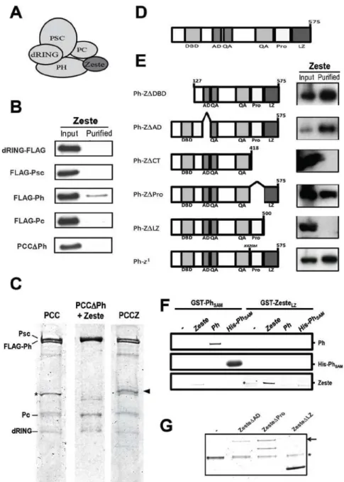
Zeste associates physically with Polyhomeotic
The transcriptional activator Zeste has been shown to be a stable
component of the PRC1 complex (82) and to associate tightly with a
constituents – PSC, Ph, Pc and dRING (101). To further dissect this
interaction, we co-expressed Flag-tagged individual PCC components
and HA-tagged full-length Zeste in Sf9 cells. Flag-tagged proteins were
purified from nuclear extracts using an M2 anti-Flag resin, the isolated
protein was separated by SDS-PAGE and the presence of Zeste
determined by anti-HA Western blotting (Figure 2.1B). Ph was capable
of retaining association with Zeste after stringent washing, unlike the
other PCC components, showing that it is sufficient for the association of
Zeste with PCC. A trimeric PSC-Pc-dRING complex (PCC∆Ph), purified
through the Flag tag on PSC, was incapable of binding Zeste [Figure
2.1B and 1C (middle)], showing that Ph is necessary for the association
of Zeste with PCC.
The Zeste protein is composed of several domains and regions with
particular features (Figure 2.1D). The most prominent regions are the
N-terminal DNA-binding domain and the C-N-terminal Proline-rich region and
Leucine-Zipper domain, with less characterized acidic Activation domain
and Glutamine/Alanine stretches in between. We cloned deletion
mutants spanning individual or multiple domains into baculovirus
expression vectors and analyzed the domain requirements for the
interaction of Zeste with Ph (Figure 2.1E). Sf9 cells were co-infected with
baculoviruses for Flag-tagged Ph and HA-tagged Zeste mutants, and
proteins were purified as described above. Deletion mutants eliminating
the C-terminal Proline-rich region and the Leucine-Zipper domain
(Z∆CT), or eliminating the Leucine-Zipper domain alone (Z∆LZ) abolish
the interaction with Ph, indicating that this protein-protein interaction
domain is required for the association. The Leucine-Zipper domain of
Zeste thus appeared to be the docking site for Ph. To confirm this
association, we expressed GST-fusions of the Leucine-Zipper of Zeste
fusion proteins to Glutathione beads and probed their capacity of
interacting with candidate partners (Figure 2.1F).
GST-PhSAM was used as a positive control for binding Ph, as it is known
to be the dimerization surface for the Ph protein (103). As expected,
GST-PhSAM is capable of associating with a purified HIS-tagged version
of itself (His-PhSAM), as well as with full-length Ph, while only very
residual association is seen with Zeste (Figure 2.1F, left); GST-ZesteLZ,
on the other hand, is capable of associating with full-length Zeste, but
not with His-PhSAM or full-length Ph (Figure 2.1F, right). These results
suggest that the Leucine-Zipper domain of Zeste is not sufficient for the
direct interaction with Ph. This observation was intriguing, given the
absolute requirement of the Leucine-Zipper for interaction with Ph
(Figure 2.1D) and the previously reported sufficiency of this domain to
retain association with the Brahma complex factor Moira (112). This
might be explained by an overall structural defect caused in Zeste upon
deletion of the Leucine-Zipper. The Zeste protein has been shown to
multimerize and its activity was suggested to be dependent on this
phenomenon (113,114). If the Leucine-Zipper deletion were to affect this
self-association process, it might impact the interaction with partner
proteins, with a functional outcome. To look directly at self-association,
we again used the baculovirus/Sf9 system and co-infected cells with
Flag-tagged deletion mutants and HA-tagged full-length Zeste.
Deletion of domains not involved in dimerization – ZesteAD, ZesteP ro –
had no effect on the recruitment of full-length Zeste, but in the absence
of the Leucine-Zipper domain the association with Zeste was completely
lost (Figure 2.1G).This observation is consistent with the GST-ZesteLZ
association with full-length Zeste (Figure 2.1F). These results show that
the PRC1 subunit Ph is the direct interaction partner of Zeste within the
complex, and suggest that an interaction surface is formed on Zeste
upon dimerization of its Leucine-Zipper domain, which allows recruitment
of Ph/PCC.
Zeste functions involved in the interaction with PCC
Functional interactions between Zeste and PcG proteins have been
extensively suggested from genetic interaction studies (115) and
biochemical studies, but the nature of this interaction with PRC1 is not
understood completely. On the one hand, Zeste binding sites on the
target chromatin substrate increase the activity of PCCZ (a reconstituted
complex with the four PCC proteins plus Zeste), but even in the absence
of such sites PCCZ inhibits chromatin remodeling more efficiently than
PCC (101). These results suggest that there is both a targeting function,
as well as structural alterations in PCC upon association with Zeste,
making the complex more efficient at silencing chromatin. We used the
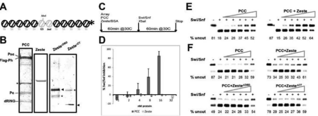
Restriction Enzyme Accessibility (REA) (83) assay to characterize these
activities further. For this biochemical assay, a 32P end-labeled
polynucleosomal template (Figure 2.2A) is pre-incubated with varying
amounts of purified PcG proteins before assaying the ability of the
human ATP-dependent chromatin remodeler complex Swi/Snf to
remodel one of the central nucleosomes. This remodeling event exposes
presence of the nucleosome. We reconstituted a nucleosomal array
substrate based on the G5E4 array (116), with a portion of the bxd PRE
replacing the E4 sequence in the central region (Figure 2.2A). C-terminal
deletion mutants of Zeste don’t form a complex with PCC, so to allow
comparison between the effects of Zeste and mutants we decided to
purify PCC, Zeste and mutants separately (Figure 2.2B), and to compare
the remodeling inhibition activities of PCC in the presence or absence of
Zeste and mutants. This strategy also allows a more rigorous
determination of the effects Zeste has in this mechanism, because the
activity of different PCC complex preparations varies considerably,
making it possible to under- or over-estimate the activity of PCCZ. By
adding Zeste separately to each PCC preparation, we can determine the
variation in activity from the basal level with PCC alone. We started by
quantifying the Swi/Snf inhibition activity of purified Zeste protein alone.
Zeste showed no Swi/Snf remodeling inhibition activity at the
concentrations used, in sharp contrast with PCC (Figure 2.2D). When
added to PCC in equimolar amounts, though, the mixture had a clearly
higher activity than PCC alone (Figure 2.2E): Increasing amounts of
PCC gradually inhibit cutting by the XbaI restriction endonuclease,
leading to the accumulation of the uncut, full-length DNA template (upper
band). The incorporation of Zeste in the PCC pre-incubation mixture
causes an increase in the Swi/Snf inhibition profile. These effects seen
with wild-type Zeste are more obvious at lower concentrations of PCC,
with nearly identical Swi/Snf inhibition profiles detected at higher PCC
concentrations. We next wanted to compare the effect of Zeste with that
of mutants lacking the N-terminal DNA-binding domain (Zeste∆DBD) or the
C-terminal protein interaction region (Zeste∆CT). Nucleosomal 5S-G5bxd
arrays were pre-incubated with 16nM Zeste or mutant protein, along with
Pre-incubation of the arrays with either mutant reduced the PCC activity
back to levels near those of PCC alone, when compared to PCC+Zeste
(Figure 2.2F).
Figure 2.2: Zeste increases the efficiency of PCC-mediated inhibition of Swi/Snf remodeling. A. Schematic of the 32P end-labeled 5S-G5bxd nucleosomal array substrate used in the remodeling assays (details on the text); B. Coomassie-stained gel of the protein preparations used in the assays. Asterisks indicate non-specific peptides co-purified and arrowheads indicate the relevant bands on the Zeste mutant proteins; C.
Experimental REA protocol for the analysis of PCC, Zeste effects on Swi/Snf remodeling;
D. Swi/Snf inhibition by PCC (1-16nM) and Zeste (2-32nM). The bars represent the average for 3 independent preparations of PCC and 2 of Zeste, and error bars are the standard error. E. Representative gel of REA assays with varying concentration of PCC (left) or PCC and an equimolar amount of Zeste (right). PCC and Zeste concentrations range from 1nM to 16nM; F. Representative gel of REA assays with varying concentration of PCC (1-16nM) or PCC and a fixed 16nM concentration of Zeste or the indicated mutants (right).
These results are consistent with a role for Zeste in mediating the
contact between PCC and its target chromatin, with both the
DNA-binding and the protein-protein interaction functions being required for
this effect. It is worth noting, though, that earlier studies have found that
C-terminal mutants of Zeste have impaired DNA-binding activity (117),
thus making it difficult to determine whether the defect in silencing
observed here is due to the lack of binding to PCC or to a DNA-binding
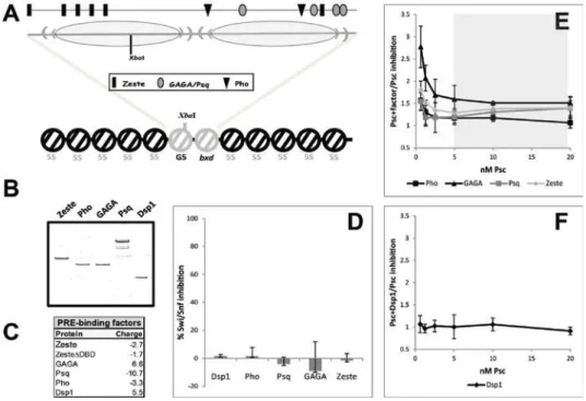
Zeste interacts functionally with Ph and PSC
Most of the in vitro chromatin compaction activity studies on PRC1 have
been done with either PCC or with PSC alone (83,118,119). This has
been the case due to the concentration of the Swi/Snf inhibition activity
of PRC1 on the PSC subunit. PRC1 activity has recently been linked to
the existence of positively charged proteins in the complex (120). ~3/4 of
the basic character of Drosophila PCC is conferred by PSC, but most of
the remaining charge is due to Ph (Figure 2.3A). The +25.8 charge of Ph
is well above the +10.4 threshold determined for active PcG proteins
(120), so we hypothesized that Swi/Snf inhibition could be carried out by
the Ph subunit and that Zeste could interact specifically with this activity,
given the physical interaction observed between these proteins. In fact,
Ph has detectable transcriptional silencing activity in vitro (84), albeit
considerably lower than that of PSC. In order to compare the effects of
Zeste on Ph and on PSC activities, we purified individual proteins
(Figure 2.3B) and used them on the REA assay. Both Ph (Figure 2.3C,
left) and PSC (Figure 2.3D, left) have activity on their own, although, as
expected, the activity of PSC is higher. To quantify the effect of Zeste on
their activities, we determined their inhibition rates in the presence or
absence of Zeste and calculated the ratio (PcG+Zeste:PcG) at each PcG
concentration point. Though Zeste leads to a more efficient inhibition of
remodeling by Ph (the average ratio is always above 1), the poor
biochemical stability of Ph leads to high variability in the assay (Figure
2.3E). Surprisingly, a clear increase in PSC activity is also seen in the
presence of Zeste, in a DNA-binding domain-dependent manner (Figure
2.3D, 2.3F). This effect appears to be less pronounced than is seen with
the whole PCC, although the trend is similar, suggesting that the strong
interaction between Ph and Zeste is not absolutely required for a
system, weaker interactions with PSC can substitute for the strong
interaction with Ph in vivo.
Figure 2.3: Functional interaction between Zeste, Ph and PSC. A. Net charge of the Drosophila PCC complex and of the individual constituent proteins; B. Recombinant purified proteins used on the assays, on coomassie-stained gel; C. Representative gel of REA assay with 1-16nM Ph (left) or 1-16nM Ph in the presence of 16nM Zeste (right); D.
Representative gel of REA assays with 0.6-20nM PSC alone (left), or in the presence of 16nM Zeste (middle), or 16nM Zeste∆DBD (right); E. Variation in Swi/Snf inhibition between Ph+Zeste and Ph. Values correspond to the ratio between the percent inhibition of Swi/Snf remodeling in the presence and absence of Zeste at each concentration point. Values above 1.0 represent an increase in the inhibition by PCC upon addition of Zeste;
The effects of Zeste on inhibition by PSC are particularly visible at lower
concentrations of PSC. If we calculate the molarity ratios between the
nucleosomal substrate and PSC molecules, it becomes obvious that the
effects of Zeste are particularly pronounced at below the 3:1 PSC:array
ratio (Figure 2.3F), the point at which the chromatin is expected to be
saturated with PSC molecules, at 1 PSC molecule per 4 nucleosomes
(118). This observation suggests a targeting effect, whereby at
sub-saturation levels of PSC, Zeste concentrates the available molecules at
the targets where Zeste DNA-binding sites are found (the central region
of the array, where the XbaI site also resides).
PSC activity is modulated by PRE-binding factors
We next wanted to know whether the apparent targeting effect could be
seen with other PRE-binding proteins as well. The engineered 5S-G5bxd
nucleosomal array template accommodates 2 central nucleosomes,
covering a sequence with 5 Gal4 binding sites and the BP fragment of
the bxd PRE (105), respectively (Figure 2.4A). The first nucleosome
occludes the XbaI site assayed by the REA assay and a cluster of Zeste
binding sites; the second nucleosome region contains binding sites for
Zeste, GAGA/Pipsqueak (Psq) and Pleiohomeotic (Pho). These
described sequences are the only regions on the template where binding
sites for these factors are found; the remaining 10 nucleosomes cover
5S positioning sequences where no binding sites are present. Also, no
binding sites for Dsp1, another PRE-binding factor are found on this
template. Previous studies have implicated some of these factors on the
We purified each of these individual proteins (Figure 2.4B) and
determined their intrinsic Swi/Snf inhibition profiles. None of these
proteins reach the +10.4 charge threshold for active silencing factors
(Figure 2.4C) and, consistently, none have appreciable levels of
remodeling inhibition at the concentrations tested (Figure 2.4D).
Figure 2.4: Influence of various PRE-binding proteins on the activity of PSC. A.
Schematic of the 5S-G5bxd nucleosomal array, with detail of the central region. The 2 central nucleosomes (grey) form over a hybrid sequence composed of the G5 region of the parental G5E4 array (left oval) and the BP fragment of the bxd PRE (105) (right oval). The XbaI site is indicated, as well as the positions of the binding sites for Zeste, GAGA/Psq and Pho (upper line); B. Coomassie-stained gel of the recombinant proteins used in these experiments; C. Net charge of the proteins used in these experiments; D.
Swi/Snf inhibition activity of the indicated proteins, at the concentrations used in the assays with PSC: 8nM Dsp1, 4nM Pho, 8nM Psq, 8nM GAGA, 16nM Zeste; E. Variation in Swi/Snf inhibition between either PSC+Pho, PSC+GAGA, PSC+Psq or PSC+Zeste and PSC. Analysis and shaded area as in Figure 3; E. Variation in Swi/Snf inhibition between PSC+Dsp1 and PSC.
When these factors were included in the pre-incubation of the 5S-G5bxd