A Transcriptome Analysis Suggests Apoptosis-Related
Signaling Pathways in Hemocytes of
Spodoptera litura
After Parasitization by
Microplitis bicoloratus
Ming Li1., Zunyu Pang1., Wei Xiao1., Xinyi Liu1., Yan Zhang1
, Dongshuai Yu1, Minjun Yang2, Yang Yang1, Jiansheng Hu1, Kaijun Luo1*
1School of Life Sciences, Yunnan University, Kunming, P. R. China; Key Laboratory for Animal Genetic Diversity and Evolution of High Education in Yunnan Province, Yunnan University, Kunming, P. R. China,2Shanghai–Ministry of Science and Technology Key Laboratory of Health and Disease Genomics, Chinese National Human Genome Center at Shanghai, Shanghai, P. R. China
Abstract
Microplitis bicoloratusparasitism induction of apoptotic DNA fragmentation of hostSpodoptera liturahemocytes has been reported. However, how M. bicoloratus parasitism regulates the host signaling pathways to induce DNA fragmentation during apoptosis remains unclear. To address this question, we performed a new RNAseq-based comparative analysis of the hemocytes transcriptomes of non-parasitized and parasitizedS. litura. We were able to assemble a total of more than 11.63 Gbp sequence, to yield 20,571 unigenes. At least six main protein families encoded by M. bicoloratus bracovirus are expressed in the parasitized host hemocytes: Ankyrin-repeat, Ben domain, C-type lectin, Egf-like and Mucin-like, protein tyrosine phosphatase. The analysis indicated that during DNA fragmentation and cell death, 299 genes were up-regulated and 2,441 genes were down-regulated. Data on five signaling pathways related with cell death, the gap junctions, Ca2+, PI3K/Akt, NF-kB, ATM/p53 revealed that CypD, which is involved in forming a Permeability Transition Pore Complex (PTPC) to alter mitochondrial membrane permeabilization (MMP), was dramatically up-regulated. The qRT-PCR also provided that the key genes for cell survival were down-regulated underM. bicoloratusparasitism, including those encoding Inx1, Inx2 and Inx3 of the gap junction signaling pathway, p110 subunit of the PI3K/Akt signaling pathway, and the p50 and p65 subunit of the NF-kB signaling pathway. These findings suggest that M. bicoloratus parasitism may regulate host mitochondria to trigger internucleosomal DNA fragmentation. This study will facilitate the identification of immunosuppression-related genes and also improves our understanding of molecular mechanisms underlying polydnavirus-parasitoid-host interaction.
Citation:Li M, Pang Z, Xiao W, Liu X, Zhang Y, et al. (2014) A Transcriptome Analysis Suggests Apoptosis-Related Signaling Pathways in Hemocytes ofSpodoptera lituraAfter Parasitization byMicroplitis bicoloratus. PLoS ONE 9(10): e110967. doi:10.1371/journal.pone.0110967
Editor:Yi Li, Wuhan Bioengineering Institute, China
ReceivedMay 19, 2014;AcceptedSeptember 19, 2014;PublishedOctober 28, 2014
Copyright:ß2014 Li et al. This is an open-access article distributed under the terms of the Creative Commons Attribution License, which permits unrestricted
use, distribution, and reproduction in any medium, provided the original author and source are credited.
Data Availability:The authors confirm that all data underlying the findings are fully available without restriction. All RNA-Seq projects are available from the DDBJ/DRA/GenBank database accession numbers DRA001149. 110967 data avaliablity.
Funding:This study was supported in part by a grant (31260448;31060251) from National Natural Science Foundation of China, a grant (2013CB127600) from Major State Basic Research Development Program and a grant (2013FA003) from Yunnan Department of Science and Technology to KL, and a grant (31360454) from National Natural Science Foundation of China to ML. The funders had no role in study design, data collection and analysis, decision to publish, or preparation of the manuscript.
Competing Interests:The authors have declared that no competing interests exist. * Email: [email protected]
.These authors contributed equally to this work.
Introduction
Polydnaviruses (PDVs) have a very special life cycle. Unlike many viruses, they are not always obligate intracellular parasites, replicating inside living host cells to produce virions that can transfer genes to other cells [1–4]. Rather, PDVs are obligate symbionts of many endoparasitic wasps in the families Braconidae (carrying bracovirus) and Ichneumonidae (carrying ichnovirus). Both viruses have similar life cycles, wherein viral DNAs are integrated into a wasp’s genome via Wasp Integration/Excision Motif (WIM) [5] and transmitted vertically to the wasp’s offspring in a proviral form. Viruses replicate in the nucleus of the calyx cell in wasp ovaries. Mature virions are stored in the lumen of the calyx and oviduct, and the suspension of virus and protein is called calyx fluid. When a female wasp finds a host, she injects calyx
fluid, venom produced by the venom gland and one or more eggs into the hemocoel of the host caterpillar. Virions infect host cells and discharge their circular dsDNA into the host nuclei, which then rapidly integrates into the host genome via the Host Integration Motif (HIM) [6]. Virulence genes are then transcribed in host cell nuclei and the cytoplasm, resulting in expression of virulence proteins. During the development of the wasp’s offspring, the host hemocoel contains innate suppressive proteins from virus gene expression. In addition, specifically among the bracoviruses, the induction of host hemocyte apoptosis causes host immunosuppression [1,3].
Apoptosis or programmed cell death (PCD), is common to all metazoan phyla, including insects. Braconidae-induced apoptosis, however, is specifically characterized by internucleosomal DNA fragmentation. Apoptotic DNA fragmentation involves a variety of
elements, including AIF, EndoG and DFF40. Every element is regulated by different signaling pathways, defined as extrinsic and intrinsic pathways. Extrinsic apoptosis pathway is triggered by the ligand-induced oligomerization of specific cell surface receptors, and this process induces the intracellular assembly of the death-inducing signaling complex for the activation of a caspase cascade initiated from caspase 8 that results in activation of caspase 3 and further cascade activation of DFF (cleavage of DFF45 releases DFF40 into the nucleus). DFF, a heterodimeric protein comprising 45 kDa and 40 kDa subunits termed ICAD/DFF45 and CAD/ DFF40 [7]. The DFF complex is localized in the cellular cytoplasm, resulting in the triggering of extrinsic apoptotic stress, and activated caspase 3 cleaves DFF45 and dissociates DFF40. Caspase 7 and Granzyme B also can cleave DFF45 but with a lower efficiency than caspase 3 [8]. Activated DFF40 translocates into the nucleus. In the nucleus, the activation of DFF40 is enhanced by interaction with the chromosomal protein Histone H1 and it cleaves chromosomal DNA at internucleosomal sites into fragments of ,200 bp. [9–11]. In contrast, the intrinsic
pathway is also controlled by mitochondria, which collects and integrates pro- and anti-apoptotic signal stimuli from other organelles as well as from the extracellular microenvironment, such as DNA damage produced by Ataxia-Telangiectasia Mutated (ATM), endoplasmic reticulum (ER) stress and calcium overload. The intrinsic pathway can mediate caspase-independent and caspase-dependent apoptosis. Following intrinsic apoptotic stress triggering, EndoG is released from the mitochondrial intermem-brane space and moves to the nucleus to produce nucleosomal DNA fragmentation, giving rise to 200,5,000 bp sized fragments
in a caspase-independent manner. AIF is another endonuclease released from the mitochondrial intermembrane space. It is a flavoprotein that produces DNA fragments up to 5,000 bp in size, and it also does not require caspase activation [12]. Releasing cytochromeccan also mediate cell death via activation of caspase 8, which triggers a caspase-dependent apoptosis.
Numerous viruses are well known to modulate the mitochon-drial apoptosis of infected host cells by altering Mitochonmitochon-drial Membrane Permeabilization (MMP) in a direct and indirect manner with viral proteins. MMP regulation is performed via the Voltage-Dependent Anion Channel (VDAC) of the outer mem-brane (OM), the Adenine Nucleotide Translocase (ANT) of the inner membrane (IM), and cyclophilin D (CypD) of matrix proteins. Viral proapoptotic proteins are direct inducers of MMP. They include viral protein R (Vpr), which directly interacts with ANT and VDAC, thereby triggering MMP associated with mitochondrial membrane potential (DYm) loss, mitochondrial
intermembrane space (IMS) protein release, and caspase cascade activation. Viral proapoptotic proteins are also indirect MMP facilitators and promote apoptosis via both p53dependent and -independent mechanisms [13]. The alteration of membrane permeability may release apoptotic-promoting factors from the mitochondria, such as AIF, EndoG, and Cyt c in the IMS, ultimately resulting in nuclear translocation. All of these signaling pathways involved in apoptotic DNA fragmentation are stimulated by intrinsic stress through the mitochondria via EndoG and AIF, in a process that is also called caspase-independent cell death, involving release of Cyt c, and extrinsic stress through caspase cascades via DFF40, which is also called caspase-dependent cell death [14].
After apoptotic stimulation, DFF40, EndoG and AIF migrate to the nucleus under the control of critical apoptosis-involved signaling pathways, including the gap junction signaling pathway, Ca2+ signaling pathway, PI3K/Akt signaling pathway, NF-kB
signaling pathway, and ATM/p53 signaling pathway. The gap
junction signaling pathway induces apoptosis via regulation of the permeability of the plasma membrane resulting in alteration of intracellular and extracellular communication via transmission of small molecules, such as apoptotic signaling ATP. Gap junction proteins are the target proteins of activated caspase 3 [15] and also Ca2+. The Ca2+signaling pathway is involved in apoptosis via
altering the permeability of the mitochondrial membrane to release apoptosis-inducing factors to trigger apoptotic dependent and -independent pathways [13]. Apoptotic caspase-dependent signaling pathways include the PI3K/Akt signaling pathway and NF-kB signaling pathway via regulation of caspase 3, and the apoptotic caspase-independent signaling pathways include regulation of the ATM/p53 signaling pathway by AIF expression [16]. The PI3K/Akt signaling pathway is crucial to many aspects of cell growth and survival, and its inhibition increases DNA fragmentation by the help of caspase 3 [17]. Baculoviruses inhibit cell apoptosis through activating the PI3K/Akt signaling pathway [18]. Nuclear Factor-kB (NF-kB) transcription factors regulate the expression of antimicrobial peptides (AMPs) and many genes involved in cell survival, such as c-IAP1/2, XIAP, and Bcl-XL. All NF-kBs are homo- or heterodimers of Rel proteins, such as p50/ p65 subunits. p53 plays an important role in suppressing tumorigenesis through inducing genomic stability via DNA repair, cell cycle arrest and apoptosis. p53 promotes AIF activity and caspase-independent cell death by binding to a p53-responsive element (p53RE) in the AIF promoter, which ultimately results in efficient induction of large-scale DNA fragmentation (5 kb) [16].
In this paper, we aimed to clarify the mechanism of parasitism induction of host hemocyte apoptosis. To test the hypothesis that parasitism regulates host apoptotic signaling pathways to produce apoptotic DNA fragmentation involved in nuclear elements to the nucleus, resulting in internucleosomal DNA fragmentation from 5 kb to 200 bp, we sequenced the RNA from hemocytes of the Oriental Leafworm Moth Spodoptera litura parasitized by the waspMicroplitis bicoloratus and compared the transcriptome of hemocytes from non-parasitized controls. Using this transcription data, we obtained an overview on howM. bicoloratusparasitism regulates apoptosis signaling pathways during the immunosup-pression and induced killing of host S. litura hemocytes. Furthermore, we proposedM. bicoloratusbracovirus products to regulate mitochondria permeability to trigger internucleosomal DNA fragmentation and block a set of key genes in the cell survival signaling pathway.
Results
Transcription sequencing and analysis
Gene expression profiling of S. litura hemocytes, both non-parasitized and non-parasitized, was achieved via sequencing with an Illumina Hiseq 2000 (Table S1). A million paired-end sequences (Table S2) from four samples, M1 and M2 from S. litura
hemocytes parasitized byM. bicoloratusand samples S1 and S2 from non-parasitizedS. liturahemocytes, were assembled into 3 different transcriptomes, M (M1+M2), S (S1+S2) and All (M1+
M2+S1+S2), using Trinity. This gave a large number of EST cluster contigs: 15,208 (M), 15,206 (S) and 20,571 (All) (Table S3). A comparison of the transcriptome pattern of the average M and average S transcriptomes indicated that 299 consensus genes were up-regulated, and 2,441 genes were down-regulated, under M. bicoloratusparasitism in host hemocytes.
MbBV Regulates Host Apoptosis
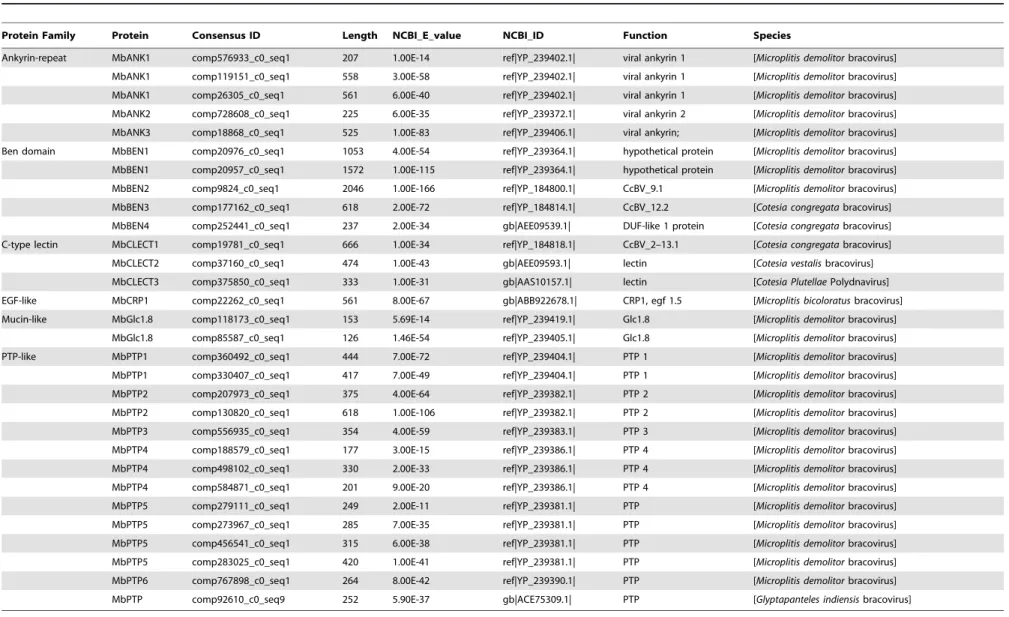
Table 1.Transcription ofM. bicoloratusbracovirus genes during development of parasitoidM. bicoloratusin host hemocytes.
Protein Family Protein Consensus ID Length NCBI_E_value NCBI_ID Function Species
Ankyrin-repeat MbANK1 comp576933_c0_seq1 207 1.00E-14 ref|YP_239402.1| viral ankyrin 1 [Microplitis demolitorbracovirus]
MbANK1 comp119151_c0_seq1 558 3.00E-58 ref|YP_239402.1| viral ankyrin 1 [Microplitis demolitor bracovirus]
MbANK1 comp26305_c0_seq1 561 6.00E-40 ref|YP_239402.1| viral ankyrin 1 [Microplitis demolitorbracovirus]
MbANK2 comp728608_c0_seq1 225 6.00E-35 ref|YP_239372.1| viral ankyrin 2 [Microplitis demolitorbracovirus]
MbANK3 comp18868_c0_seq1 525 1.00E-83 ref|YP_239406.1| viral ankyrin; [Microplitis demolitorbracovirus]
Ben domain MbBEN1 comp20976_c0_seq1 1053 4.00E-54 ref|YP_239364.1| hypothetical protein [Microplitis demolitorbracovirus]
MbBEN1 comp20957_c0_seq1 1572 1.00E-115 ref|YP_239364.1| hypothetical protein [Microplitis demolitorbracovirus]
MbBEN2 comp9824_c0_seq1 2046 1.00E-166 ref|YP_184800.1| CcBV_9.1 [Microplitis demolitorbracovirus]
MbBEN3 comp177162_c0_seq1 618 2.00E-72 ref|YP_184814.1| CcBV_12.2 [Cotesia congregatabracovirus]
MbBEN4 comp252441_c0_seq1 237 2.00E-34 gb|AEE09539.1| DUF-like 1 protein [Cotesia congregatabracovirus]
C-type lectin MbCLECT1 comp19781_c0_seq1 666 1.00E-34 ref|YP_184818.1| CcBV_2–13.1 [Cotesia congregatabracovirus]
MbCLECT2 comp37160_c0_seq1 474 1.00E-43 gb|AEE09593.1| lectin [Cotesia vestalisbracovirus]
MbCLECT3 comp375850_c0_seq1 333 1.00E-31 gb|AAS10157.1| lectin [Cotesia PlutellaePolydnavirus]
EGF-like MbCRP1 comp22262_c0_seq1 561 8.00E-67 gb|ABB922678.1| CRP1, egf 1.5 [Microplitis bicoloratusbracovirus]
Mucin-like MbGlc1.8 comp118173_c0_seq1 153 5.69E-14 ref|YP_239419.1| Glc1.8 [Microplitis demolitorbracovirus]
MbGlc1.8 comp85587_c0_seq1 126 1.46E-54 ref|YP_239405.1| Glc1.8 [Microplitis demolitorbracovirus]
PTP-like MbPTP1 comp360492_c0_seq1 444 7.00E-72 ref|YP_239404.1| PTP 1 [Microplitis demolitorbracovirus]
MbPTP1 comp330407_c0_seq1 417 7.00E-49 ref|YP_239404.1| PTP 1 [Microplitis demolitorbracovirus]
MbPTP2 comp207973_c0_seq1 375 4.00E-64 ref|YP_239382.1| PTP 2 [Microplitis demolitorbracovirus]
MbPTP2 comp130820_c0_seq1 618 1.00E-106 ref|YP_239382.1| PTP 2 [Microplitis demolitorbracovirus]
MbPTP3 comp556935_c0_seq1 354 4.00E-59 ref|YP_239383.1| PTP 3 [Microplitis demolitorbracovirus]
MbPTP4 comp188579_c0_seq1 177 3.00E-15 ref|YP_239386.1| PTP 4 [Microplitis demolitorbracovirus]
MbPTP4 comp498102_c0_seq1 330 2.00E-33 ref|YP_239386.1| PTP 4 [Microplitis demolitorbracovirus]
MbPTP4 comp584871_c0_seq1 201 9.00E-20 ref|YP_239386.1| PTP 4 [Microplitis demolitorbracovirus]
MbPTP5 comp279111_c0_seq1 249 2.00E-11 ref|YP_239381.1| PTP [Microplitis demolitorbracovirus]
MbPTP5 comp273967_c0_seq1 285 7.00E-35 ref|YP_239381.1| PTP [Microplitis demolitorbracovirus]
MbPTP5 comp456541_c0_seq1 315 6.00E-38 ref|YP_239381.1| PTP [Microplitis demolitorbracovirus]
MbPTP5 comp283025_c0_seq1 420 1.00E-41 ref|YP_239381.1| PTP [Microplitis demolitorbracovirus]
MbPTP6 comp767898_c0_seq1 264 8.00E-42 ref|YP_239390.1| PTP [Microplitis demolitorbracovirus]
MbPTP comp92610_c0_seq9 252 5.90E-37 gb|ACE75309.1| PTP [Glyptapanteles indiensisbracovirus]
doi:10.1371/journal.pone.0110967.t001
MbBV
Regulates
Host
Apoptosis
PLOS
ONE
|
www.ploson
e.org
3
October
2014
|
Volume
9
|
Issue
10
|
Table 2.The differential expression of genes regulated byM. bicoloratusbracovirus in the host gap junction signaling pathway.
M/S
A_ID Function read_M RPKM_M read_S RPKM_S
log2(Fold_change)
normalized p-value Result S_ID M-ID
comp95316_c0_seq3 adenylate cyclase 8 414 1.538851011 5365 20.66052119 23.746951184 0 down comp59135_c1_seq10 comp18779_c0_seq1
comp96543_c0_seq4 classical protein kinase C
1502 6.301173776 3740 16.25542916 21.367229143 4.6108E-232 down comp30329_c0_seq1 comp20807_c0_seq1
comp97909_c0_seq6 guanine nucleotide-binding protein G(s) subunit alpha
41 0.622358814 430 6.762398457 23.441716531 3.85048E-83 down comp59076_c0_seq5 /
comp88846_c0_seq1 gap junction 628 3.260154915 1623 8.73E+00 21.420902221 2.19E-107 down comp57755_c2_seq1 comp19421_c1_seq1
comp65035_c0_seq1 gap junction 1808 16.23951848 11125 1.04E+02 22.672414439 0.00E+00 down comp45671_c0_seq1 comp10397_c0_seq1
comp99381_c0_seq1 gap junction 3994 22.25932132 36919 213.1714802 23.259532924 0 down comp59804_c0_seq1 comp30941_c0_seq1
comp121018_c0_seq1 gap junction 36 0.410377626 935 1.10E+01 24.749973239 7.01E-217 down comp59264_c0_seq1 comp10397_c0_seq1
comp96275_c0_seq13 inositol 1,4,5-triphosphate receptor type 1
2068 3.72067313 5214 9.718904704 21.385230083 0 down comp59099_c0_seq4 comp94669_c0_seq1
comp106866_c0_seq1 protein kinase A 2326 9.047590091 7695 31.01038396 21.777145916 0 down comp65026_c0_seq1 comp17984_c0_seq1
comp97791_c0_seq2 phosphatidylinosito l phospholipase C, beta
366 2.438775601 738 5.09474476 21.062852854 8.52689E-32 down comp55943_c2_seq1 comp8084_c0_seq1
comp95574_c0_seq5 protein kinase, cGMP-dependent
1795 5.701522537 4620 15.20350361 21.414984693 1.1324E-301 down comp58204_c1_seq7 comp16873_c0_seq1
comp63482_c0_seq3 tubulin alpha 20547 224.0520545 53712 606.8022006 21.437392362 0 down comp41562_c0_seq1 comp14668_c0_seq1
comp94424_c0_seq3 adenylate cyclase 1 53 0.61819881 151 1.824754972 21.561559971 1.42371E-12 comp29410_c0_seq1 comp128727_c0_seq1
comp94556_c0_seq4 adenylate cyclase 5 23 0.17008429 341 2.612558705 23.941141659 7.22672E-73 comp55791_c2_seq2 /
comp69534_c0_seq1 adenylate cyclase 9 17 0.46329195 29 0.81880247 20.82159384 0.069978649 comp55534_c1_seq1 comp4228_c0_seq1
comp76441_c0_seq1 cyclin-dependent kinase 1
1207 7.410567531 2282 14.51560537 20.969948801 1.51194E-82 comp27910_c0_seq1 comp16516_c0_seq1
comp93202_c0_seq1 epidermal growth factor receptor
6514 18.3968606 6149 17.99184785 0.032116227 0.238280263 comp72419_c0_seq1 comp33128_c0_seq1
comp83895_c0_seq2 guanine
nucleotide-binding protein G(q) subunit alpha
4274 19.02930474 7980 36.81006701 20.951877524 5.7673E-277 comp55254_c0_seq5 comp17311_c0_seq3
comp103695_c0_seq1 growth factor receptor-binding protein 2
4553 31.55832529 7467 53.62135235 20.764786958 6.7036E-179 comp59056_c4_seq1 comp101386_c0_seq1
comp112119_c0_seq1 GTPase KRas 2904 11.39420805 3605 14.6544025 20.363033492 2.01055E-23 comp46714_c0_seq1 comp156998_c0_seq1
comp81191_c0_seq1 mitogen-activated protein kinase kinase 1
1619 6.495897191 2927 12.16719112 20.905395446 3.63546E-94 comp174562_c0_seq1 comp20958_c0_seq1
MbBV
Regulates
Host
Apoptosis
PLOS
ONE
|
www.ploson
e.org
4
October
2014
|
Volume
9
|
Issue
10
|
M. bicoloratusbracovirus genes transcribed in the hemocytes of parasitized host
It is well known that polydnaviruses manipulate host cell physiology [19]. Bracoviruses encode at least 20 gene families identified from 5 species of bracoviruses, Cotesia congregata
bracovirus (CcBV) [20],Microplitis demolitorbracovirus (MdBV) [21],Glyptapanteles indiensisbracovirus (GiBV) [22], Glyptapan-teles flavicoxis bracovirus (GfBV) [5], and Cotesia vestalis
bracovirus (CvBV) [23]. In the present study, genes belonging to at least 6 conserved gene families were found to be expressed in the host hemocytes parasitized by M. bicoloratus including 1) Ankyrin-repeat, 2) BEN domain, 3) C-type lectin, 4) Epidermal growth factor-like (EGF-like), 5) Mucin-like, and 6) protein tyrosine phosphatases (PTPs) (Table 1). Some of the proteins encoded by these genes are likely to be involved in regulating host cell death.
Gap junction signaling pathway regulation byM. bicoloratusparasitism
Gap junction proteins form gap junction channels connecting cells for cell-cell communication and form hemichannels facilitat-ing extracellular and intracellular communication includfacilitat-ing between ER and mitochondria to exchange small molecular, such as ATP and Ca2+, to trigger apoptosis [24]. In the insect
circulating hemocytes, gap junction proteins form hemichannels to allow communication between the cell and environment. Under lipopolysaccharide (LPS) immunochallenge, hemichannel dye uptake decreases [25]. Typically, the decrease of the transcription level of hemichannel components and the decrease in opening of hemichannels on the cell surface result in the decrease of dye uptake. Gap junction proteins, Spli-Inx2 and Inx3, have been characterized and functioned [26] and in this study,Spli-inx1and
inx4also were detected from hemocytes (Fig. S1 and Table S4). Comparisons with S and M transcriptome data indicated that all 26 elements of the gap junction signaling pathway existed in the hemocytes. During immune challenge byM. bicoloratus parasit-ization, 2 genes (Spli-GNAS, ADCY5) were not expressed in the parasitized host hemocytes. To determine the differential expres-sion of genes, all transcriptome were assembled into a combined pool, and S1, S2, M1, and M2 were mapped using this pool to obtain reads and the RPM values of S and M. Furthermore, the analysis obtained the fold change and p-value between parasitized and non-parasitized. These analyses indicated that 12 genes (ADCY8, CPKC, GNAS, INX1, INX2, INX3, INX4, ITPR1, PKA, PLCB, PRKG, and TUBA) were down-regulated (Table 2). The qRT-PCR results indicate that the parasitization down-regulated 3 key molecules, Inx1, Inx2, Inx3, on the cell membrane, not Inx4 (Fig. 1). These molecules are involved in forming hemichannels and gap junctions, suggesting that there might be disruptions of intracellular between ER, mitochondria and extracellular molecular exchanges.
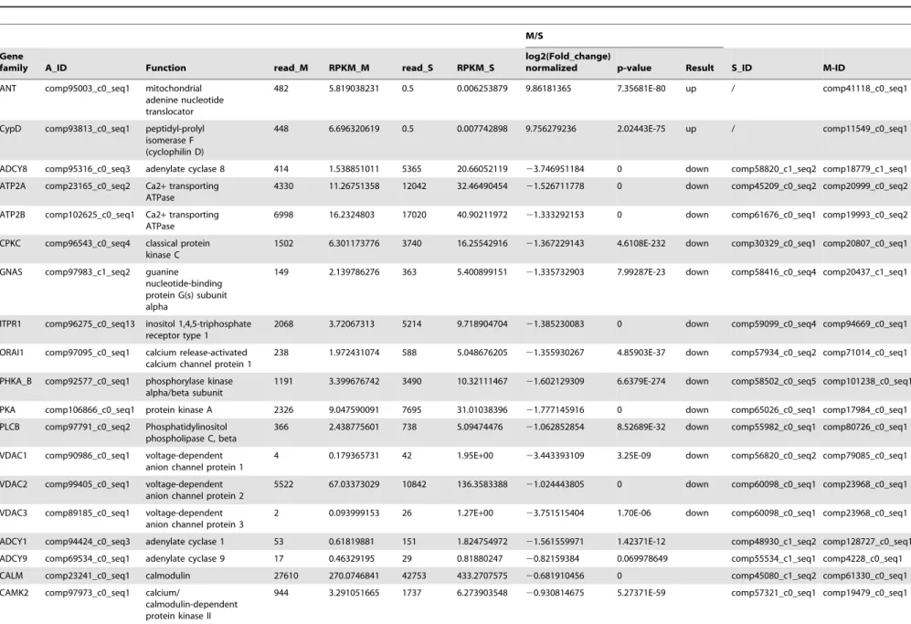
Ca2+signaling pathway regulation by M. bicoloratus parasitism with respect to apoptosis
Calcium ions (Ca2+) control every aspect of cells as cellular
messengers. Ca2+ ions also can become death signals when
delivered at physiologically aberrant conditions. Mitochondria eventually decide whether Ca2+signals are life or death signals via
regulation of the mitochondrial membrane proteins Bcl-2 and Bax/Bak [27]. Comparisons of the transcription data from the S and M pools indicate that all 31 elements of the Ca2+signaling
pathway existed in the examined hemocytes. UnderM. bicoloratus
parasitism, 3 genes (Spli-ANT, CypD, PLCG2) increased in
Table 2. Cont. M/S A_ID Function read_M RPKM_M read_S RPKM_S log2(Fold_chang e) normalized p -value Result S _ID M-ID comp23161_c0_seq1 m itogen-activated protein kinase 1/3 5679 21.44919695 7831 3 0.64302995 2 0.514635322 3.1947E-93 comp28328_c0_seq1 comp9963_c0_seq1 comp96783_c0_seq1 1 son o f sevenless 74 1.123281762 195 3.066669068 2 1.448952634 2.40343E-14 comp47573_c1_seq1 comp17032_c0_seq1 comp97420_c0_seq2 tyrosine-protein kinase Src 126 1.148163839 460 4.342766357 2 1.919285813 2 .09308E-47 comp56420_c1_seq1 comp106968_c0_se q1 comp97925_c2_seq1 tight junction p rotein 1 7 1 0.575127537 454 3.810105311 2 2.727877054 8.33036E-71 comp58674_c2_seq3 comp93783_c0_seq1 comp92127_c0_seq2 tubulin beta 63513 551.0471151 102657 9 22.7626088 2 0.74378387 0 comp118372_c0_se q1 comp18397_c1_seq1 doi:10.1371/journal.pone. 0110967.t002
MbBV Regulates Host Apoptosis
expression, and 1 genes (Spli-PDE1) was not expressed in the parasitized hemocytes. The other 13 genes (ADCY8, ATP2A, ATP2B, CPKC, GNAS, ITPR1, ORAI1, PHKA_B, PKA, PLCB, VDAC1, VDAC2 and VDAC3) had been down-regulated (Table 3). The qRT-PCR results indicate that the parasitism up-regulated a key molecule, CypD, in the mitochondria (Fig. 1). This molecule is involved in forming a permeability transition pore complex (PTPC), suggesting that the M. bicoloratus alters Ca2+
signaling pathway to promote apoptosis.
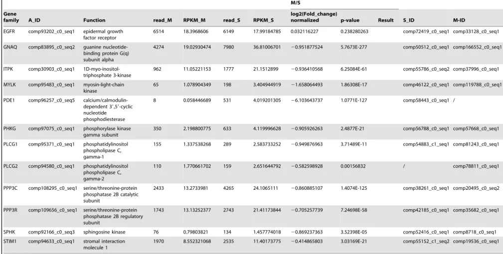
PI3K/Akt signaling pathway regulation byM. bicoloratus parasitism
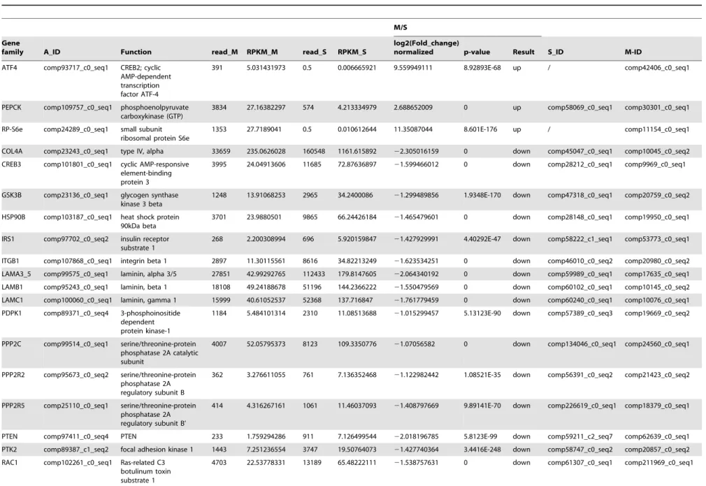
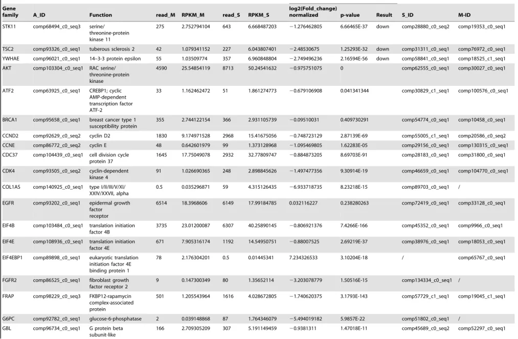
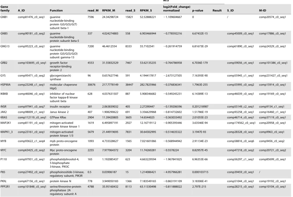
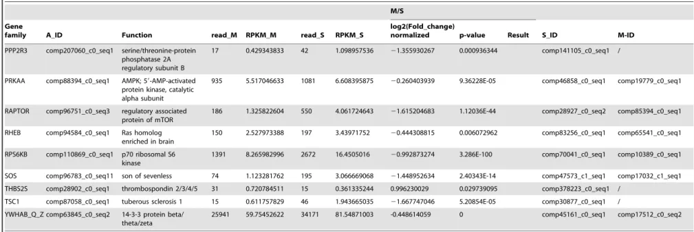
The PI3K/Akt signaling pathway is involved in multiple different pathways, including cell survival, apoptosis, cell cycle, and DNA repair, through different downstream molecules. A comparison of the transcription data from the S and M pools revealed that all 65 elements of the PI3K/Akt signaling pathway existed in the hemocytes. Under immune challenge, 4 genes (ATF4, RP-S6e, EIF4EBP1, and GNB1) were expressed in the parasitized hemocytes, and 7 genes (COL1AS, FGFR2, G6PC, p85, PPP2R3, THBS2S, and TSC1) were not expressed in the parasitized host hemocytes (Table 4). Another 19 genes (COL4A, CREB3, HSP90B, IRS1, ITGB1, LAMA3_5, LAMB1, LAMC1, PDPK1, PPP2C, PPP2R2, PPP2R5, PTEN, PTK2, RAC1, STK11, TSC2 and YWHAE) were down-regulated, (Table 4). The qRT-PCR results indicated that the parasitism down-regulated a key molecule, the p110 subunit, in the PI3K/Akt signaling pathway, suggesting that the disruption of cell survival signaling pathway by the parasitism may promote cell apoptosis (Fig. 1).
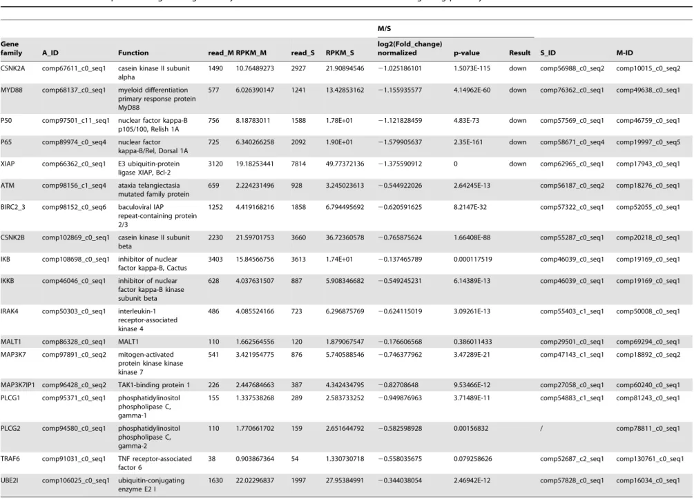
NF-kB signaling pathway regulation byM. bicoloratus parasitism
The NF-kB signaling pathway regulates gene expression via regulation of nuclear transcription factor. Comparison of the transcription data from the S and M pools indicates that all 18 elements of NF-kB signaling pathway existed in the hemocytes.
Under M. bicoloratus parasitism, 1 gene (Spli-PLCG2) was expressed in the parasitized host hemocytes, and 5 genes (Spli-CSNK2A, MYD88, P50, P65 and XIAP) were down-regulated (Table 5). The qRT-PCR results indicate that the parasitism down-regulated two key molecules, the p50 (Relish) and p65 (Dorsal) subunits in the NF-kB signaling pathway, suggesting the disruption of the cell survival signaling pathway (Fig. 1).
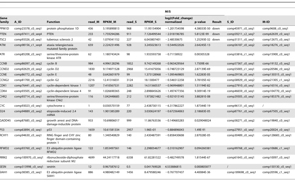
ATM/p53 signaling pathway regulation byM. bicoloratus parasitism
The ATM/p53 signaling pathway plays an important role in cell cycle control and apoptosis. In normal cells, the p53 protein level is low. DNA damage and stress signaling may trigger an increase of p53 protein levels, which has three major functions: cell cycle arrest, DNA repair and apoptosis. The cell cycle arrest prevents replication of proteins involved in DNA repair. Apoptosis avoids proliferation of cells containing abnormal DNA. p53 is a transcriptional activator that regulates the expression of MDM2. A comparison of the transcription data from the S and M pools indicate that all 21 elements of the ATM/p53 signaling pathway existed in the hemocytes. UnderM. bicoloratusparasitism, 1 gene (Spli-SESN), was expressed in the parasitized host hemocytes, and 1 gene (CYC) was not expressed in the parasitized host hemocytes. Another 3 genes (Spli-PPM1D, PTEN, and TSC2) were down-regulated (Table 6). The qRT-PCR results indicate that the parasitism increased expression of a key molecule, p53, in the ATM/p53 signaling pathway (Fig. 1).
Discussion
M. bicoloratus parasitism regulated host hemocyte apoptosis, resulting in DNA fragmentation. In this study, we examined the impacts of both the apoptotic caspase-dependent and -indepen-dent signaling pathways on the host hemocytes based on transcriptome data. Our results demonstrated that bracovirus proteins are expressed in the host hemocytes, suggesting their roles in DNA fragmentation by regulating key signaling pathways,
Figure 1. qRT-PCR detected key genes in five signaling pathways with hemocytes 5 days post-parasitization (p.p.).
doi:10.1371/journal.pone.0110967.g001
MbBV Regulates Host Apoptosis
Table 3.The differential expression of genes regulated byM. bicoloratusbracovirus in the host Ca2+signaling pathway.
M/S
Gene
family A_ID Function read_M RPKM_M read_S RPKM_S
log2(Fold_change)
normalized p-value Result S_ID M-ID
ANT comp95003_c0_seq1 mitochondrial
adenine nucleotide translocator
482 5.819038231 0.5 0.006253879 9.86181365 7.35681E-80 up / comp41118_c0_seq1
CypD comp93813_c0_seq1 peptidyl-prolyl isomerase F (cyclophilin D)
448 6.696320619 0.5 0.007742898 9.756279236 2.02443E-75 up / comp11549_c0_seq1
ADCY8 comp95316_c0_seq3 adenylate cyclase 8 414 1.538851011 5365 20.66052119 23.746951184 0 down comp58820_c1_seq2 comp18779_c1_seq1
ATP2A comp23165_c0_seq2 Ca2+transporting ATPase
4330 11.26751358 12042 32.46490454 21.526711778 0 down comp45209_c0_seq2 comp20999_c0_seq2
ATP2B comp102625_c0_seq1 Ca2+transporting ATPase
6998 16.2324803 17020 40.90211972 21.333292153 0 down comp61676_c0_seq1 comp19993_c0_seq2
CPKC comp96543_c0_seq4 classical protein kinase C
1502 6.301173776 3740 16.25542916 21.367229143 4.6108E-232 down comp30329_c0_seq1 comp20807_c0_seq1
GNAS comp97983_c1_seq2 guanine
nucleotide-binding protein G(s) subunit alpha
149 2.139786276 363 5.400899151 21.335732903 7.99287E-23 down comp58416_c0_seq4 comp20437_c1_seq1
ITPR1 comp96275_c0_seq13 inositol 1,4,5-triphosphate receptor type 1
2068 3.72067313 5214 9.718904704 21.385230083 0 down comp59099_c0_seq4 comp94669_c0_seq1
ORAI1 comp97095_c0_seq1 calcium release-activated calcium channel protein 1
238 1.972431074 588 5.048676205 21.355930267 4.85903E-37 down comp57934_c0_seq2 comp71014_c0_seq1
PHKA_B comp92577_c0_seq1 phosphorylase kinase alpha/beta subunit
1191 3.399676742 3490 10.32111467 21.602129309 6.6379E-274 down comp58502_c0_seq5 comp101238_c0_seq1
PKA comp106866_c0_seq1 protein kinase A 2326 9.047590091 7695 31.01038396 21.777145916 0 down comp65026_c0_seq1 comp17984_c0_seq1
PLCB comp97791_c0_seq2 Phosphatidylinositol phospholipase C, beta
366 2.438775601 738 5.09474476 21.062852854 8.52689E-32 down comp55982_c0_seq1 comp80726_c0_seq1
VDAC1 comp90986_c0_seq1 voltage-dependent anion channel protein 1
4 0.179365731 42 1.95E+00 23.443393109 3.25E-09 down comp56820_c0_seq2 comp79085_c0_seq1
VDAC2 comp99405_c0_seq1 voltage-dependent anion channel protein 2
5522 67.03373029 10842 136.3583388 21.024443805 0 down comp60098_c0_seq1 comp23968_c0_seq1
VDAC3 comp89185_c0_seq1 voltage-dependent anion channel protein 3
2 0.093999153 26 1.27E+00 23.751515404 1.70E-06 down comp60098_c0_seq1 comp23968_c0_seq1
ADCY1 comp94424_c0_seq3 adenylate cyclase 1 53 0.61819881 151 1.824754972 21.561559971 1.42371E-12 comp48930_c1_seq2 comp128727_c0_seq1
ADCY9 comp69534_c0_seq1 adenylate cyclase 9 17 0.46329195 29 0.81880247 20.82159384 0.069978649 comp55534_c1_seq1 comp4228_c0_seq1
CALM comp23241_c0_seq1 calmodulin 27610 270.0746841 42753 433.2707575 20.681910456 0 comp45080_c1_seq2 comp61330_c0_seq1
CAMK2 comp97973_c0_seq1 calcium/
calmodulin-dependent protein kinase II
944 3.291051665 1737 6.273903548 20.930814675 5.27371E-59 comp57321_c0_seq1 comp19479_c0_seq1
MbBV
Regulates
Host
Apoptosis
PLOS
ONE
|
www.ploson
e.org
7
October
2014
|
Volume
9
|
Issue
10
|
Table 3.Cont.
M/S
Gene
family A_ID Function read_M RPKM_M read_S RPKM_S
log2(Fold_change)
normalized p-value Result S_ID M-ID
EGFR comp93202_c0_seq1 epidermal growth factor receptor
6514 18.3968606 6149 17.99184785 0.032116227 0.238280263 comp72419_c0_seq1 comp33128_c0_seq1
GNAQ comp83895_c0_seq2 guanine nucleotide-binding protein G(q) subunit alpha
4274 19.02930474 7980 36.81006701 20.951877524 5.7673E-277 comp50512_c0_seq1 comp166552_c0_seq1
ITPK comp30903_c0_seq1 1D-myo-inositol-triphosphate 3-kinase
962 11.05221153 1777 21.1512899 20.936410568 6.25084E-61 comp55786_c0_seq2 comp37996_c0_seq1
MYLK comp95483_c0_seq1 myosin-light-chain kinase
65 1.078904349 198 3.404944919 21.658064493 1.86308E-17 comp46122_c0_seq1 comp119788_c0_seq1
PDE1 comp96257_c0_seq5 calcium/calmodulin-dependent 39,59-cyclic nucleotide
phosphodiesterase
8 0.058446689 531 4.019201305 26.103643737 1.0771E-127 comp58443_c0_seq1 /
PHKG comp97075_c0_seq1 phosphorylase kinase gamma subunit
350 2.198800775 633 4.119996628 20.905926263 2.4877E-21 comp56788_c0_seq1 comp57668_c0_seq1
PLCG1 comp95371_c0_seq1 phosphatidylinositol phospholipase C, gamma-1
155 1.337538268 289 2.583733252 20.949876963 3.71489E-11 comp54883_c1_seq1 comp81243_c0_seq1
PLCG2 comp94580_c0_seq1 phosphatidylinositol phospholipase C, gamma-2
110 1.770661702 159 2.651644792 20.582598928 0.00156832 / comp78811_c0_seq1
PPP3C comp108295_c0_seq1 serine/threonine-protein phosphatase 2B catalytic subunit
2433 13.2733981 4265 24.1065111 20.860885107 1.4074E-125 comp38261_c0_seq1 comp20495_c0_seq2
PPP3R comp109656_c0_seq1 serine/threonine-protein phosphatase 2B regulatory subunit
1743 13.13252377 2743 21.41173844 20.705257739 7.24698E-58 comp42185_c0_seq1 comp35682_c0_seq1
SPHK comp92166_c0_seq3 sphingosine kinase 76 0.79803821 134 1.457774018 20.869237363 3.52398E-05 comp52416_c0_seq1 comp8718_c0_seq1
STIM1 comp94633_c0_seq1 stromal interaction molecule 1
1970 8.552321068 2535 11.40173775 20.414865803 3.03169E-21 comp55152_c1_seq2 comp19536_c0_seq1
doi:10.1371/journal.pone.0110967.t003 MbBV
Regulates
Host
Apoptosis
PLOS
ONE
|
www.ploson
e.org
8
October
2014
|
Volume
9
|
Issue
10
|
Table 4.The differential expression of genes regulated byM. bicoloratusbracovirus in the host PI3K/Akt signaling pathway.
M/S
Gene
family A_ID Function read_M RPKM_M read_S RPKM_S
log2(Fold_change)
normalized p-value Result S_ID M-ID
ATF4 comp93717_c0_seq1 CREB2; cyclic
AMP-dependent transcription factor ATF-4
391 5.031431973 0.5 0.006665921 9.559949111 8.92893E-68 up / comp42406_c0_seq1
PEPCK comp109757_c0_seq1 phosphoenolpyruvate carboxykinase (GTP)
3834 27.16382297 574 4.213334979 2.688652009 0 up comp58069_c0_seq1 comp30301_c0_seq1
RP-S6e comp24289_c0_seq1 small subunit ribosomal protein S6e
1353 27.7189041 0.5 0.010612644 11.35087044 8.601E-176 up / comp11154_c0_seq1
COL4A comp23243_c0_seq1 type IV, alpha 33659 235.0626028 160548 1161.615892 22.305016159 0 down comp45047_c0_seq1 comp10045_c0_seq2
CREB3 comp101801_c0_seq1 cyclic AMP-responsive element-binding protein 3
3995 24.04913606 11685 72.87636897 21.599466012 0 down comp28212_c0_seq1 comp9969_c0_seq1
GSK3B comp23136_c0_seq1 glycogen synthase kinase 3 beta
1248 13.91068253 2965 34.2400086 21.299489856 1.9348E-170 down comp47318_c0_seq1 comp20759_c0_seq2
HSP90B comp103187_c0_seq1 heat shock protein 90kDa beta
3701 23.9880501 9865 66.24426184 21.465479601 0 down comp28148_c0_seq1 comp19950_c0_seq1
IRS1 comp97702_c0_seq2 insulin receptor substrate 1
268 2.200308994 696 5.920159847 21.427929991 4.40292E-47 down comp58222_c1_seq1 comp53773_c0_seq1
ITGB1 comp107868_c0_seq1 integrin beta 1 2897 11.30115561 8616 34.82213249 21.623534251 0 down comp46010_c0_seq2 comp20980_c0_seq2
LAMA3_5 comp99575_c0_seq1 laminin, alpha 3/5 27851 42.99292765 112433 179.8147605 22.064340192 0 down comp59989_c0_seq1 comp17635_c0_seq1
LAMB1 comp95243_c0_seq1 laminin, beta 1 18108 49.24188678 51196 144.2366222 21.550479569 0 down comp60102_c0_seq1 comp10145_c0_seq2
LAMC1 comp100060_c0_seq1 laminin, gamma 1 15999 40.61052537 52368 137.716847 21.761779459 0 down comp60240_c0_seq1 comp10076_c0_seq1
PDPK1 comp89371_c0_seq4 3-phosphoinositide dependent protein kinase-1
1184 5.484101314 2310 11.08513688 21.015299457 5.13123E-90 down comp57389_c0_seq3 comp19669_c0_seq2
PPP2C comp99514_c0_seq1 serine/threonine-protein phosphatase 2A catalytic subunit
4007 52.05795373 8123 109.3350776 21.07056582 0 down comp134046_c0_seq1 comp24560_c0_seq1
PPP2R2 comp95673_c0_seq2 serine/threonine-protein phosphatase 2A regulatory subunit B
362 3.276611055 761 7.136352468 21.122982442 1.08521E-35 down comp56391_c0_seq2 comp21423_c0_seq2
PPP2R5 comp25110_c0_seq1 serine/threonine-protein phosphatase 2A regulatory subunit B’
414 4.316267161 1061 11.46037093 21.408797669 9.89141E-70 down comp226619_c0_seq1 comp18379_c0_seq1
PTEN comp97411_c0_seq4 PTEN 233 1.759294286 911 7.126499544 22.018196785 5.8123E-99 down comp59211_c2_seq7 comp62639_c0_seq1
PTK2 comp89387_c1_seq2 focal adhesion kinase 1 1443 7.251236554 3747 19.50764073 21.427740364 3.4416E-248 down comp58747_c0_seq2 comp20857_c0_seq2
RAC1 comp102261_c0_seq1 Ras-related C3 botulinum toxin substrate 1
4703 22.53778331 13189 65.48222111 21.538757631 0 down comp61307_c0_seq1 comp211969_c0_seq1
MbBV
Regulates
Host
Apoptosis
PLOS
ONE
|
www.ploson
e.org
9
October
2014
|
Volume
9
|
Issue
10
|
Table 4.Cont.
M/S
Gene
family A_ID Function read_M RPKM_M read_S RPKM_S
log2(Fold_change)
normalized p-value Result S_ID M-ID
STK11 comp68494_c0_seq3 serine/
threonine-protein kinase 11
275 2.752794104 643 6.668487203 21.276462805 6.66465E-37 down comp28880_c0_seq2 comp19353_c0_seq1
TSC2 comp93326_c0_seq1 tuberous sclerosis 2 42 1.079341152 227 6.043807401 22.48530675 1.25293E-32 down comp31311_c0_seq1 comp76972_c0_seq1
YWHAE comp96021_c0_seq1 14–3-3 protein epsilon 55 1.03509774 357 6.960848804 22.749496236 2.16594E-56 down comp58841_c0_seq1 comp18525_c1_seq1
AKT comp103304_c0_seq1 RAC serine/
threonine-protein kinase
4590 25.54854119 8713 50.24541632 20.975751075 0 comp62555_c0_seq1 comp30027_c0_seq1
ATF2 comp63925_c0_seq1 CREBP1; cyclic
AMP-dependent transcription factor ATF-2
33 1.162462472 51 1.861274773 20.679106908 0.041341344 comp30829_c1_seq1 comp100576_c0_seq1
BRCA1 comp95658_c0_seq1 breast cancer type 1 susceptibility protein
355 2.744122154 366 2.931105739 20.09510031 0.409730291 comp54774_c0_seq1 comp10458_c0_seq1
CCND2 comp92629_c0_seq2 cyclin D2 1830 9.174971528 2968 15.41675056 20.748723129 2.87139E-69 comp55005_c1_seq1 comp20586_c0_seq2
CCNE comp86772_c0_seq2 cyclin E 48 0.642601979 99 1.373128968 21.095469805 1.62283E-05 comp29156_c0_seq1 comp130315_c0_seq1
CDC37 comp104439_c0_seq1 cell division cycle protein 37
1645 17.75049078 2932 32.77809747 20.884873205 8.69703E-91 comp28183_c0_seq1 comp31800_c0_seq1
CDK4 comp93505_c0_seq2 cyclin-dependent
kinase 4
91 1.026690365 248 2.898845626 21.497477356 9.30914E-19 comp46659_c0_seq1 comp104770_c0_seq1
COL1AS comp140925_c0_seq1 type I/II/III/V/XI/ XXIV/XXVII, alpha
0.5 0.035296871 59 4.315126435 26.933718735 8.23218E-15 comp89703_c0_seq1 /
EGFR comp93202_c0_seq1 epidermal growth
factor receptor
6514 18.3968606 6149 17.99184785 0.032116227 0.238280263 comp72419_c0_seq1 comp33128_c0_seq1
EIF4B comp103484_c0_seq1 translation initiation factor 4B
3735 23.01200087 6307 40.25890145 20.806921376 7.4266E-166 comp45352_c0_seq1 comp9966_c0_seq1
EIF4E comp108936_c0_seq1 translation initiation factor 4E
671 7.905316174 1192 14.54950751 20.88007525 2.69219E-37 comp38976_c0_seq1 comp18053_c0_seq1
EIF4EBP1 comp89898_c0_seq1 eukaryotic translation initiation factor 4E binding protein 1
78 2.176304201 0.5 0.01445341 7.234326533 3.10204E-18 / comp65767_c0_seq1
FGFR2 comp86525_c0_seq1 fibroblast growth factor receptor 2
9 0.147300349 80 1.35652114 23.203078779 1.50516E-15 comp134334_c0_seq1 /
FRAP comp98229_c0_seq3 FKBP12-rapamycin
complex-associated protein
501 1.205543964 1616 4.028672805 21.740620375 3.1793E-143 comp57729_c1_seq1 comp19045_c1_seq1
G6PC comp92782_c0_seq1 glucose-6-phosphatase 2 0.039148868 87 1.764346079 25.494019182 5.9857E-22 comp51802_c0_seq1 /
GBL comp96734_c0_seq1 G protein beta
subunit-like
166 2.709305209 307 5.191149459 20.9381311 1.47018E-11 comp45689_c0_seq2 comp52297_c0_seq1
MbBV
Regulates
Host
Apoptosis
PLOS
ONE
|
www.ploson
e.org
10
October
2014
|
Volume
9
|
Issue
10
|
Table 4.Cont.
M/S
Gene
family A_ID Function read_M RPKM_M read_S RPKM_S
log2(Fold_change)
normalized p-value Result S_ID M-ID
GNB1 comp81476_c0_seq1 guanine
nucleotide-binding protein G(I)/G(S)/G(T) subunit beta-1
7596 24.34298724 15821 52.52888221 21.109604667 0 / comp20574_c0_seq1
GNB5 comp90181_c0_seq2 guanine
nucleotide-binding protein subunit beta-5
337 4.024274883 558 6.903466944 20.778592216 6.67432E-15 comp45009_c0_seq1 comp17886_c0_seq1
GNG13 comp95223_c0_seq1 guanine
nucleotide-binding protein G(I)/G(S)/G(O) subunit gamma-13
7200 46.4612554 8333 55.7102541 20.261914759 6.81673E-29 comp61890_c0_seq1 comp24329_c0_seq1
GRB2 comp103695_c0_seq1 growth factor
receptor-binding protein 2
4553 31.55832529 7467 53.62135235 20.764786958 6.7036E-179 comp59056_c4_seq1 comp101386_c0_seq1
GYS comp95471_c0_seq2 glycogen(starch)
synthase
96 0.657627746 591 4.194417817 22.673127505 7.16393E-90 comp55945_c1_seq1 comp231427_c0_seq1
HSP90A comp23248_c1_seq1 molecular chaperone HtpG
30676 217.7778149 38447 282.7825966 20.376836341 1.7963E-255 comp55995_c0_seq2 comp15914_c0_seq1
IKBKB comp46046_c0_seq1 inhibitor of nuclear factor kappa-B kinase subunit beta
628 4.037631507 887 5.908346682 20.549245231 6.14389E-13 comp46039_c0_seq1 comp19169_c0_seq1
INSR comp97941_c0_seq7 insulin receptor 381 2.063839032 405 2.27290447 20.139206596 0.201218987 comp55149_c2_seq1 comp9154_c1_seq1
JAK2 comp98009_c1_seq1 Janus kinase 2 407 1.908290622 691 3.356629968 20.814732602 1.51786E-19 comp93258_c0_seq1 comp16460_c1_seq1
KRAS comp112119_c0_seq1 GTPase KRas 2904 11.39420805 3605 14.6544025 20.363033492 2.01055E-23 comp46714_c0_seq1 comp37119_c0_seq1
MAP2K1 comp81191_c0_seq1 mitogen-activated protein kinase kinase 1
1619 6.495897191 2927 12.16719112 20.905395446 3.63546E-94 comp174562_c0_seq1 comp20958_c0_seq1
MAPK1_3 comp23161_c0_seq1 mitogen-activated protein kinase 1/3
5679 21.44919695 7831 30.64302995 20.514635322 3.1947E-93 comp28328_c0_seq1 comp9963_c0_seq1
MYB comp93622_c1_seq4 myb proto-oncogene
protein
1093 4.733328627 1565 7.021601066 20.568944942 2.91134E-23 comp58816_c0_seq6 comp9456_c0_seq2
MYC comp63425_c0_seq2 Myc proto-oncogene
protein
2255 7.977064372 3204 11.74260281 20.5578224 8.82957E-45 comp47218_c0_seq2 comp20721_c0_seq2
P110 comp97931_c0_seq1 phosphatidylinositol-4, 5-bisphosphate 3-kinase, PIK3C
165 1.192085437 623 4.663229594 21.967841825 6.96353E-66 comp56297_c1_seq1 comp85699_c0_seq1
P85 comp27492_c0_seq1 phosphoinositide-3-kinase, regulatory subunit, PIK3R
0.5 0.03906187 15 1.214086421 24.957966281 0.000103715 comp39459_c0_seq1 /
PKN; comp67156_c0_seq1 protein kinase N 778 3.949050183 1366 7.183549165 20.863191109 3.18306E-41 comp51044_c0_seq1 comp19192_c0_seq1
PPP2R1 comp101848_c0_seq1 serine/threonine-protein phosphatase 2A regulatory subunit A
4788 35.95160432 8113 63.11330498 20.811888022 2.797E-215 comp28215_c0_seq1 comp10104_c0_seq1
MbBV
Regulates
Host
Apoptosis
PLOS
ONE
|
www.ploson
e.org
11
October
2014
|
Volume
9
|
Issue
10
|
Table 4.Cont.
M/S
Gene
family A_ID Function read_M RPKM_M read_S RPKM_S
log2(Fold_change)
normalized p-value Result S_ID M-ID
PPP2R3 comp207060_c0_seq1 serine/threonine-protein phosphatase 2A regulatory subunit B
17 0.429343833 42 1.098957536 21.355930267 0.000936344 comp141105_c0_seq1 /
PRKAA comp88394_c0_seq1 AMPK; 59-AMP-activated protein kinase, catalytic alpha subunit
935 5.517046633 1081 6.608395875 20.260403939 9.36228E-05 comp46858_c0_seq1 comp19779_c0_seq1
RAPTOR comp96751_c0_seq3 regulatory associated protein of mTOR
186 1.325822604 550 4.061724643 21.615204683 1.12036E-44 comp28927_c0_seq2 comp85394_c0_seq1
RHEB comp94584_c0_seq1 Ras homolog
enriched in brain
150 2.527973388 197 3.43971752 20.444308815 0.006072962 comp83256_c0_seq1 comp65541_c0_seq1
RPS6KB comp110869_c0_seq1 p70 ribosomal S6 kinase
1391 8.265982996 2672 16.4505016 20.992873274 3.286E-100 comp70041_c0_seq1 comp10389_c0_seq1
SOS comp96783_c0_seq11 son of sevenless 74 1.123281762 195 3.066669068 21.448952634 2.40343E-14 comp47573_c1_seq1 comp17032_c1_seq1
THBS2S comp28902_c0_seq1 thrombospondin 2/3/4/5 31 0.720784511 15 0.361335244 0.996230029 0.029739095 comp378223_c0_seq1 /
TSC1 comp87058_c0_seq1 tuberous sclerosis 1 15 0.611757829 46 1.943665035 21.667747046 5.20854E-05 comp30877_c0_seq1 /
YWHAB_Q_Z comp63845_c0_seq2 14-3-3 protein beta/ theta/zeta
25941 59.75452622 34171 81.54871003 -0.448614059 0 comp45161_c0_seq1 comp17512_c0_seq2
doi:10.1371/journal.pone.0110967.t004
MbBV
Regulates
Host
Apoptosis
PLOS
ONE
|
www.ploson
e.org
12
October
2014
|
Volume
9
|
Issue
10
|
Table 5.The differential expression of genes regulated byM. bicoloratusbracovirus in the host NF-kB signaling pathway.
M/S
Gene
family A_ID Function read_M RPKM_M read_S RPKM_S
log2(Fold_change)
normalized p-value Result S_ID M-ID
CSNK2A comp67611_c0_seq1 casein kinase II subunit alpha
1490 10.76489273 2927 21.90894546 21.025186101 1.5073E-115 down comp56988_c0_seq2 comp10015_c0_seq2
MYD88 comp68137_c0_seq1 myeloid differentiation primary response protein MyD88
577 6.026390147 1241 13.42853162 21.155935577 4.14962E-60 down comp76362_c0_seq1 comp49638_c0_seq1
P50 comp97501_c11_seq1 nuclear factor kappa-B p105/100, Relish 1A
756 8.18783011 1588 1.78E+01 21.121828459 4.83E-73 down comp57569_c0_seq1 comp46759_c0_seq1
P65 comp89974_c0_seq4 nuclear factor
kappa-B/Rel, Dorsal 1A
725 6.340266258 2092 1.90E+01 21.579905637 2.35E-161 down comp58671_c0_seq4 comp19997_c0_seq5
XIAP comp66362_c0_seq1 E3 ubiquitin-protein ligase XIAP, Bcl-2
3120 19.18253441 7814 49.77372136 21.375590912 0 down comp62965_c0_seq1 comp17943_c0_seq1
ATM comp98156_c1_seq4 ataxia telangiectasia mutated family protein
659 2.224231496 928 3.245023613 20.544922026 2.64245E-13 comp56187_c0_seq2 comp18276_c0_seq1
BIRC2_3 comp98152_c0_seq6 baculoviral IAP repeat-containing protein 2/3
1252 4.419168216 1858 6.794495692 20.620591625 8.2147E-32 comp57322_c0_seq1 comp52055_c0_seq1
CSNK2B comp102869_c0_seq1 casein kinase II subunit beta
2230 21.59701753 3660 36.72360578 20.765875624 1.66408E-88 comp55287_c0_seq1 comp20218_c0_seq1
IKB comp108698_c0_seq1 inhibitor of nuclear factor kappa-B, Cactus
3403 15.84566756 3613 1.74E+01 20.137465789 0.000117519 comp46039_c0_seq1 comp19169_c0_seq1
IKKB comp46046_c0_seq1 inhibitor of nuclear factor kappa-B kinase subunit beta
628 4.037631507 887 5.908346682 20.549245231 6.14389E-13 comp46039_c0_seq1 comp19169_c0_seq1
IRAK4 comp50303_c0_seq1 interleukin-1 receptor-associated kinase 4
486 4.085524166 723 6.296875769 20.624115019 3.09261E-13 comp55403_c1_seq1 comp50008_c0_seq1
MALT1 comp86328_c0_seq1 MALT1 110 1.662564556 120 1.879067547 20.176606568 0.386011433 comp29501_c0_seq1 comp69294_c0_seq1
MAP3K7 comp97891_c0_seq2 mitogen-activated protein kinase kinase kinase 7
541 3.421954775 876 5.740588546 20.746377962 3.47289E-21 comp47143_c1_seq1 comp18892_c0_seq2
MAP3K7IP1 comp96428_c0_seq2 TAK1-binding protein 1 226 2.447684663 387 4.342434795 20.82708648 9.53466E-12 comp27058_c0_seq1 comp60240_c0_seq1
PLCG1 comp95371_c0_seq1 phosphatidylinositol phospholipase C, gamma-1
155 1.337538268 289 2.583733252 20.949876963 3.71489E-11 comp54883_c1_seq1 comp81243_c0_seq1
PLCG2 comp94580_c0_seq1 phosphatidylinositol phospholipase C, gamma-2
110 1.770661702 159 2.651644792 20.582598928 0.00156832 / comp78811_c0_seq1
TRAF6 comp91031_c0_seq1 TNF receptor-associated factor 6
38 0.903867364 54 1.330730718 20.558035675 0.079258626 comp52687_c2_seq1 comp130761_c0_seq1
UBE2I comp106025_c0_seq1 ubiquitin-conjugating enzyme E2 I
1630 22.02296837 1997 27.95384991 20.344038054 2.46942E-12 comp57828_c0_seq1 comp16034_c0_seq1
doi:10.1371/journal.pone.0110967.t005
MbBV
Regulates
Host
Apoptosis
PLOS
ONE
|
www.ploson
e.org
13
October
2014
|
Volume
9
|
Issue
10
|
Table 6.The differential expression of genes regulated byM. bicoloratusbracovirus in the host ATM/p53 signaling pathway.
M/S
Gene
family A_ID Function read_M RPKM_M read_S RPKM_S
log2(Fold_change)
normalized p-value Result S_ID M-ID
PPM1D comp23378_c0_seq1 protein phosphatase 1D 436 5.195890813 968 11.95154904 21.201754598 4.38033E-50 down comp45971_c0_seq1 comp9608_c0_seq1
PTEN comp97411_c0_seq4 PTEN 233 1.759294286 911 7.126499544 22.018196785 5.8123E-99 down comp59211_c2_seq7 comp62639_c0_seq1
TSC2 comp93326_c0_seq1 tuberous sclerosis 2 42 1.079341152 227 6.043807401 22.48530675 1.25293E-32 down comp31311_c0_seq1 comp12675_c0_seq1
ATM comp98156_c1_seq4 ataxia telangiectasia mutated family protein
659 2.224231496 928 3.245023613 20.544922026 2.64245E-13 comp56187_c0_seq2 comp18276_c0_seq1
ATR comp85208_c0_seq1 serine/threonine-protein kinase ATR
62 1.180743424 98 1.933593758 20.71158922 0.00305328 comp53816_c1_seq1 comp15900_c0_seq1
CCNB comp86097_c0_seq2 cyclin B 984 4.996128296 1852 9.742149268 20.963429564 1.7509E-66 comp57367_c0_seq1 comp19132_c0_seq2
CCND2 comp92629_c0_seq2 cyclin D2 1830 9.174971528 2968 15.41675056 20.748723129 2.87139E-69 comp55005_c1_seq1 comp20586_c0_seq2
CCNE comp86772_c0_seq2 cyclin E 48 0.642601979 99 1.373128968 21.095469805 1.62283E-05 comp29156_c0_seq1 comp130315_c0_seq1
CCNG2 comp81700_c0_seq1 cyclin G2 2216 12.41316031 3124 18.1300477 20.546512258 3.78105E-42 comp58929_c0_seq2 comp21305_c1_seq2
CDK1 comp76441_c0_seq1 cyclin-dependent kinase 1 1207 7.410567531 2282 14.51560537 20.969948801 1.51194E-82 comp27910_c0_seq1 comp16516_c0_seq1
CDK4 comp93505_c0_seq2 cyclin-dependent kinase 4 91 1.026690365 248 2.898845626 21.497477356 9.30914E-19 comp46659_c0_seq1 comp104770_c0_seq1
CHK2 comp93714_c0_seq1 serine/threonine-protein kinase Chk2
116 1.042448685 212 1.973821482 20.921015145 3.88281E-08 comp29305_c0_seq1 comp185379_c0_seq1
CYC comp93023_c0_seq1 cytochrome c 1 0.030570159 77 2.438730115 26.317862227 1.87344E-19 comp96131_c0_seq1 /
EI24 comp94889_c0_seq2 etoposide-induced 2.4 mRNA
143 1.901385289 220 3.030624197 20.672564063 2.18683E-05 comp87761_c0_seq1 comp67505_c0_seq1
GADD45 comp87685_c0_seq2 growth arrest and DNA-damage-inducible protein
933 10.69806017 999 11.86763536 20.149683283 0.029048024 comp58271_c0_seq2 comp19840_c0_seq1
P53 comp63894_c0_seq1 p53 1659 10.61581334 2957 1.96E+01 20.884896043 1.49E-91 comp27951_c0_seq1 comp20024_c0_seq1
RCHY1 comp94630_c0_seq1 RING finger and CHY zinc finger domain-containing protein 1
80 1.340540829 140 2.430487591 20.858430608 2.87028E-05 comp30489_c0_seq1 comp128683_c0_seq1
RFWD2 comp93760_c0_seq1 E3 ubiquitin-protein ligase RFWD2
122 1.853497561 146 2.298054677 20.310162907 0.094260381 comp99768_c0_seq1 comp10686_c1_seq1
RRM2 comp100970_c0_seq1 ribonucleoside-diphosphate reductase subunit M2
4699 44.24117718 6338 61.82281522 20.482749578 1.81544E-67 comp61643_c0_seq1 comp10097_c0_seq1
SESN comp313998_c0_seq1 sestrin 12 0.967587612 0.5 0.041769028 4.533886815 0.000805877 / comp150130_c0_seq1
SIAH1 comp38385_c0_seq1 E3 ubiquitin-protein ligase SIAH1
886 4.980482149 1456 8.479580246 20.767707437 4.40084E-36 comp189698_c0_seq1 comp20596_c1_seq1
doi:10.1371/journal.pone.0110967.t006
MbBV
Regulates
Host
Apoptosis
PLOS
ONE
|
www.ploson
e.org
14
October
2014
|
Volume
9
|
Issue
10
|
resulting in the triggering of caspase-dependent and -independent pathways.
First, we found thatM. bicoloratusparasitism regulated genes involved in forming the PTPC, which control mitochondrial apoptosis. FollowingM.bicoloratusparasitization, Spli-CypD was dramatically up-regulated (Table 3, Fig. 1). PTPC, which is a large multiprotein structure assembled at the contact sites between outer membrane (OM) and inner membrane (IM) of mitochon-dria, regulates MMP. PTPC activation provokes a sudden increase in the IM permeability to solutes of low molecular weight, causing the unregulated entry of water and osmotic swelling of the mitochondrial matrix. Numerous studies suggest that the PTPC is assembled by ANT (in the IM), VDAC (in the OM) and mitochondrial matrix protein cyclophilin D (Cyp D) [13]. According to our data, under M. bicoloratus parasitism, PTPC can form in the mitochondria of host hemocytes. Some DNA viral proteins may be direct inducers of MMP, and some may be indirect MMP facilitators, resulting in the activation of the mitochondrial apoptosis pathway [13]. This suggests an inducing condition of PTPC. M. bicoloratus parasitism may promote cell death via regulation of PTPC formation to release factors involved in DNA fragmentation from mitochondria into nuclei to cleave DNA.
PTPC formed suddenly during immunochallenge, AIF, EndoG, and Cyt c in the mitochondria were released from the inter-mitochondrial space into the cytoplasm. EndoG and AIF directly move into the nucleus to digest DNA [28,29]. In mammals, the endonuclease DFF40 initiates DNA fragmentation. A recent report found that inCaenorhabditis elegans, there is an unexpected connection between Dicer and DNA degradation during cell death [30]. The Dicer-family RNase III enzymes include helicase, PAZ, RNaseIII, and dsRNA-binding domains [30]. CED-3 cleaves DCR-1, the C. elegans Dicer orthologue, as a candidate, at a specific position to yield a short isoform termed tDCR-1, which lacks the helicase and PAZ domain, and gains the capacity to cleave DNA into fragments [31]. Once DNA suffers double-strand breaks, the ATM signaling pathway activates and interacts with many different proteins to induce cell cycle arrest, increase DNA repair, and inhibit apoptosis, which involves the p53 signaling pathway, NF-kB signaling pathway and PI3K/Akt signaling pathway via the activation of IKKband p53 [32]. Typically, the activated ATM signaling pathway should inhibit host cell apoptosis for cell survival [33,34].
At this point, we wish to examine how the parasitism inhibited the ATM-triggered DNA repair and cell survival signaling pathways. During DNA damage in the host hemocytes, ATM is expressed (Table 6). The ATM signaling pathway is responsible for DNA repair via activation of the related cell survival signaling pathway [35]. DNA damage may activate protein kinases, such as ATM, to phosphorylate p53 at one of these three residues, which thereby increases the p53 level. After the DNA damage is repaired, the ATM kinase is no longer active. p53 will be quickly dephosphorylated and destroyed by the accumulated MDM2 [36]. p53 is conserved across eukaryotic organisms, and the decrease of transcriptional levels of genes regulated by p53 leads to a subdued resistance to pathogens infections. InC. elegans, p53/ CEP-1 are inhibited by the nucleolar protein NOL-6, a nucleolar RNA-associated protein, causing innate immune suppression [37]. It is well known that PI3K/Akt signaling pathway regulates cell survival and apoptosis. PI3K is composed of heterodimers of inhibitory adaptor/regulatory (p85) and a catalytic (p110) subunits. p85 binds and integrates signals from various cellular proteins, including transmembrane tyrosine kinase-linked recep-tors and intracellular proteins, providing an integration point for
activation of p110. Akt, which contains a PH domain in the N-terminal region, is the primary downstream mediator of the effects of PI3K. The PH domain of Akt interacts with 39 -phosphoino-sitides, contributing to recruitment of Akt to the plasma membrane. Recruitment to the membrane results in a conforma-tion change, contributing to exposure of two crucial phosphory-lation sites, serine 473 and threonine 308, for activation. An unexpected finding is that p85 was not expressed under M. bicoloratus parasitism (Table 4). HSV-1, herpes simplex virus, induces the phosphorylation of Akt during infection of oral epithelial cells, leading to anti-apoptosis, and inhibition of HSV-1-induced PI3K activity increases DNA fragmentation [17]. Insect baculovirus AcMNPV activates PI3K/Akt signaling pathway antiapoptosis to replicate itself in the host cell via enhancing phosphorylation of Ser 473 of Akt [18]. In our laboratory, overexpression of the gap junction proteins Inx2 and Inx3 caused dramatic apoptosis in Sf9 and Spli221 cells but no phosphorylation of Akt in Hi5 cell lines, which reveals an anti-apoptosis function [26].
NF-kB signaling pathway regulates cell survival and apoptosis. In innate immunosuppression in invertebrates, it is well known that PDV protein vankyrins, which lack the phosphorylation and ubiquitination domains, function as IkB mimics via completion for the NF-kB site with IkB [38]. This results in retention of NF-kB in the cytoplasm, which inhibits immune gene expression for products such as antimicrobial peptides (AMPs) [39]. Three vankyrin genes were expressed in the host hemocytes (Table 1). NF-kB is constituted of p50 and p65 subunits. Normally, the p50/ p65 complex is released from IkB and translocated to the nucleus to activate the transcription of genes involved in cell survival. During the immunochallenge, p50 and p65 were down-regulated byM. bicoloratusparasitism (Table 5, Fig. 1) suggesting thatM. bicoloratusblocked the critical signaling pathway to promote cell apoptosis.
Ca2+ overload from the ER to mitochondria is required for
initiation of programmed cell death. An unexpected result concerns Ca2+ loading between the endoplasmic reticulum and
mitochondria. Previously, we proposed that innexin hemichannels on the ER can be Ca2+ channels, providing a pannexin 3-like
function in the mammal to deliver Ca2+[24,31]. In such a case,inx
genes should be up-regulated to produce more hemichannels, but 3inxgenes were been down-regulated, onlyinx4was up-regulated (Table 2, Fig. 1). This suggests a disruption in hemichannel activation underM. bicoloratusparasitism.
In Table 1 and Fig. 1, we show six types of gene transcriptions in the parasitized host hemocytes related to the Ankyrin-repeat, PTP, C-type lectin, Ben domain, Mucin-like and EGF-like families. Recent research indicates that C-type lectin (SIGN-R1) enhances uptake and the processing of circulating apoptotic cells in the spleen [40]. CpBV-lectin encoded byC. plutellaebracovirus is secreted into plasma and binds to the surface of parasitoid eggs to induce host immunosuppression via inhibition of host hemocyte non-self recognition [41]. In our research system, considering the interaction between M. bicoloratus bracovirus proteins and apoptosis, whether MbBV-lectin provides a relative contribution to apoptotic cell clearance, similar to SIGN-R1, requires further examination. However, it is reasonable to indicate that most important genes displayed less transcription in the host hemocytes during apoptosis. The Ben domain-containing proteins are well known to be involved in the transcriptional repression through its interaction with histone deacetylase, and overexpression causes cell cycle arrest [42]. The ankyrin-repeat protein family acts as inhibitors of nuclear transcription factors via binding of NF-kB homodimers [39]. Protein tyrosine phosphatases are the largest
MbBV Regulates Host Apoptosis
family encoded by bracovirus, and PTPs are well known as a regulator of apoptosis in human [43], such as PTP-1B regulation of the PI3K/Akt cascade to influence the nuclear localization of FOXO1, a transcription factor that regulates the expression of several pro-apoptotic genes [44], and SHP-1 that disrupts anti-apoptotic pathways through the regulation of the p85 subunit of PI3K [45], and TC-PTP also regulates p53 expression during apoptosis [46]. PTP-H2 from MdBV is a functional tyrosine phosphatase [47] and induces apoptosis of Sf21 cells [48]. MbCrp (egf-like) disrupts the cytoskeleton of host hemocytes [49].
In conclusion, our findings demonstrated that M. bicoloratus
parasitism could regulate critical signaling pathways of host hemocytes to promote apoptosis to suppress host cellular immunity. Bracovirus may regulate proteins to form a PTPC structure that altered mitochondrial permeability, resulting in the release of DNA fragmentation elements, causing DNA damage and keeping ATM expression. This might have implications for better understanding of the mechanism of innate immunosup-pression via the apoptosis pathway. However, analysis of the bracovirus proteins regulation of the critical signaling pathway may involve three levels in the cell, as a ligand binding to receptor on the cell surface, as a mini-protein to compete with scaffold proteins, as a nuclear factor to promote gene expression, as a host translation inhibitory factor to inhibit host protein translation or utilization of an RNAi mechanism [50] to inhibit gene expression on the mRNA level. The proteins responsible for specific signaling molecules in host hemocytes remain to elucidated.
Materials and Methods
Insect rearing and experimental animals
TheS. lituracolony was reared on an artificial diet (formulated according to [51]) at 2761uC, RH 60–80%, and under a 12:12 h photoperiod regimen. The parasitoid M. bicoloratuscolony was maintained onS. lituralarvae reared in the laboratory according to established methods [52]. Adults were also provided with honey as a dietary supplement.
Isolation of hemocytes from larvae ofS. litura
Hemocytes were collected 5 days post-parasitization from parasitized S. litura larvae (more than 1,000) (when immature parasitoids in the host developed to the second larvae [52], approximately 21% hemocytes underwent apoptosis [1]) and named ‘M’ (parasitized by M. bicoloratus) in this paper. The fourth instar S. litura larvae were used to collect hemocytes to serve as the control group, named ‘S’ (non-parasitized S. litura
hemocytes) in this paper.
Total RNA extraction
Total RNA was isolated from hemocytes using an RNeasy Plus Universal Mini Kit (QIAGEN, Maryland, USA), which is specific for genome DNA elimination, according to the manufacturer’s instructions. The concentration of each RNA sample was determined by measuring OD at A260/A280 using the NanoDrop 2000 and running 1 x TBE agarose gel. High quality samples (with an A260/A280 ratio .2.0, A260/A230.2.0, concentration.
500 ng/ul) showing 28S and 18S RNA bands clearly were stored at 280uC until use. RNA was prepared from at least two biological replicates and used for independent library prepara-tions.
Transcription mRNA sequencing, assemble, gene predicted
Sequencing libraries were prepared using a RNA-Seq sample preparation kit from Illumina following the manufacturer’s instructions. The transcription sequences were sequenced using an Illumina Hiseq2000, and the total base number was more than 26.3 Gb per sample. There were two replications for the M1, M2, S1, and S2 pools. RNA-seq de novo assembly was performed using Trinity [53]. GetORF in EBOSS were used to find protein from contigs [54].
Gene Ontology (GO) and KEGG data
GO Slim test were assigned to the NR-annotated transcripts using a local Blast2GO pipelineb2g4pipe [55] with access to a local GO MySQL database (version of April 2013). The Kyoto Encyclopedia of Genes and Genomes (KEGG) was used for analysis of molecular networks [56].
Definition of up- or down-regulated genes based on fold change
Clean reads were mapping to assembled contigs, to get RPM value based on reads number [57]. Statistical analysis of data was performed using DESeq [58]. Transcript abundances for each gene were expressed as a weighted mean of counts from each replicate normalized to the overall library size (known as ‘base mean’).p-values (adjusted for false discovery rate) were generated for each gene in pair-wise comparisons between different conditions. In our analyses, we used an adjusted p-value of 0.001 as a criteria for identifying significant differences in gene expression.
Total RNA isolation, cDNA synthesis and qRT-PCR
Total RNA was isolated from hemocytes of parasitizedS. litura
larvae 5 days post-parasitization using RNAiso Plus (TaKaRa, Dalian, China), according to manufacturer’s instructions, includ-ing DNase treatment. The concentration and purity of each RNA sample was determined by measuring OD at A260/A280 using NanoDrop 2000. Samples with an A260/A280 ratio.2.0) were used to synthesize cDNA using Oligo d (T) 18 primers following manufacturer’s instruction (TaKaRa, Dalian, China). All cDNA samples were stored at 280uC for preservation. qRT-PCR was performed using SYBR PCR Kit (Takara, Dalin, China) with the ABI 7500 system following the cycling parameters: 50uC, 2 min; 95uC, 10 mim; 95uC, 5 sec, 60uC, 34 sec, 40 cycles; 95uC, 15 sec; 60uC, 1 min; 95uC, 30 sec; 60uC, 15 sec. The 2-DDCT method was used to get the relative mRNA levels [59]. 18S rDNA gene was used as the housekeeping genes for normalization. Three replications have been carried out for per sample.
GenBank accession numbers
The whole RNA-Seq project was deposited into DDBJ/DRA/ GenBank under the accession DRA001149.
Supporting Information
Figure S1 Completed ORF and short qRT-PCR prod-ucts.
(TIF)
Table S1 Sample information.
(XLS)
Table S2 Sequencing output and quality.
(XLSX)
MbBV Regulates Host Apoptosis