and Tumorigenicity in the DLD1 Colorectal Cancer Cell
Line
Shawna L. Organ1,2, Josephine Hai1,3, Nikolina Radulovich1,2, Christopher B. Marshall1, Lisa Leung1,3, Takehiko Sasazuki4, Senji Shirasawa5, Chang-Qi Zhu1, Roya Navab1, Mitsuhiko Ikura1,3,
Ming-Sound Tsao1,2,3*
1Princess Margaret Cancer Centre, Toronto, Ontario, Canada,2Department of Laboratory Medicine and Pathobiology, University of Toronto, Toronto, Ontario, Canada, 3Department of Medical Biophysics, University of Toronto, Toronto, Ontario, Canada,4Department of Pathology, Research Institute, International Medical Center of Japan, Tokyo, Japan,5Department of Cell Biology, School of Medicine, Fukuoka University, Fukuoka, Japan
Abstract
KRASis mutated in,40% of colorectal cancer (CRC), and there are limited effective treatments for advancedKRASmutant CRC. Therefore, it is crucial that downstream mediators of oncogenic KRAS continue to be studied. We identified p190RhoGAP as being phosphorylated in the DLD1 CRC cell line, which expresses a heterozygousKRASG13D allele, and not in DKO4 in which the mutant allele has been deleted by somatic recombination. We found that a ubiquitous binding partner of p190RhoGAP, p120RasGAP (RasGAP), is expressed in much lower levels in DKO4 cells compared to DLD1, and this expression is regulated by KRAS. Rescue of RasGAP expression in DKO4 rescued Rho pathway activation and partially rescued tumorigenicity in DKO4 cells, indicating that the combination of mutant KRAS and RasGAP expression is crucial to these phenotypes. We conclude that RasGAP is an important effector of mutant KRAS in CRC.
Citation:Organ SL, Hai J, Radulovich N, Marshall CB, Leung L, et al. (2014) p120RasGAP Is a Mediator of Rho Pathway Activation and Tumorigenicity in the DLD1 Colorectal Cancer Cell Line. PLoS ONE 9(1): e86103. doi:10.1371/journal.pone.0086103
Editor:Juliet Spencer, University of San Francisco, United States of America
ReceivedOctober 28, 2013;AcceptedDecember 5, 2013;PublishedJanuary 21, 2014
Copyright:ß2014 Organ et al. This is an open-access article distributed under the terms of the Creative Commons Attribution License, which permits unrestricted use, distribution, and reproduction in any medium, provided the original author and source are credited.
Funding:This work is supported in parts by grants from the Canadian Institutes of Health Research MOP-64345 (MST), Cancer Research Society of Canada (MI), and the Ontario Ministry of Health and Long Term Care. SLO is supported by the CIHR Banting and Best Doctoral Research Award. MI holds the Canada Research Chair in Cancer Structural Biology. MST is the M. Qasim Choksi Chair in Lung Cancer Translational Research. The UHN NMR facility is supported by the Canada Foundation for Innovation. The funders had no role in study design, data collection and analysis, decision to publish, or preparation of the manuscript.
Competing Interests:The authors have declared that no competing interests exist.
* E-mail: ming.tsao@uhn.on.ca
Introduction
In North America, colorectal cancer (CRC) is the third most prevalent form of cancer in both men and women. In 2013, it is estimated that over 100,000 new cases will be diagnosed in the United States, resulting in over 50,000 deaths [1]. Although the rate of death from colorectal cancer has declined by 3% over the past ten years [1], metastatic disease, most prominently to the liver, will develop in 30% to 40% of CRC patients, and 50% will die of CRC recurrence [2]. Surgical resection is the standard for treatment of early stage CRC, but limited effective therapies are available for advanced patients [3]. The development of CRC involves a multistep process with the accumulation of both genetic and epigenetic changes, including alterations of the KRAS pathway [4].KRAS activating mutations occur in approximately
40–50% of CRC, with the most common mutations being found in codon 12 (,80%) and codon 13 (,20%).
Currently, the newest approved treatments for CRC are with the targeted epidermal growth factor receptor (EGFR) inhibitors, such as cetuximab and panitumumab, in combination with chemotherapy. However, only patients with wild-type KRAS
derive significant clinical benefit from this treatment, as those withKRASmutations do not show a significant survival benefit [5].
Therefore, current studies are aimed at finding novel downstream
effectors of mutant KRAS that can be used in combination to
inhibit signaling from this pathway.
The activity of wild-type RAS is closely controlled by families of GTP-ase activating proteins (GAPs), which inactivate RAS by facilitating the hydrolysis of bound GTP, and GTP exchange factors (GEFs), which facilitate the release of GDP so that RAS can once again bind GTP[6]. Of the large family of RasGAPs that are now known, one of the earliest identified and most extensively studied is p120RasGAP, or simply RasGAP, the product of the
RASA1gene [7,8]. Disruption of theRASA1gene in mice results in
embryonic lethality at E10.5, due to aberrant cardiovascular system development [9]. Transgenic mouse embryos created from RNAi-mediatedRASA1knockdown in ES cells demonstrated that
the severity of vascular defects correlated with the level of residual RasGAP expression, and mosaic embryos develop localized defects [10]. Consistent with these mouse studies, mutations in theRASA1gene have been linked with familial capillary venous
malformation syndromes which can present with a wide range of phenotypes, most commonly that known as a ‘‘port wine stain’’ [11,12,13,14,15]. Recent proteomic analysis of these skin lesions showed consistent decreased expression of RasGAP compared to surrounding normal tissue [16]. This together suggests thatRASA1
found in several neoplasms including chronic myelogenous leukemia [17], astrocytoma [18], trophoblastic tumors [19], prostate cancer [20], liver cancer [21], and basal cell carcinoma [22], protein levels have not necessarily been found to be correlated with RAS activity or cancer severity [22,23]. Therefore, the role of RasGAP in cancer remains to be clarified.
The SH2-SH3-SH2 domain configuration in the N-terminal region of RasGAP has long suggested to researchers that RasGAP could play a role as a signaling adaptor protein, by contributing to, as well as being independent of, its GAP activity [7,24]. Importantly, these domains were found to bind to tyrosine phosphorylated p190RhoGAP (here referred to as RhoGAP) in response to upstream kinase activity and cell adhesion [25,26,27]. This finding provided the first mechanistic evidence for a link between RAS activation and Rho pathway signaling. Our group has recently found that RhoGAP becomes tyrosine phosphorylat-ed downstream of c-MET signaling in the DLD1 mutant KRAS
CRC cell line [28]. We therefore sought to determine the role of active KRAS in the RhoGAP-RasGAP interaction, and the effect of this interaction in CRC tumor cells.
Experimental Procedures
Cell culture
DLD1 (ATCC, Manassas, VA) is a colorectal cancer cell line that is heterozygous for the G13D KRASmutation. DKO4 cells
were derived from DLD1 by disruption of the mutantKRASallele
by somatic recombination [29]. Both cell lines were routinely grown in Dulbecco’s Modified Eagle’s Media (DMEM) supple-mented with 10% fetal bovine serum (FBS).
Modulation of gene expression
The RASA1 overexpressing lentiviral vector was constructed using the Gateway Recombination System (Invitrogen, Life Technologies, Carlsbad, CA). Entry vector containing either CMV promoter or RASA1 ORF (OpenBiosystems, Thermo-Scientific, Ottawa, ON) were recombined with pLenti-CMV-GFP-DEST vector (Addgene plasmid 19732) creating pLentiCMV and pLentiRASA1. HEK293T cells were transfected with these pLenti vectors and lentiviral packaging vectors as described previously [30]. Viral supernatants were collected, filtered, and used to infect target cells in the presence of 4mg/mL polybrene (Sigma-Aldrich, St. Louis, MO). 72 hours after transduction, cells were sorted for GFP expression. For KRAS mutant overexpres-sion, retroviruses were generated by transfecting Phoenix eco-tropic packaging cells with the retroviral vector pBabepuro containing either wild-type KRAS-4B, the mutant KRAS
con-structs, or empty vector using FuGENE 6 transfection reagent (Promega, Madison, WI). Retroviral supernatants were collected as above, and cells were selected with 0.5mg/mL puromycin (ICN Biomedicals, Irvine, CA) until no untransfected control cells were left alive.
Immunoprecipitation and Western Blotting
Cells were lysed in a 1% Triton-X 100 buffer (1% Triton X-100, 10% glycerol, 50 mM Hepes, 150 mM NaCl, 1.5 mM MgCl2, 10 mM sodium pyrophosphate, 100 mM NaF, 10 mM Na4P2O4, 1 mM EDTA, 1 mM sodium orthovanadate plus a
cOmplete mini EDTA-free protease inhibitor tablet (Roche, Laval, QC), allowed to rest on ice for 10 minutes and then cleared by centrifugation at 14,000 g at 4uC for 30 mins. Protein concentration standardization was performed using Bradford protein assay reagent (Bio-Rad, Hercules, CA). For immunopre-cipitations, protein concentration was equalized among samples,
and lysates were combined with 2–5ml of antibody and allowed to rock overnight at 4uC. 30ml of Protein G PLUS-agarose beads (Santa Cruz Biotechnology, Santa Cruz, CA) were combined with the lysates and rocked for 1 h at 4uC. Immunoprecipitated proteins were then washed 3x with lysis buffer, eluted with 20mL of 2xSDS sample buffer and boiled for 5 minutes. Whole cell lyates were normalized for protein concentration, combined with 6xSDS sample buffer and boiled for 5 minutes as well. Lysates were then loaded onto a 4–20% gradient SDS polyacrylamide gel (Bio-Rad) and run at 120 V. Gels were transferred onto PVDF membranes before being blocked with either 5% dry skim milk in TBST or 5% BSA in TBST for 1 hour at room temperature and then probed overnight with appropriate antibody. Westerns were probed with appropriate secondary antibody, reacted with ECL prime (GE Healthcare, Piscataway, NJ) and exposed to XRAY film for the appropriate amount of time. Antibodies were used as directed: RasGAP clone B4F8 and anti-phosphotyrosine clone 4G10 (EMD Millipore, Billerica, MA), p190A-RhoGAP (Cell Signaling, Danvers, MA), GAPDH and KRAS 4B clone F234 (Santa Cruz Biotechnology).
Quantitative Real-Time PCR
Total RNA (1–2mg) was isolated from cell lines and tissues using a Qiagen RNeasy kit (Qiagen, Hilden, Germany) and was reverse-transcribed using with SuperScript III reverse transcriptase (Invitrogen, Life Technologies, Burlington, ON). A 10 ng equiv-alent of cDNA was used for each quantitative PCR (qPCR) assay performed with the Stratagene Mx3000p Sequence Detection System using SYBR green 26master mix. Primers used are:
GAPDH F – CCCCCACCACACTG, GAPDH R – GCCCCTCCCCTCTTCAAG RPS13 F - GTTCTGTTCGAAAGCATTG RPS13 R – AATATCGAGCCAAACGGTGAA RASA1 F – GGACGAAGGTGACTCTCTGGAT RASA1 R – GGAGGAGCGGTCAACGGTAT KRASF – CAGGCTCAGGACTTAGCAAGAAG KRASR-TGTTTTCGAATTTCTCGAACTAATGTA Predicted PCR product sequences were verified by using BLAST for recognition of target and non-target sequences. Results were analyzed using the delta-delta Ct method, normal-izing against the average of two housekeeping genes.
Cell-based assays
Cell counting. 56103cells were plated in full serum media, in triplicate, for 5 days of counting in a 24 well plate. Beginning at 72 hours after plating, 3 wells of each cell line are trypsinized and then counted using a Beckman Coulter Z2 cell counter (Beckman-Coulter, Brea, CA).
Cell adhesion assay. 16105cells were seeded onto a 24-well dish coated with 0.01% PureCol collagen (Sigma-Aldrich) for 15 minutes. The wells were stained with 0.2% crystal violet and lysed with 0.1% Triton X-100. The lysate was read at 590 nm on a Tecan XFlour4 plate reader (Mannedorf, Switzerland).
areas with large pixel intensity variations (cells) appear light, whereas smooth areas of the image (wound) appear dark. The filtered image was then converted to a binary image by applying a pixel threshold and the wound area was determined by counting the sum of pixels assigned. The pixel sum was then expressed as percent wound closure, where zero pixels in the wound represents 100% closure.
Immunofluorescence
Glass chamber slides (BD Biosciences, San Jose, CA) were coated overnight at 4uC with 0.01% collagen in PBS. Trypsanised cells were resuspended in DMEM+5% BSA, plated on slides and allowed to adhere overnight. Slides were then washed once with PBS and fixed with paraformaldehyde for 20 mins at room temperature. Cells were permeabilized with 0.01% Tween-20 in PBS for 20 mins, blocked with 3% BSA in PBS and stained with 1:300 rhodamine phalloidin for 1 h at room temperature. Glass coverslips were applied with Vectashield containing DAPI (Vector Laboratories, Burlingame, CA) for nuclear staining. Images presented here were taken at 436 magnification with an oil immersion lens on a Zeiss LSM 700 confocal microscope.
CRC sample collection
Patient tissue samples were obtained from the UHN snap-frozen tissue bank following approval by the UHN Research Ethics Board. All tissues were collected within 30 min of resection and snap-frozen in liquid nitrogen as well as being formalin fixed and paraffin embedded (FFPE), and their quality has been verified by histology. Tissues included 63 primary colorectal cancers and 30 metastatic colorectal cancers. Metastatic tumors were from liver (22 cases) and lung (8 cases) metastasectomy specimens.
RASA1 mutation analysis
cDNA was isolated from patient tissues cell lines as described above from total RNA.RASA1cDNA was first amplified with 6
sets of primers to cover the length of the gene, and sequencing was performed on the amplified DNA with these same corresponding primers, which are as follows:
F1- CTCAGCCTGGGGAGCTGAAGG
R1 - TGGAGGAGCGGTCAACGGTATG (bp 2–649) F2 - GGCCTCGGGACAGTGGACGA
R2 - GGGCCTCACAAGAAAACTGCAGAC (bp 563–1252) F3-AGGTGGGCCGGGAAGAAGATCC
R3-TCCAATCCTCTGCTTGTTCTGGAGT (bp 1131–1823) F4-TGGCAGGCCAAACTGTTTTCAGA
R4–TGCTGGCCAGTAGTGTTCGGT (bp 1723–2381) F5-CCGAACACTACTGGCCAGCATCC
R5 - TGACACCTTCCATGTAGGGCTCC (bp 2362–2987) F6-CGACTCATCTGTCCTGCCATCCT
R6 - CTGGGGCGAAGGCTGCTACC (bp 2825–3277) For PCR amplification, 0.3 ul each of the forward and reverse primers (50 uM) were added to 6 ul of cDNA (20 ng/ul), 12.5 ul of 2x Taq select DNA polymerase, 0.2 ul of 25 mM dNTP and ultra-pure water (Sigma-Aldrich) to a total volume 25 ul for each reaction. The cycling conditions were: 95uC for 10 min, followed by 39 cycles, with denaturing at 95uC for 450, annealing at 64uC for 450, and extension at 72uC for 1 min, a 10 minute incubation at 72uC followed by a 4uC. 5 ul of PCR product was checked on a 2% agarose gel. The PCR product was cleaned up with ExoSAP-IT (Affymatrix, Santa Clara, CA).
To validate sequencing in genomic DNA, the same methods were used as above, using the following primers to amplify and sequence exon 16:
F – CGCTGCCAGTTGAGCCGATTACA
R - CTCTGGCATCATTGTGCTACTAAGC
Real-Time NMR GAP activity assay
Labeled GTPase domain of RAS 1-171 were expressed from pET15b vectors inE. coliin M9 media supplemented with 15N
ammonium chloride and purified by Ni-NTA affinity chromatog-raphy. His tags were removed by thrombin cleavage and monomeric GTPases were further purified by gel filtration chromatography (Superdex 75) [32,33]. Samples were concen-trated, and if necessary exchanged into NMR buffer (e.g., 25 mM HEPES pH 7.0, 100 mM NaCl, 5 mM MgCl2, 1 mM DTT, and
10% D2O).
To assay intrinsic GTP hydrolysis, a GTP-loaded sample was prepared by incubating the RAS protein (,10 min at 37uC or longer at room temperature) in the presence of 10-fold molar excess GTP and 10 mM EDTA [34]. Following the exchange, MgCl2is added to a final concentration of 20 mM to stabilize the
newly bound nucleotide, and the sample passed through a gel filtration or desalting column (PD MidiTrapTM G-25 (GE Healthcare) equilibrated with NMR buffer to remove excess nucleotide and the eluted sample was then quickly concentrated and snap frozen.
Cells were harvested by scraping in a minimal volume (150ml for a 10 cm plate) of lysis buffer as described for Western blotting, then cleared by brief centrifugation (16,000 g for 30 s) and the total cellular protein in the supernatant was analyzed using the Bradford assay reagent (BioRad) to standardize the amount of protein used in each assay. A concentration of 10–20mg/ml total protein in the lysate was achieved and 35 ug in 3.5ml was added to the purified GTP-bound RAS fragment.
Data collection was subsequently initiated as rapidly as possible. Half lives of reactions were initially estimated by visual inspection of spectra, then, the fraction of GDP-bound GTPase present at each time point was assessed from several pairs of peaks and the data was fitted to a single-phase exponential decay function to obtain the exchange/hydrolysis rates [35].
RAS activity assay
Cells were serum starved for 24 hours and then assayed for active RAS using the RAS activation kit (Millipore) as directed. Briefly, cells were lysed in 1x MLB lysis buffer and equal protein amounts were mixed with the RAS-binding domain of RAF1 fused to glutathione-S-transferase and coupled to
glutathione-sepharose beads. After rocking at 4uC for 1 h, the beads were washed in the same lysis buffer and resuspended in 2x SDS sample buffer. Western Blotting proceeded as described above.
Subcutaneous tumorigenicity assay
Ethics Statement. All manipulations were done to minimize animal suffering, in accordance with protocols approved by the Ontario Cancer Institute (OCI) Animal Care Committee under the animal use protocol number AUP 736.9.
Severe combine immunodeficient (SCID) mice were bred on site and obtained from the Ontario Cancer Institute (OCI, Toronto, ON). One million cells were injected subcutaneously in the right shoulder region of 4- to 6-week-old male SCID mice (n = 5–8 per cell line). Once tumors were palpable they were measured every 3 days until humane endpoint was reached, which was either when the tumors reached 1.5 cm, or when they became ulcerated to the point of animal distress. Tumor volume was measured using the formula (length x width2) xp/6. Mice were
euthanized using CO2, as approved by the OCI Animal Care
formalin for paraffin embedding and immunohistochemical staining. For statistical analysis, a linear mixed effects (LME) model was used to incorporate the high correlation occurring among measurements taken on the same mouse. All of the tumor volume measurements were square-root transformed to stabilize the variance, and Wald p-value was used to indicate significance.
Results
Loss of RasGAP leads to loss of RhoGAP phosphorylation
To further study the role of RhoGAP phosphorylation in DLD1 cells, we investigated its interaction with RasGAP, one of its major binding partners. At the same time, we wanted to know if the loss of mutantKRASin the DKO4 isogenic derivative cell line would
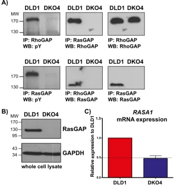
have an effect on this interaction. We found that RhoGAP could only be phosphorylated in DLD1 cells, not in DKO4, while total levels of RhoGAP remain unchanged. This phosphorylated RhoGAP co-immunoprecipitated with RasGAP (Figure 1A). In addition, we found that expression of RasGAP was not detectable in the DKO4 cell line (Figure 1A). It has been reported that RasGAP phosphorylation frequently occurs downstream of receptor tyrosine kinase signaling [18,36,37,38,39]. However, we found that the major tyrosine phosphorylated band that appears after immunoprecipitation of RasGAP was actually at,190 kDa, most likely representing RhoGAP (Figure 1A), indicating that RasGAP itself is not highly phosphorylated in these cells.
RasGAP expression is lost in DKO4 cells
We found that while the DKO4 cell line expressed little to no RasGAP protein compared to DLD1, the mRNA levels of the
RASA1gene remained at 50% of the parent cell line (Figure 1, B
& C). We performed SNP arrays to examine potential copy number alterations between these isogenic cell pairs. We found very little difference between the two cell lines (data not shown). A similar result was recently found in a series of alternate clones (DKO3 and DKO1) derived from the DLD1 model [40]. Importantly, no copy number differences were seen in chromo-some 5q13.3, showing that the decrease in mRNA level in DKO4 is not due to chromosomal loss.
RasGAP mutation in CRC
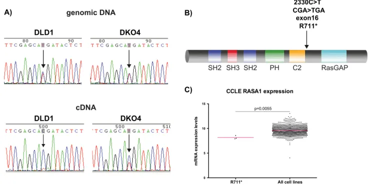
We then looked for mutations that could explain the differences in RasGAP expression level. We sequenced both the genomic DNA and the cDNA from DLD1 and DKO4 cells. We found a heterozygous point mutation in the genomic DNA of both cell lines. This C.T transition is a nonsense mutation, encoding a R709* change (Figure 2, A & B), located between the C2 and RasGAP domains of RasGAP. If the mutated gene was translated into a truncated protein, a ,77 kDa band should have been detectable in Western blots, using a RasGAP antibody that recognizes the N-terminal portion of the protein. However, we did not see a band of this size in any cell conditions.
Interestingly, cDNA sequencing in the DLD1 and DKO4 cell lines showed that the DLD1 cell line had almost completely lost expression of the mutated gene, expressing only the wild-type, while the DKO4 cell line maintained 50% of each of the wild-type and mutantRASA1gene product (Figure 2A).
We looked for the presence of the C2330T mutation in primary tumor and metastases tissues from CRC patients, but were unable to identify this mutation in any of our samples. However, we were able to find this mutation in three cell lines from the Broad Institute Cancer Cell Line Encyclopedia (http://www. broadinstitute.org/ccle) [41]: the colorectal cancer cell lines HRT18 and HCT15, as well as the urinary tract cancer cell line 639 V. This implies that although the mutation is rare, it is present in certain types of cancer. This mutation is also not found in the human SNP database, implying that it is not found in the population at large.
To further investigate our hypothesis that the mutatedRASA1
transcript is unstable and possibly degraded, we compared the mRNA expression levels of RASA1 in the three cell lines that
contain the C2330T mutation to the remainder of the cancer cell lines in the Cell Line Encyclopedia. These three cell lines express significantly lessRASA1than the majority of other cancer cell lines
in the database (Figure 2C). Although not conclusive, this appears to indicate that this truncating mutation could result in decreased mRNA gene expression.
These findings suggest several levels of regulation of RasGAP, all possibly as a result of the loss of active KRAS in DKO4. First, the complete lack of mutant p120RasGAP mRNA in the DLD1 cell line as detected by sequencing indicates that either the mutant allele is not being transcribed into mRNA in this cell line, or that the mutant mRNA is very unstable and degraded immediately upon being transcribed. Interestingly, although the mutant mRNA was present in the DKO4 cell line, the 50% decrease in overall
RASA1mRNA levels as detected by real time qPCR suggest that
this mutant mRNA is also degraded, but possibly not as quickly or as efficiently as in DLD1.
In all, it appears that the loss of active KRAS in DKO4 decreases the pressure on the cell to stabilize RasGAP expression at both the mRNA and protein level; however, the mechanism by which the protein levels of RasGAP are regulated in these cell lines in still unknown.
Figure 1. RhoGAP phosphorylation and RasGAP expression in DLD1 and DKO4 cell lines. A) RhoGAP and RasGAP were immunoprecipitated from DLD1 and DKO4 cells. Immunoprecipitates were subjected to Western blotting and probed for total phosphotyr-osine, RasGAP and RhoGAP. Western Blotting of whole cell lysates (A) and rt-QPCR (C) were used to determine total protein and mRNA levels respectively in these cell lines.
Regulation of RasGAP mRNA expression by mutant KRAS
To clarify the role that loss of mutantKRASin DKO4 plays in
the expression of RasGAP, we stably overexpressed the pBabepuro (pBp) empty vector, vector containing full length wild-typeKRAS,
or vector containing KRAS with point mutations in codon 12
(G12V or G12D) or codon 13 (G13D), in DKO4 cells. We hypothesized that the G13D mutation would have the largest effect in stabilizing and rescuing RasGAP expression in DKO4, due to this mutation being the one that originally occurred in this cell line. However, after repeated attempts, we were only able to overexpress the KRASG12V mutant gene in this cell line
(Figure 3A). Overexpression of G12V was accompanied by an overall increase in GTP-KRAS, as expected (Figure 3C). Interestingly, transient transfection of these constructs showed that KRAS was able to be overexpressed up to 7 days post transfection with all mutants, indicating that long-term expression of KRAS constructs other than G12V were not sustained in DKO4. Overexpression ofKRASG12Vcaused a significant increase
in RASA1 mRNA; however, this increase was not seen at the
protein level (Figure 3, B & C). Interestingly, although we could not detect anyKRAS overexpression with the G13D mutant, we
still noted a slight increase inRASA1mRNA expression, although
was not significant (p-value = 0.074).
To further probe the role of active KRAS in RasGAP expression, we transiently transfected DLD1 cells with 11 separate shRNAs againstKRAS. We found a significant correlation between
the amount of KRAS knockdown and the decrease in RASA1
mRNA expression compared to the non-specific shRNA control (Figure 3D). However, these changes were not observed at the protein level (Figure 3E). To ensure that KRAS shRNA knockdown did cause any significant changes in non-specific genes,Figure S1Ashows levels of two housekeeping genes that are not affected by the knockdown. Figure S1B shows the Western blots from which Figure 3E was derived.
Together, these results suggest that loss of active KRAS played a partial role in the stability and/or expression ofRASA1mRNA,
although additional mechanisms are present that regulate the protein expression in this cell line.
RasGAP overexpression rescues RasGAP activity
To determine if any of the phenotypes attributed to loss of active KRAS in the DLD1 isogenic cell lines could be explained by the loss of RasGAP protein expression, we overexpressed RasGAP in the DKO4 cell line (Figure 4A). Despite our ability to get.100 fold mRNA overexpression of RasGAP in the DKO4 cell line, the resulting protein levels were similar to endogenous expression in DLD1 cells (Figure 4D).
To determine if overexpression of RasGAP had any effect on KRAS activity, a RAS activity assay was performed, using Raf-RBD-linked agarose beads to immunoprecipitate active KRAS (Figure 4D). As has been shown previously [40], KRAS overall was less active in the DKO4 cells compared to the DLD1 cells. We saw a slight but significant decrease in KRAS activity in DKO4 cells afterRasGAPoverexpression, indicating that RasGAP is able
to regulate the wild-type KRAS in DKO4. To further clarify the role of RasGAP in these cells, we used a real-time NMR-based assay to determine RasGAP activity. We found that the levels of RasGAP activity were concordant with RasGAP protein expres-sion (Figure 4, B & C). Extracts of DLD1 cells accelerated RAS GTP hydrolysis,1.8 fold, whereas DKO4 extracts, matched for total protein content elicited a modest 1.2 fold hydrolysis rate increase. These results were consistent with the presence of basal activity from other RasGAPs. RasGAP overexpression in DKO4 raised this rate back to ,1.8 fold. This indicated that the ectopically expressed RasGAP is functional, as well as suggesting that RasGAP may be an important mediator of overall GAP activity in the DLD1 cell line
Figure 2. Identification of a novel truncating mutation in RASA1.A) Chromatogram showing relative intensities of each base pair after Sanger sequencing in both the genomic and cDNA derived from the cell lines. B) Illustration of location of the mutation at the RasGAP protein level. C) RASA1 expression data derived from all cell lines in the Broad Institute Cancer Cell Line Encyclopedia. Bars represent mean.
RasGAP overexpression rescues RhoGAP phosphorylation and Rho-mediated phenotypes
As we showed earlier, RhoGAP phosphorylation was lost in DKO4 cells. Here we see that rescue of RasGAP expression in DKO4 cells was able to restore RhoGAP phosphorylation in these cells, as well as binding of phosphorylated RhoGAP to RasGAP (Figure 4E).
RhoGAP is a key regulator of the Rho pathway, affecting phenotypes such as cell proliferation, cell adhesion to the extracellular matrix, and cell motility. These same phenotypes are differentially demonstrated in the DLD1 cell line compared to its isogenic derivatives [29]. Therefore, we were interested to know if the rescue of RhoGAP phosphorylation could also rescue these phenotypes in DKO4 cells. We found that modulating RasGAP expression did not change cell proliferation (Figure 5A). How-ever, RasGAP overexpression did rescue DKO4 cell adhesion to a collagen substrate, motility, and stress fiber formation (Figure 5 B–D). Together, these results indicate that rescue of RasGAP expression in DLD1 can also rescue phenotypes generally associated with Rho pathway activation.
RasGAP and mutant KRAS together are required for full tumorigenicity of DLD1 cells
To determine if the phenotypes rescued by RasGAP overex-pression in DKO4 cells could be recapitulatedin vivo, we injected 1
million cells subcutaneously into SCID mice and measured tumor growth. Although overexpression of RasGAP did increase tumor growth significantly compared to DKO4 cells alone (Figure 5, E
& F), it was not able to fully attain the growth rate of the DLD1 parent cell line, indicating that RasGAP alone is not sufficient to rescue tumorigenicity of cells that have lost active KRAS. The
mRNA extracted from the xenografts showed that RasGAP expression remained consistent with the cells as they were prior to injection (Figure 5G).
Discussion
In this study, we described a role for RasGAP as an important mediator of Rho signaling and tumorigenicity in a colorectal cancer cell line, and identified mutantKRASas a key contributor
to this pathway (Figure 6). While RasGAP can act as a suppressor of RAS function by enhancing GTP hydrolysis, [6] a GAP-independent effector function has also been proposed, by virtue of its multiple binding partners [42,43,44,45].
Although we were able to transiently overexpress both wild-type and various activated KRAS mutants in DKO4 cells, only the
G12V mutation could be stably expressed. It was previously reported that Kelleret al.failed to overexpress KRAS G12V in
DKS-8, another clone of DLD1 with knockout of the KRAS mutant allele, due in part to proteasomal degradation of the mutant [46]. However, HRAS or NRAS bearing this mutation could be expressed in this cell line, suggesting that the removal of a powerful oncogene may have a myriad of effects of a cell line, causing irreversible changes that cannot be rescued by simple re-introduction [46,47,48]. In our hands, attempts to stably express wild-type or any codon 12 or 13 KRAS mutant (other than Figure 3. RasGAP expression is mediated in part by KRAS.Wild-type (WT) or mutant KRAS was overexpressed in DKO4 cells. mRNA was extracted from cells and quantified using rt-qPCR to measure KRAS (A) or RASA1 (B). C) Western blotting showing levels of these proteins, along with activation status of KRAS. Correlation of mRNA (D) and protein expression using densitometry analysis of Western blotting (E) of KRAS and RASA1 after knockdown of KRAS using 11 different shRNAs. For protein correlation, outliers over 3 standard deviations from the mean were excluded. All quantification is relative to empty vector. Statistical analysis of expression using unpaired t-test, ***p,0.001, *p,0.05.
KRASG12V) failed in DKO4 cells, while transient transduction of
these constructs resulted in high expression, which is consistent with previous results using DKO1 cells (another clone of DKO4) [49]. Recent work has shown that different KRAS mutants are
associated with significantly different clinical outcomes, and yet the biological basis of these differences is just beginning to be explored [50,51,52]. For instance,KRASG12Vconfers greater resistance to
EGFR inhibitors in colorectal cancer, while KRASG13D is
associated with worse overall survival of colorectal cancer patients treated with standard chemotherapy [53].
In addition, biochemical differences may contribute to the differential expression ofKRASmutations in the DKO4 cell line.
Using NMR to probe the GTPase activity of oncogenic RAS proteins, Smith et al. [52] showed that the G12V mutant exhibited similar intrinsic nucleotide exchange to WT, but was completely resistant to GAP-mediated GTP hydrolysis. On the other hand, intrinsic exchange of the G13D mutant was 15-fold faster than WT - this mutation retained some sensitivity to GAP-mediated hydrolysis. These results are the first steps to understanding both the pathways underlying the biology of the different RAS mutations, and the clinical differences between them.
The question of how KRAS can modulate expression ofRASA1
is an important one (Figure 4). Tools transcription factor prediction tool PSCAN [54] suggested that the transcription factor SPI1 may activateRASA1gene transcription (p = 0.02). SPI1
transcription is activated downstream of RAS-dependent AKT activation [55], and may be one mechanism by which KRAS can stabilizeRASA1expression.
The phosphorylated tyrosines on RhoGAP responsible for RasGAP binding have not been definitively identified. One report
found that phosphorylation of both tyrosines on RhoGAP, Y1087 and Y1105 [56], are responsible for binding to the tandem SH2 domains of RasGAP, while another suggested that just one site (Y1105) is sufficient [57]. However, it is generally agreed that Y1105 is the major site of tyrosine phosphorylation on RhoGAP, and the major determinant of RasGAP binding [57]. Our phospho-proteomics screen showed that Y1105 responded more strongly than Y1087 to HGF stimulation in the DLD1 cell line, although both were basally phosphorylated after serum starvation [28]. Western blots for total phosphotyrosine in RhoGAP immunoprecipitated from DLD1 detected a strong band during normal growth conditions, which did not change appreciably after HGF stimulation. Similar results were seen in mouse embryonic fibroblasts (MEF) derived fromRASA1knockout mice, in which no
phosphorylation of RhoGAP was observed in vitro, even after
stimulation with PDGF [58]. Constitutive phosphorylation of other tyrosines may obscure changes in pY1105 by Western blot, which uses an antibody against total phospho-tyrosine. We are also not able to detect any increase in RhoGAP phosphorylation in DKO4 after HGF stimulation, suggesting that in this cell line, RasGAP is required for RhoGAP phosphorylation, both basally and in response to growth factor stimulation.
Phosphorylation of RhoGAP, and its binding to RasGAP has been shown to have conflicting roles in Rho signaling and cell migration, which likely reflect the localization of these proteins to different areas of spreading or migrating cells. In newly-adhered cells, integrin engagement leads to Src-dependent phosphorylation of RhoGAP [59] and transiently inactivation of Rho to allow Rac/ Cdc42-mediated membrane protrusion at the leading edge. Later stages of migration and/or cell adhesion involve the maturation of Figure 4. Overexpression of RasGAP expression in DLD1 cells rescues RhoGAP phosphorylation and overall GAP activity.A) mRNA expression of RasGAP after overexpression in DKO4 cells compared to the GFP vector control. B) Real-time NMR analysis of RasGAP activity, showing mean rate of GTP hydrolysis (B) and GAP activity over time (C). Each curve in (C) is derived from a single representative experiment. Error bars in (B) denote standard error of the mean (SEM). D) RAS activity assay showing levels of active KRAS after RasGAP overexpression. Numbers denote densitometry values from this blot, which is representative of three biological replicates. E) RhoGAP was immunoprecipitated from cell lines, subjected to Western blotting, then probed for total phosphotyrosine, RasGAP and RhoGAP.
focal adhesions and the formation of stress fibers, which are regulated by the restoration of Rho-GTP at the cell periphery of stably adherent cells, or at the leading edge of migrating cells [59]. Early studies suggested that RhoGAP phosphorylation leads to its sequestration away from Rho, allowing Rho activation and cell adhesion [25,60], which may be a mechanism by which RasGAP mediates Rho signaling in DLD1 cells. To clarify this question, we assayed Rho in these cell lines, but could not detect any differences between DLD1 and DKO4 parental lines, nor between DKO4 cells with or without overexpressed RasGAP. In addition to having high basal levels of RhoGAP phosphorylation, the DLD1 cell line also exhibits high basal Rho activity [61], which could limit the sensitivity of the assay to detect changes. It is important to note that this method assays levels of total activated RhoA in the cell, but may be insensitive to changes in the activity or localization of a single RhoGAP against the background of many cellular
RhoGAPs. Nevertheless, these events produce spatiotemporally controlled bursts of GAP activity that functionally regulate discrete sub-populations of RhoA.
The correct localization of the RhoGAP/RasGAP complex has been shown to be crucial for the proper polarization of migrating cells. MEF
RASA1-/-cells showed major defects in wound healingin vitro, which could be partially rescued by expression of a RasGAP
variant lacking the GAP domain [62]. These cells did not migrate as efficiently as wild-type MEFs, but were able to move in a fully coordinated manner, indicating that the initial polarization of cell motility requires RasGAP but not RAS, and this polarization was also dependent on p190 binding to RasGAP [62]. MEFs from RhoGAP knockout cells also showed a defect in directional cell migration [63]. In this study, we see a decrease in cell migration and cell adhesion in DKO4 cells compared with DLD1, and a concomitant increase in these phenotypes when RasGAP is re-Figure 5. RasGAP overexpression modulates cell adhesion, cell motility, stress fiber formation and tumorigenicity.A) Cell counting assay in RasGAP overexpressing and knockdown cells. B) Cell adhesion to collagen. Statistical significance was determined by t-test: *p,0.05, ***p,0.001 C) Wound healing assay. D) Rhodamine-phalloidin staining of actin filaments after overnight adhesion to collagen. E) Tumor volume and (F) final excised tumor weight of xenograft tumors in SCID mice derived from subcutaneous injection of DLD1 empty vector (GFP), DKO4 GFP, or DKO4 overexpressing RasGAP. Number of mice used is indicated on graph. p-value calculated as indicated in materials and methods section. G) rt-qPCR analysis of RASA1 gene expression derived from xenograft tumors after excision.
expressed. This is consistent with a previous study that further showed that siRNA knockdown of RhoA decreased in cell migration in DLD1 but not DKO4 cells [61], further demon-strating the requirement for RasGAP, as well as active KRAS, for Rho signaling in these cells.
To explain the lack of RasGAP expression in DKO4 cells, we identified a nonsense mutation in the RASA1 gene that likely results in the decay of the messenger RNA. This specific mutation has been identified in two colon cancer cell lines, as well as urinary tract cancer cells. Interestingly, all three of these cell lines contain an activating mutation in KRAS. It is not surprising that we did not find this mutation in any of our tissue samples; for a rare mutation, our sample size was likely too small. It could be that that this mutation, although rare, is an important factor in the
destabilization ofRASA1mRNA expression in tumors. However,
mutation of a neighboring arginine to a stop codon (R709*) was also recently identified in a lung carcinoma sample (COSMIC mutation 738997, obtained from the Sanger Institute Catalogue Of Somatic Mutations In Cancer web site, http://www.sanger.ac. uk/cosmic)[64] and was previously identified in two families presenting with capillary venous malformation syndrome [14]. Further analysis of these truncating mutations, and their roles in cancer and other developmental diseases, will further elaborate on the role of RasGAP in cancer.
In conclusion, this study has provided new insights into the complexity of RasGAP and KRAS signaling, and reveals a novel role for RasGAP as an effector of KRAS and Rho pathway activity in colorectal cancer. Our study also identified a novel Figure 6. Summary of findings and proposed mechanism.In DLD1 cells, active KRAS stabilizes RasGAP expression, which in turn binds to and stabilizes RhoGAP phosphorylation. This complex then activates Rho pathway activation, either by sequestration of RhoGAP away from Rho, or by increasing Rho turnover. In DKO4 cells, RasGAP is not expressed, due in part to a truncating mutation and in part to lack of expression downstream of active KRAS. In this situation, RhoGAP is not phosphorylated, and so Rho pathway phenotypes are inactive. When RasGAP is overexpressed in DKO4, RhoGAP is once again phosphorylated and Rho pathway is active- however, lack of stabilization and/or contributing signaling pathways from active RAS means that tumorigenicity does not attain the same level as DLD1. The bottom of the figure summarizes the main characteristics of RhoGAP and RasGAP in these cell lines.
genomic aberration with potentially significant effects on signaling studies involving the commonly used colorectal cancer cell line DLD1 and its derivatives.
Supporting Information
Figure S1 Controls for shRNA-mediated KRAS knock-down in DLD1 cells.A) rt-qPCR results showing no significant expression changes in two housekeeping genes after transient KRAS knockdown. Genes are those used as loading controls in
Figure 3D. B) Western blot of Figure 3E, showing protein levels after KRAS knockdown.
(EPS)
Author Contributions
Conceived and designed the experiments: SLO JH NR CM MI. Performed the experiments: SLO JH NR LL CM. Analyzed the data: SLO LL CQZ CM. Contributed reagents/materials/analysis tools: SLO JH NR CM RN MI. Wrote the paper: SLO CM MI MST. Provided DLD1 and DKO4 cell lines: TS SS.
References
1. Siegel R, Naishadham D, Jemal A (2013) Cancer statistics, 2013. CA: A Cancer Journal for Clinicians 63: 11–30.
2. Sinclair P, Singh A, Riaz AA, Amin A (2012) An unsolved conundrum: the ideal follow-up strategy after curative surgery for colorectal cancer. Gastrointest Endosc 75: 1072–1079.
3. Konopke R, Roth J, Volk A, Pistorius S, Folprecht G, et al. (2012) Colorectal liver metastases: an update on palliative treatment options. J Gastrointestin Liver Dis 21: 83–91.
4. Vogelstein B, Fearon ER, Hamilton SR, Kern SE, Preisinger AC, et al. (1988) Genetic alterations during colorectal-tumor development. N Engl J Med 319: 525–532.
5. Allegra CJ, Jessup JM, Somerfield MR, Hamilton SR, Hammond EH, et al. (2009) American Society of Clinical Oncology provisional clinical opinion: testing for KRAS gene mutations in patients with metastatic colorectal carcinoma to predict response to anti-epidermal growth factor receptor monoclonal antibody therapy. J Clin Oncol 27: 2091–2096.
6. Siderovski DP, Willard FS (2005) The GAPs, GEFs, and GDIs of heterotrimeric G-protein alpha subunits. Int J Biol Sci 1: 51–66.
7. Trahey M, Wong G, Halenbeck R, Rubinfeld B, Martin GA, et al. (1988) Molecular cloning of two types of GAP complementary DNA from human placenta. Science 242: 1697–1700.
8. Vogel US, Dixon RA, Schaber MD, Diehl RE, Marshall MS, et al. (1988) Cloning of bovine GAP and its interaction with oncogenic ras p21. Nature 335: 90–93.
9. Henkemeyer M, Rossi DJ, Holmyard DP, Puri MC, Mbamalu G, et al. (1995) Vascular system defects and neuronal apoptosis in mice lacking ras GTPase-activating protein. Nature 377: 695–701.
10. Kunath T, Gish G, Lickert H, Jones N, Pawson T, et al. (2003) Transgenic RNA interference in ES cell-derived embryos recapitulates a genetic null phenotype. Nature Biotechnology 21: 559–561.
11. Eerola I, Boon LM, Mulliken JB, Burrows PE, Dompmartin A, et al. (2003) Capillary malformation-arteriovenous malformation, a new clinical and genetic disorder caused by RASA1 mutations. Am J Hum Genet 73: 1240–1249. 12. Boon LM, Mulliken JB, Vikkula M (2005) RASA1: variable phenotype with
capillary and arteriovenous malformations. Current Opinion in Genetics and Development 15: 265–269.
13. Hershkovitz D, Bergman R, Sprecher E (2008) A novel mutation in RASA1 causes capillary malformation and limb enlargement. Archives of Dermatolog-ical Research 300: 385–388.
14. Revencu N, Boon LM, Mulliken JB, Enjolras O, Cordisco MR, et al. (2008) Parkes Weber syndrome, vein of Galen aneurysmal malformation, and other fast-flow vascular anomalies are caused by RASA1 mutations. Human Mutation 29: 959–965.
15. de Wijn RS, Oduber CE, Breugem CC, Alders M, Hennekam RC, et al. (2012) Phenotypic variability in a family with capillary malformations caused by a mutation in the RASA1 gene. Eur J Med Genet 55: 191–195.
16. Kadam SD, Gucek M, Cole RN, Watkins PA, Comi AM (2012) Cell proliferation and oxidative stress pathways are modified in fibroblasts from Sturge-Weber syndrome patients. Archives of Dermatological Research 304: 229–235.
17. Skorski T, Kanakaraj P, Ku DH, Nieborowska-Skorska M, Canaani E, et al. (1994) Negative regulation of p120GAP GTPase promoting activity by p210bcr/ abl: implication for RAS-dependent Philadelphia chromosome positive cell growth. Journal of Experimental Medicine 179: 1855–1865.
18. Hecker TP, Ding Q, Rege TA, Hanks SK, Gladson CL (2004) Overexpression of FAK promotes Ras activity through the formation of a FAK/p120RasGAP complex in malignant astrocytoma cells. Oncogene 23: 3962–3971. 19. Stahle-Backdhal M, Inoue M, Zedenius J, Sandstedt B, DeMarco L, et al. (1995)
Decreased expression of Ras GTPase activating protein in human trophoblastic tumors. American Journal of Pathology 146: 1073–1078.
20. Davidson B, Agulansky L, Goldberg I, Friedman E, Ramon J, et al. (1998) Immunohistochemical analysis of rasGTPase activating protein (rasGAP) in prostate cancer. Pathology, Research and Practice 194: 399–404.
21. Calvisi DF, Ladu S, Conner EA, Seo D, Hsieh JT, et al. (2011) Inactivation of Ras GTPase-activating proteins promotes unrestrained activity of wild-type Ras in human liver cancer. Journal of Hepatology 54: 311–319.
22. Barshack I, Goldberg I, Davidson B, Ravid A, Schiby G, et al. (1998) Expression of rasGTPase activating protein in basal cell carcinoma of the skin. Modern Pathology 11: 271–275.
23. Mitsudomi T, Friedman E, Gejman PV, McCormick F, Gazdar AF (1994) Genetic analysis of the catalytic domain of the GAP gene in human lung cancer cell lines. Human Genetics 93: 27–31.
24. Moran MF, Koch CA, Anderson D, Ellis C, England L, et al. (1990) Src homology region 2 domains direct protein-protein interactions in signal transduction. Proceedings of the National Academy of Sciences of the United States of America 87: 8622–8626.
25. Sharma SV (1998) Rapid recruitment of p120RasGAP and its associated protein, p190RhoGAP, to the cytoskeleton during integrin mediated cell-substrate interaction. Oncogene 17: 271–281.
26. Roof RW, Haskell MD, Dukes BD, Sherman N, Kinter M, et al. (1998) Phosphotyrosine (p-Tyr)-dependent and -independent mechanisms of p190 RhoGAP-p120 RasGAP interaction: Tyr 1105 of p190, a substrate for c-Src, is the sole p-Tyr mediator of complex formation. Mol Cell Biol 18: 7052–7063. 27. Chang JH, Gill S, Settleman J, Parsons SJ (1995) c-Src regulates the
simultaneous rearrangement of actin cytoskeleton, p190RhoGAP, and p120Ras-GAP following epidermal growth factor stimulation. Journal of Cell Biology 130: 355–368.
28. Organ SL, Tong J, Taylor P, St-Germain JR, Navab R, et al. (2011) Quantitative Phospho-Proteomic Profiling of Hepatocyte Growth Factor (HGF)-MET Signaling in Colorectal Cancer. Journal of Proteome Research 10: 3200– 3211.
29. Shirasawa S, Furuse M, Yokoyama N, Sasazuki T (1993) Altered growth of human colon cancer cell lines disrupted at activated Ki-ras. Science 260: 85–88. 30. Radulovich N, Qian JY, Tsao MS (2008) Human pancreatic duct epithelial cell
model for KRAS transformation. Methods in Enzymology 439: 1–13. 31. Hai J, Zhu CQ, Bandarchi B, Wang YH, Navab R, et al. (2012) L1 cell adhesion
molecule promotes tumorigenicity and metastatic potential in non-small cell lung cancer. Clinical Cancer Research 18: 1914–1924.
32. Marshall CB, Ho J, Buerger C, Plevin MJ, Li GY, et al. (2009) Characterization of the intrinsic and TSC2-GAP-regulated GTPase activity of Rheb by real-time NMR. Sci Signal 2: ra3.
33. Scheidig AJ, Franken SM, Corrie JE, Reid GP, Wittinghofer A, et al. (1995) X-ray crystal structure analysis of the catalytic domain of the oncogene product p21H-ras complexed with caged GTP and mant dGppNHp. Journal of Molecular Biology 253: 132–150.
34. John J, Sohmen R, Feuerstein J, Linke R, Wittinghofer A, et al. (1990) Kinetics of interaction of nucleotides with nucleotide-free H-ras p21. Biochemistry 29: 6058–6065.
35. Marshall CB, Meiri D, Smith MJ, Mazhab-Jafari MT, Gasmi-Seabrook GM, et al. (2012) Probing the GTPase cycle with real-time NMR: GAP and GEF activities in cell extracts. Methods 57: 473–485.
36. Cailliau K, Browaeys-Poly E, Vilain JP (2001) RasGAP is involved in signal transduction triggered by FGF1 in Xenopus oocytes expressing FGFR1. FEBS Letters 496: 161–165.
37. Druker B, Okuda K, Matulonis U, Salgia R, Roberts T, et al. (1992) Tyrosine phosphorylation of rasGAP and associated proteins in chronic myelogenous leukemia cell lines. Blood 79: 2215–2220.
38. Yue Y, Lypowy J, Hedhli N, Abdellatif M (2004) Ras GTPase-activating protein binds to Akt and is required for its activation. Journal of Biological Chemistry 279: 12883–12889.
39. Moran MF, Polakis P, McCormick F, Pawson T, Ellis C (1991) Protein-tyrosine kinases regulate the phosphorylation, protein interactions, subcellular distribu-tion, and activity of p21ras GTPase-activating protein. Molecular and Cellular Biology 11: 1804–1812.
40. Vartanian S, Bentley C, Brauer MJ, Li L, Shirasawa S, et al. (2013) Identification of Mutant K-Ras-dependent Phenotypes Using a Panel of Isogenic Cell Lines. Journal of Biological Chemistry 288: 2403–2413. 41. Barretina J, Caponigro G, Stransky N, Venkatesan K, Margolin AA, et al. (2012)
The Cancer Cell Line Encyclopedia enables predictive modelling of anticancer drug sensitivity. Nature 483: 603–607.
43. Settleman J, Narasimhan V, Foster LC, Weinberg RA (1992) Molecular cloning of cDNAs encoding the GAP-associated protein p190: implications for a signaling pathway from ras to the nucleus. Cell 69: 539–549.
44. Tocque B, Delumeau I, Parker F, Maurier F, Multon MC, et al. (1997) Ras-GTPase activating protein (GAP): a putative effector for Ras. Cellular Signalling 9: 153–158.
45. Koehler JA, Moran MF (2001) Regulation of extracellular signal-regulated kinase activity by p120 RasGAP does not involve its pleckstrin homology or calcium-dependent lipid binding domains but does require these domains to regulate cell proliferation. Cell Growth and Differentiation 12: 551–561. 46. Keller JW, Franklin JL, Graves-Deal R, Friedman DB, Whitwell CW, et al.
(2007) Oncogenic KRAS provides a uniquely powerful and variable oncogenic contribution among RAS family members in the colonic epithelium. Journal of Cellular Physiology 210: 740–749.
47. Jain M, Arvanitis C, Chu K, Dewey W, Leonhardt E, et al. (2002) Sustained loss of a neoplastic phenotype by brief inactivation of MYC. Science 297: 102–104. 48. Habets GG, Knepper M, Sumortin J, Choi YJ, Sasazuki T, et al. (2001) cDNA array analyses of K-ras-induced gene transcription. Methods in Enzymology 332: 245–260.
49. Plattner R, Gupta S, Khosravi-Far R, Sato KY, Perucho M, et al. (1999) Differential contribution of the ERK and JNK mitogen-activated protein kinase cascades to Ras transformation of HT1080 fibrosarcoma and DLD-1 colon carcinoma cells. Oncogene 18: 1807–1817.
50. Andreyev HJ, Norman AR, Cunningham D, Oates J, Dix BR, et al. (2001) Kirsten ras mutations in patients with colorectal cancer: the ’RASCAL II’ study. British Journal of Cancer 85: 692–696.
51. Andreyev HJ, Norman AR, Cunningham D, Oates JR, Clarke PA (1998) Kirsten ras mutations in patients with colorectal cancer: the multicenter ‘‘RASCAL’’ study. Journal of the National Cancer Institute 90: 675–684. 52. Smith MJ, Neel BG, Ikura M (2013) NMR-based functional profiling of
RASopathies and oncogenic RAS mutations. Proc Natl Acad Sci U S A 110: 4574–4579.
53. Tejpar S, Celik I, Schlichting M, Sartorius U, Bokemeyer C, et al. (2012) Association of KRAS G13D Tumor Mutations With Outcome in Patients With Metastatic Colorectal Cancer Treated With First-Line Chemotherapy With or Without Cetuximab. J Clin Oncol 30: 3570–3577.
54. Zambelli F, Pesole G, Pavesi G (2009) Pscan: finding over-represented transcription factor binding site motifs in sequences from regulated or co-expressed genes. Nucleic Acids Res 37: W247–252.
55. Rieske P, Pongubala JM (2001) AKT induces transcriptional activity of PU.1 through phosphorylation-mediated modifications within its transactivation domain. Journal of Biological Chemistry 276: 8460–8468.
56. Hu KQ, Settleman J (1997) Tandem SH2 binding sites mediate the RasGAP-RhoGAP interaction: a conformational mechanism for SH3 domain regulation. EMBO Journal 16: 473–483.
57. Roof RW, Haskell MD, Dukes BD, Sherman N, Kinter M, et al. (1998) Phosphotyrosine (p-Tyr)-dependent and -independent mechanisms of p190 RhoGAP-p120 RasGAP interaction: Tyr 1105 of p190, a substrate for c-Src, is the sole p-Tyr mediator of complex formation. Molecular and Cellular Biology 18: 7052–7063.
58. van der Geer P, Henkemeyer M, Jacks T, Pawson T (1997) Aberrant Ras regulation and reduced p190 tyrosine phosphorylation in cells lacking p120-Gap. Molecular and Cellular Biology 17: 1840–1847.
59. Arthur WT, Burridge K (2001) RhoA inactivation by p190RhoGAP regulates cell spreading and migration by promoting membrane protrusion and polarity. Molecular Biology of the Cell 12: 2711–2720.
60. Chen JC, Zhuang S, Nguyen TH, Boss GR, Pilz RB (2003) Oncogenic Ras leads to Rho activation by activating the mitogen-activated protein kinase pathway and decreasing Rho-GTPase-activating protein activity. Journal of Biological Chemistry 278: 2807–2818.
61. Makrodouli E, Oikonomou E, Koc M, Andera L, Sasazuki T, et al. (2011) BRAF and RAS oncogenes regulate Rho GTPase pathways to mediate migration and invasion properties in human colon cancer cells: a comparative study. Mol Cancer 10: 118.
62. Kulkarni SV, Gish G, van der Geer P, Henkemeyer M, Pawson T (2000) Role of p120 Ras-GAP in directed cell movement. Journal of Cell Biology 149: 457–470. 63. Jiang W, Betson M, Mulloy R, Foster R, Levay M, et al. (2008) p190A RhoGAP is a glycogen synthase kinase-3-beta substrate required for polarized cell migration. Journal of Biological Chemistry 283: 20978–20988.