enterocolitica
O:3, the Most Frequent Cause of Human
Yersiniosis
Frank Uliczka1,2, Fabio Pisano1, Julia Schaake1,2, Tatjana Stolz1,2, Manfred Rohde3, Angelika Fruth4, Eckhard Strauch5, Mikael Skurnik6, Julia Batzilla7, Alexander Rakin7, Ju¨rgen Heesemann7, Petra Dersch1,2*
1Department of Molecular Infection Biology, Helmholtz-Zentrum fu¨r Infektionsforschung, Braunschweig, Germany,2Institut fu¨r Mikrobiologie, Technische Universita¨t Braunschweig, Braunschweig, Germany,3Department of Medical Microbiology, Helmholtz-Zentrum fu¨r Infektionsforschung, Braunschweig, Germany,4Robert Koch-Institut, Wernigerode, Germany,5Bundesinstitut fu¨r Risikoforschung, Berlin, Germany,6Department of Bacteriology and Immunology, The Haartman Institute, University of Helsinki and Helsinki University Central Hospital Laboratory Diagnostics, Helsinki, Finland,7Max von Pettenkofer Institut, Ludwigs-Maximilians-Universita¨t, Mu¨nchen, Germany
Abstract
Many enteric pathogens are equipped with multiple cell adhesion factors which are important for host tissue colonization and virulence.Y. enterocolitica, a common food-borne pathogen with invasive properties, uses the surface proteins invasin and YadA for host cell binding and entry. In this study, we demonstrate unique cell adhesion and invasion properties ofY. enterocoliticaserotype O:3 strains, the most frequent cause of human yersiniosis, and show that these differences are mainly attributable to variations affecting the function and expression of invasin in response to temperature. In contrast to other enteric Yersinia strains, invasin production in O:3 strains is constitutive and largely enhanced compared to other Y. enterocoliticaserotypes, in whichinvAexpression is temperature-regulated and significantly reduced at 37uC. Increase of invasin levels is caused by (i) an IS1667 insertion into theinvApromoter region, which includes an additional promoter and RovA and H-NS binding sites, and (ii) a P98S substitution in theinvAactivator protein RovA rendering the regulator less susceptible to proteolysis. Both variations were shown to influence bacterial colonization in a murine infection model. Furthermore, we found that co-expression of YadA and down-regulation of the O-antigen at 37uC is required to allow efficient internalization by the InvA protein. We conclude that even small variations in the expression of virulence factors can provoke a major difference in the virulence properties of closely related pathogens which may confer better survival or a higher pathogenic potential in a certain host or host environment.
Citation:Uliczka F, Pisano F, Schaake J, Stolz T, Rohde M, et al. (2011) Unique Cell Adhesion and Invasion Properties ofYersinia enterocoliticaO:3, the Most Frequent Cause of Human Yersiniosis. PLoS Pathog 7(7): e1002117. doi:10.1371/journal.ppat.1002117
Editor:Jorge E. Gala´n, Yale University School of Medicine, United States of America
ReceivedDecember 17, 2010;AcceptedApril 27, 2011;PublishedJuly 7, 2011
Copyright:ß2011 Uliczka et al. This is an open-access article distributed under the terms of the Creative Commons Attribution License, which permits unrestricted use, distribution, and reproduction in any medium, provided the original author and source are credited.
Funding:This work was supported by the BMBF (Consortium FBI-Zoo) and the Deutsche Forschungsgemeinschaft (SFB621, Project B10), as well as the Fonds der Chemischen Industrie. The funders had no role in study design, data collection and analysis, decision to publish, or preparation of the manuscript.
Competing Interests:The authors have declared that no competing interests exist.
* E-mail: petra.dersch@helmholtz-hzi.de
Introduction
Yersinia enterocoliticais a common gram-negative zoonotic pathogen that is able to grow in the environment and cause enteric diseases (Yersiniosis), ranging from enteritis, severe diarrhea, mesenteric lymphadenitis, hepatic or splenic abscesses to postinfectious extrain-testinal sequelae such as reactive arthritis and erythema nodosum [1]. Infection by Y. enterocolitica is usually initiated through uptake of contaminated food or water. Following ingestion, the bacteria first colonize the lumen and transmigrate through antigen-sampling M cells across the epithelial lining of the small intestine, resulting in the colonization of the underlying lymphoid tissues (Peyer’s patches) [2,3]. Subsequently,Y. enterocoliticacan spread via the lymph and/or blood into the mesenteric lymph nodes or to extraintestinal sites such as liver and spleen [4,5,6]. Alternatively, the bacteria may bypass colonization of the Peyer’s patches and spread directly from the intestine to the systemic tissues, similar to what has been observed for enteropathogenicYersinia pseudotuberculosis[7,8].
Adhesion, invasion and survival in deeper tissues depend on several Yersinia virulence factors encoded on the Yersinia
DNA-binding activity and its susceptibility to the proteolytic degradation by ATP-dependent proteases [18].
After the initiation of the infection, the YadA and Ail proteins seem to be the predominant adhesins in infected tissues. Both virulence factors mediate serum resistance and promote tight adherence to extracellular matrix proteins, such as fibronectin and/or collagen, but their contribution to bacterial uptake is relatively small [19,20,21,22,23,24,25]. TheyadAgene is located on pYV and its expression, together with the plasmid-encoded type III secretion system (Ysc proteins) and the antiphagocytic effector proteins (Yops) is controlled by the VirF(LcrF) activator. VirF-dependent induction of yadA, yop and ysc expression occurs exclusively at 37uC [26,27]. Ail is also predominantly expressed at 37uC, and regulated by pH and oxygen content, but the control mechanisms are still unclear [24].
Besides the classical pathogenicity factors, other surface factors also contribute or are required for full virulence. Lipopolysaccha-rides (LPS) ofY. enterocolitica serotypes O:3 and O:8 are required for successful colonization of the gut and play an important role in the outer membrane integrity of the bacteria [28,29,30]. LPS O polysaccharide (O-antigen) mutants were attenuated in virulence and impaired in their ability to colonize the Peyer’s patches, liver and spleen [30,31]. Production of the O-antigen is also temperature-regulated with maximal expression at moderate temperatures [32,33]. A complex network regulates O-antigen expression at the transcriptional level and the RosA/RosB efflux pump/potassium antiporter system and Wzz, the O-antigen chain length determinant, are indirectly involved in the temperature-dependent control process [33]. In addition, flagella-temperature-dependent motility is required to initiate host cell invasion by ensuring migration and cell contact of the bacteria [34].
Most studies on Y. enterocolitica virulence factors and their contribution to virulence were performed using highly mouse-virulent bioserogroup 1B/O:8 strains, in particularY. enterocolitica
8081v. However, several other human pathogenic Y. enterocolitica
strains which are less virulent in mice (e.g. serotypes O:3, O:9 and O:5,27) were also frequently isolated from patients [1]. Among these strains,Y. enterocolitica bioserotype 4/O:3 is by far the most frequent cause of human yersiniosis in Europe and Japan (80– 90%).Y. enterocoliticainfections are less common in North America.
However, since the 1980s, serogroup O:3 strains have emerged as an occasional cause of foodborne outbreaks and replaced O:8 as the predominant serotype of Y. enterocolitica reported to CDC [35,36,37,38]. They mainly originate from domestic pigs (preva-lence of 0–65% in fattening pig herds), which are often asymptotic carriers, and in which they commonly colonize the lymphoid tissue of the gut and oropharynx [39,40]. As only very little is known about the pathogenicity ofY. enterocoliticabioserotype 4/O:3, we compared host cell interactions of different human-, pig- and food-derived Y. enterocolitica isolates and found that expression and function of surface-exposed virulence factors of serotype O:3 strains differ significantly from otherY. enterocoliticaserotypes. This may reflect an adaptation ofY. enterocoliticaO:3 to the intestine of pigs which make them also highly pathogenic for humans.
Results
Y. enterocoliticaO:3 interaction with epithelial cells differs significantly from otherY. enterocoliticaserotypes
In order to obtain information about interactions of Y. enterocoliticaserotype O:3 (YeO:3) strains with host cells, we first investigated the adhesion and invasion efficiency of two reference strains Y11 and YeO3 and 25 different YeO:3 strains isolated from human patients, animals or food between 2005 and 2008 in Germany (TableS1). None of the serotype O:3 isolates was able to efficiently bind and invade into cultured human epithelial cells when the bacteria were grown at standard culture conditions and similar patterns of host-cell associated bacteria (adhesion and invasion) were obtained when infection was performed at 22–25uC or 37uC (Fig. 1, S1, data not shown). A prolongation of the infection time from 30 min to 3 hours and/or use of other human, porcine and murine epithelial cell lines did not significantly enhance the efficiency of cell adherence (data not shown), indicating that low-efficiency of adhesion and invasion is independent of the cell line and host species. In contrast, all other tested Y. enterocolitica isolates (serotypes O:5,27, O:8 and O:9) adhered very efficiently and were able to enter all tested epithelial cell lines after 30 min with a frequency ranging from 20–30% depending on the serotype and the isolate (Fig.1, data not shown).
Amotility ofY. enterocoliticaO:3 affects cell invasion efficiency
It is known that motility is an important factor enhancing the invasion efficiency of yersiniae [34]. We tested motility of the YeO:3 strains and found that none of the isolates was motile on swimming and swarming agar plates in contrast to other Y. enterocolitica serotypes, e.g.Y. enterocolitica YeO:8 8081v (Fig. 2A, data not shown). Transmission electron microscopy further revealed that YeO:3 strains are not flagellated (Fig.2B, data not shown), indicating that flagella synthesis is abolished or does not occur under used growth conditions (LB, 25uC). This phenotype was also observed with YeO:3 strains isolated from liver and spleen of BALB/c mice three days post infection (data not shown). Notably, 30–40% of the bacteria isolated from the intestine were flagellated (Fig. S2) and motile after in vitro cultivation for 24 h (data not shown). However, none of them remained motile and flagellated after 48 h, indicating that the bacteria are motile within the intestinal tract and rapidly repress flagella synthesis when grown on agar plates.
It has been assumed that motility enhances the frequency of bacteria-cell interaction and/or provides an additional force for active cell entry. To investigate whether non-invasiveness of the serotype O:3 strains was caused by a reduction of host cell contacts due to amotility, we performed adhesion and invasion assays with
Author Summary
or without centrifugation of the bacteria onto host cells (Fig.3A). When we pre-grew the bacteria at 25uC, adhesion and internalization was slightly increased after centrifugation, but the overall efficiency was still significantly lower compared to YeO:8 8081v. This demonstrated that amotility of the bacteria reduced host cell contact and invasion of YeO:3 grown at 25uC. However, this does not fully explain the observed differences. In this context, we also analyzed host cell adhesion and invasion of bacteria grown at 37uC (Fig.3B). Without centrifugation, the number of adherent YeO:8 8081v was significantly reduced and no invasion of the bacteria was detectable at 37uC. In contrast, YeO:8 8081v adhered tightly to HEp-2 cells after bacteria were artificially brought into cell contact by centrifugation, but they were not internalized (Fig. 3B). This is consistent with previous studies showing that synthesis of the flagella and the primary internali-zation factor invasin is repressed at 37uC inY. enterocolitica8081v, whereas production of the major adhesion factor YadA is induced at 37uC but not at moderate growth temperatures [15,41]. As shown in Fig.3B, pre-growth at 37uC and artificially induced host cell contact led to a significant raise of cell adhesion of all tested O:3 strains. Notably, only under these conditions efficient host cell invasion of YeO:3 strains was as efficient as cell uptake obtained with YeO:8 8081v grown at 25uC (20–30% of adherent bacteria). Since efficient cell adhesion and internalization of YeO:3 strains was only achieved after artificial host cell contact, in all following experiments bacteria were centrifugated onto host cells.
Internalization ofY. enterocoliticaO:3 into human epithelial cells
Based on the previous experiments it seemed possible that an additional thermo-regulated internalization mechanism is responsible for host cell invasion at 37uC. To compare the invasion mechanism used by YeO:3 and YeO:8 strains, we monitored cell entry of YeO:8 8081v grown at 25uC and YeO:3 Y1 grown at 37uC into HEp-2 (Fig.4A) and Caco-2 (data not shown) cells by scanning electron microscopy. We found that adherence and invasion of both Y. enterocoliticaserotypes showed very common features and were not cell type specific. After host cell binding, the cell surface in the vicinity of the microbes seems to be slightly drawn down, pseudopodia and lamellipodia are formed and the eukaryotic cell membrane then seems to enclose and surround the bacteria into a membrane-bound vacuole. In contrast, no cell adherence of YeO:3 strain Y1 was observed when grown at 25uC, and only simple attachment, but no formation of membrane protrusions was detectable when YeO:8 8081v was precultivated at 37uC (data not shown). This suggested that the internalization mechanism initiated byY. enterocoliticaserotype O:3 and O:8 strains is similar but expressed at different temperatures.
Expression analysis ofY. enterocoliticaO:3 invasin In order to test this hypothesis, we analyzed the amount of invasin in bothY. enterocoliticaserotypes and found that high amounts of the primary invasion factor invasin were present in all tested YeO:3 Figure 1.Y. enterocoliticaO:3 interaction with epithelial cells.Ten differentY. enterocoliticaserotype O:3 isolates from human patients or pigs,
Y. enterocoliticaO:8 strain 8081v,Y. enterocoliticaO:9 strain 4620 andY. enterocoliticaO:5,27 strain 3056 were grown at 25uC overnight in LB medium. About 5?104HEp-2 cells were infected with 5?105bacteria and incubated at 22–25
uC to monitor cell association or 37uC to determine the internalization efficiency of the bacteria by the gentamicin protection assay.E. coliK-12 was used as negative control. Data are presented as means6 standard deviations of three independent experiments performed in duplicate.
strains at 25uC and 37uC, whereas in YeO:8 8081v invasin was only detectable at 25uC, but not at 37uC (Fig. 5AB). Production of invasin in YeO:3 strains at 37uC explains why the invasion rate is significantly enhanced at this growth temperature. However, this also raised the question why no internalization of the bacteria was observed when the bacteria were grown at 25uC, although similar amounts of the invasin protein were produced (Fig.3,5).
Y. enterocoliticaO:3 O-antigen blocks InvA-mediated invasion at 25uC
To decipher the differences in the host cell invasion properties between theY. enterocoliticaO:3 and O:8 serotype, we first performed adhesion and invasion experiments withE. coliK-12 expressing the
invAO:3andinvAO:8genes and found a similar ability of both invasin proteins to promote cell attachment (25%) and entry (5%) (data not shown). This led to the hypothesis that a temperature-regulated surface structure might block invasin function of YeO:3 strains at moderate growth temperatures. Indeed, composition of the outer membrane and particularly make-up of LPS was shown to be strongly temperature-dependent in Y. enterocolitica [32]. Both the branched outer core hexasaccharide (OC) and the homopolymeric O-antigen (O-Ag) of the unique YeO:3 LPS are maximally produced below 30uC, whereas only very reduced levels of these LPS components are displayed on the bacterial surface at 37uC [32]. In order to investigate whether they sterically block the access of invasin to host cells, we used different mutant strains ofY. enterocolitica O:3 Figure 2. Motility and flagellation ofY. enterocoliticaO:3 and O:8.(A) Swimming ofY. enterocoliticaO:3 (Y1) and O:8 (8081v). Aliquots of 2ml of the bacterial culture were inoculated onto LB swimming plates. The plates were incubated at 25uC for 48 h. (B) Transmission electron microscopy ofY. enterocoliticaO:3 (Y1) and O:8 (8081v) grown to stationary phase. Bars indicate 2mm and 1mm, respectively.
strain YeO3 deficient in O-Ag formation (YeO3-R2), OC biosyn-thesis (YeO3-OC), or both (YeO3-OCR) [42]. As shown in Fig.6A, both O-Ag deficient mutant strains (YeO3-R2, YeO3-OCR) have an increased capacity to interact and enter human epithelial cells, whereas no difference was detectable with the OC knock-out mutant (YeO3-OC). When the adhesion and uptake assays were performed at 37uC, the overall adhesion and invasion levels of the YeO3 wild-type strain were significantly increased and identical to that of YeO:8 8081v grown at 25uC, and no significant differences were observed in the absence of the O-Ag or the OC (Fig.6A). Notably, differences in host cell interactions did not result from differences ininvAoryadA
expression as identical amounts of invasin and YadA were detectable in theY. enterocolitica O:3 wild-type YeO3 and the O-Ag and OC mutants grown at 25uC and 37uC (Fig.6B). Taken together, these data strongly suggest that the YeO:3 O-Ag reduces host cell interactions at 25uC, most likely through steric hindrance of adhesin/invasin host cell receptor binding. However, this does not seem to be the only reason why YeO3 is less invasive at 25uC than YeO8 8081v, as invasion of the O-Ag mutant strains was still lower compared to invasion of theY. enterocoliticaO:8 strain (Fig.6A).
Co-expression of invasin and YadA is necessary for efficient invasion at 37uC
Besides invasin, also the virulence plasmid-encoded YadA protein promotes tight adhesion of Y. enterocolitica to host cells
[43]. We first investigated expression of theyadAgene in response to temperature and found that similar amounts of YadA are produced in all testedY. enterocoliticaO:8 and O:3 strains at 37uC (Fig.5B,6B) whereas no synthesis could be detected at 25uC (data not shown). Furthermore, we analyzed cell adhesion and internalization ofY. enterocolitica O:3 strain Y1 grown at 25uC or 37uC in the presence and absence of invasin or YadA (Fig.7A), and confirmed production or loss of adhesins in the equivalentY. enterocoliticastrains (Fig.7B). Deletion of theinvAgene had no effect on host cell binding, but eliminated the ability of the YeO:3 strains to invade human epithelial cells independently from growth temperature. This phenotype was fully complemented by aninvA
expression plasmid. In contrast, loss of YadA had no effect on host cell invasion and cell adherence at 25uC. However, host cell binding and invasion were significantly reduced when yadA -deficient bacteria were grown at 37uC. Overexpression of theyadA
gene under control of an inducible promoter (PBAD) complemented
this phenotype and increased cell binding and entry levels at 37uC. Even more strikingly, it promoted highly efficient cell adhesion and invasion of bacteria grown at moderate temperature, similar to YeO:8 8081v (Fig.7A). Thus, co-expression of both adhesins is required to permit efficient cell binding and internalization of serotype O:3 strains into host cells: YadA is needed to maximize adhesion whereas invasin is necessary to initiate the internalization process.
Figure 4.Y. enterocoliticaO:3 and O:8 interaction with epithelial cells.Y. enterocoliticaO:3 strain Y1 was pregrown at 37uC andY. enterocolitica
O:8 strain 8081v was grown at 25uC. The bacteria were added to HEp-2 and incubated for 30 min at 37uC after centrifugation of the bacteria onto the monolayer. Different stages of the internalization process are shown (initial binding, filopodia and lamellipodia formation). Bars indicate 1mm. doi:10.1371/journal.ppat.1002117.g004
Figure 3. Host cell interaction ofY. enterocoliticaO:3 by InvA at 256C is less efficient due to amotility.AmotileY. enterocoliticaO:3 strains YeO3, Y11, and Y1 and motileY. enterocoliticastrain O:8 8081v were grown at 25uC (A) or 37uC (B) overnight. About 5?104HEp-2 cells were infected
with 5?105bacteria and incubated with or without centrifugation of the bacteria onto the monolayer to monitor cell association (adhesion+invasion) or the internalization efficiency of the bacteria by the gentamicin protection assay.E. coliK-12 was used as negative control. Data are presented as means6standard deviations of three independent experiments performed in duplicate.
Analysis ofinvAexpression inY. enterocoliticaO:3 Our previous experiments clearly demonstrated that the absence of YadA results in low invasiveness of YeO:3 strains at 25uC, despite the presence of invasin. Yet, invasin expression at 37uC is a special feature of serotype O:3 strains, as it is not produced in other previously characterized Yersinia strains, e.g. YeO:8 8081v (Fig.5) [15] andY. pseudotuberculosis[44]. Therefore, we started to elucidate the molecular mechanisms underlying such differences. First, theinvA coding and regulatory region of allY. enterocoliticaO:3 isolates used in this study were sequenced and an IS1667 element inserted at position2143 of theinvA promoter was identified (Fig. 8A). To address whether presence of the IS1667 insertion is restricted to strains of the same geographic region isolated over a relatively short timeframe, we also
sequenced the invA locus of 22 additional Y. enterocolitica O:3 isolates collected from all over the world between 1973 and 2008 (TableS1). All tested isolates contained the IS1667 element at the same position within the invA regulatory region. To test the influence of the inserted IS element, we compared the activities of theinvApromoter of YeO:8 8081v (PinvO:8) and YeO:3 Y1 wild-type (PinvO:3) or after deletion of the IS1667 insertion (PinvO:3DIS). We found that integration of the mobile element is accompanied with a much stronger expression of theinvApromoter. As shown in Fig. 8B, expression of the PinvO:3DIS::luxCDABEand the PinvO:8::
luxCDABEfusions were very similar and significantly lower than PinvO:3::luxCDABEexpression at 37uC. This result is consistent with a western blotting analysis showing that invasin production is considerably higher in the YeO:3 strains than in YeO:8 strain Figure 5. Expression analysis ofY. enterocoliticaO:3 invasin, YadA, and RovA.Y. enterocoliticaO:3 strains and the serotype O:8 reference strain 8081v were grown overnight at 25uC (A) and 37uC (B). Whole cell extracts for analysis of the DNA-binding protein RovA and the adhesins InvA and YadA were prepared, separated on SDS-polyacrylamid gels and analyzed by western blotting using polyclonal antibodies directed against RovA, InvA and YadA. A molecular marker the PageRuler Prestained Protein Ladder was loaded on the left.
8081v at 37uC (Fig.5). As theluxCDABEreporter generates a non-linear and often stronger signal than the relative change in transcription, we also performed a quantitative RT-PCR analysis and observed a 6.5-fold reduction of relativeinvAmRNA levels in theDIS1667 mutant YE15 compared to the wild-type strain (Fig. S3).
To find out whether higher activation of theinvO:3 promoter was due to the insertional inactivation of inhibitory sequences (e.g. H-NS binding sites) or to the presence of specific IS sequences different portions of the invA upstream region were deleted and transcription of the PinvO:3::luxCDABEfusion in the Y1 wild-type strain was analyzed. High expression of the PinvO:3::luxCDABE fusion was obtained with deletion constructs harboring sequences upstream of position2448, whereas PinvO:3promoter activity was severely reduced with the fusions starting at or downstream from position 2248 (Fig. 8C). This demonstrated that the PinvO:3 activity cannot solely be caused by insertional inactivation of inhibitory sequences, and indicated that an IS-encoded function contributes to PinvO:3activation. In fact, insertion of the IS1667 sequences from position 2448 and 2144 upstream of the promoterless luxCDABE operon resulted in strong expression of the fusion construct, indicating that an additional promoter (PIS1667) oriented outward of the IS element drives invAO:3 expression (Fig. 8B). In fact, primer extension analyses revealed a strong IS-encoded promoter (PIS1667) with a235 region located
upstream and the 210 region downstream of position 2248. PIS1667initiated transcription from position2219 with respect to
the transcriptional start site of a second promoter (PinvA) located
within theinvAregulatory region (Fig.8D, S4). PinvAwas equal to
the invA promoter of Y. enterocolitica O:8 [15] and exhibited a similar activity when the inserted IS1667 element was deleted (Fig.8B).
Since the IS1667 is inserted into the 39-end of the binding site I of the transcriptional activator protein RovA (Fig.8A) [45,46], we also analyzed whetherinvAexpression inY. enterocoliticaO:3 strain Y1 is still dependent on RovA. We found thatinvAmRNA levels and the activity of all highly activated PinvO:3::luxCDABEfusions starting from position21830,21169 and2448 were significantly reduced in the absence of therovAgene, demonstrating that strong enhancement ofinvAexpression by the IS-encoded promoter still requires the function of the transcriptional activator protein (Fig.8C, S3).
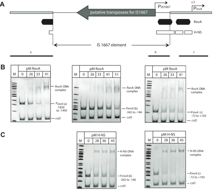
Previous footprint analysis revealed that RovA interacts with two distinct binding sites of theY. pseudotuberculosis invApromoter [46], and sequence homology as well as RovA band shift analysis indicated that similar binding sites are also recognized by RovA in theinvAregulatory region of YeO:8 8081v [45]. RovA-binding site I was partially destroyed by the insertion of the IS1667 element in theinvAO:3promoter (Fig.9A). However, band shift analysis with purified recombinant RovA and different DNA fragments of the
invA promoter region demonstrated that RovA still interacts specifically with RovA sequences upstream of the IS1667 element containing major parts of binding site I (Fig.9B). Interestingly, RovA was also found to specifically interact with fragments harboring the 39-end (2342 to2146) of the integrated mobile
element, although a slightly higher concentration was required for RovA-DNA complex formation. This demonstrated that this portion of the IS1667 element includes sequences, which are also preferentially recognized by RovA.
It is very likely that RovA is needed to alleviate H-NS-mediated repression at sites located downstream of the IS1667 insertion (Fig.9A) to permit maximal transcription of theinvApromoter. To test this hypothesis, we also studied the interaction of H-NS with different fragments of theinvO:3 promoter region. As shown in Fig.9C, H-NS was able to preferentially interact with a fragment harboring the 39-portion of the IS1667 element (2342 to2146), but the affinity was slightly lower compared to H-NS binding to theinvApromoter fragment (272 to+103). This strongly suggests that RovA is still required to eliminate H-NS mediated repression to allow optimal expression of invasin by the PIS1667and the PinvO:3
promoter.
Enhanced RovA production inY. enterocoliticaO:3 strains Requirement of RovA forinvA transcription in Y. enterocolitica
O:3 at 37uC was unexpected as it has been shown that rovA
expression inY. enterocoliticaO:8 andY. pseudotuberculosis strains is strongly thermoregulated and only expressed at moderate temperatures [44,47]. The RovA protein was found to act as a thermosensor which undergoes a conformational change upon a temperature shift from 25uC to 37uC. This thermo-induced conformational change reduces the DNA binding activity of the regulatory protein and renders it more susceptible to proteolysis by the Lon protease [18]. As a result, RovA activation of invA
expression is abolished at 37uC. Although RovA was shown to activateinvA expression inY. enterocolitica O:3 (Fig.8C), invasin expression does not appear to be strongly temperature-regulated compared to otherYersiniastrains (Fig.5,6B[15,44]).
To better understand the different control mechanisms, rovA
expression in the differentY. enterocoliticaisolates was analyzed. We found that all YeO:3 isolates produced very high levels of RovA at 25uC and 37uC; whereas no RovA was detected at 37uC in other
Yersinia strains, e.g. YeO:8 strain 8081v and Y. pseudotuberculosis
(Fig. 5, data not shown [44,47]). Expression analysis of the ProvAO:3::lacZ and ProvAO:8::lacZ fusions revealed that both rovA promoters are not auto-activated and are either not or only very weakly dependent on the temperature (Fig.10A, S6). Next, we addressed whether thermo-sensing and proteolysis varies between the RovAO:8 and RovAO:3 proteins. We introduced low-copy plasmids carrying therovAO:3 gene of YeO:3 Y1 or therovAO:8 gene of YeO:8 8081v into aY. enterocoliticaO:3rovAmutant strain (YE12) and compared RovA levels after growth at 25uC and 37uC (Fig. 10B). Almost identical levels of the RovA proteins were detected at 25uC. However, significantly lower amounts of the RovAO:8protein were visible at 37uC, while RovAO:3 concentra-tions remained almost the same (Fig.10B). This strongly suggested that post-transcriptional mechanisms controlling RovA levels must be different in YeO:3 strains. To test this hypothesis, we sequenced therovAlocus of all 49 availableY. enterocoliticaO:3 strains (Table S1). We found that the rovA genes of 45 strains, including all isolates tested in this study vary fromrovAof otherY. enterocolitica
Figure 6. Influence of theY. enterocoliticaO:3 O-antigen on host cell invasion.Y. enterocoliticawildtype strains YeO3 and 8081v, and outer core and/or O-antigen deficient derivatives (YeO3-OC, YeO3-R2, YeO3-OCR) were grown at 25uC and 37uC. (A) About 56104HEp-2 cells were infected
with 56105bacteria. After centrifugation of the bacteria onto the monolayer, cell association (adhesion+invasion) was monitored and internalization efficiency of the bacteria was determined by the gentamicin protection assay.E. coliK-12 was used as negative control. Data are presented as means 6standard deviations of three independent experiments performed in duplicate. Data were analyzed by the students t test. Stars indicate the results that differed significantly from those of YeO3 with * (P,0.05), ** (P,0.01), and *** (P,0.001) (B) Whole cell extracts were prepared from overnight cultures, separated on SDS-polyacrylamide gels and analyzed by western blotting using polyclonal antibodies directed against InvA and YadA. As a molecular marker the PageRuler Prestained Protein Ladder was loaded on the left.
serotypes by a single point mutation in codon 98, resulting in a P98S change in the amino acid sequence of the translated regulatory protein. To find out whether this substitution affects function of RovA as a thermosensor, we overexpressed and purified RovA of YeO:8 8081v and YeO:3 Y1 and compared their DNA-binding capacity at 25uC and 37uC (Fig. S5). However, interaction of both RovA variants with DNA fragments of theinvA
regulatory region was still temperature-dependent. Significantly more of both RovA proteins was required at 37uC for RovA-DNA complex formation, indicating that the thermosensing function is not severely affected by the P98S exchange. Next, we addressed thermo-dependent susceptibility of the RovA variants to degrada-tion by the Lon protease. To this aim, we reintegrated a copy of the rovAO:3 or rovAO:8 gene into the genome of a rovA deficient YeO:3 strain (YE12) and performed stability assays. Identical amounts of RovAO:3 were still visible 90 min after protein biosynthesis was stopped (Fig. 10C). In contrast, the RovAO:8 protein was rapidly degraded at 37uC, and significantly lower amounts of the regulatory protein were detectable 90 min after cessation of protein synthesis.
Influence of higher RovA and invasin levels in YeO:3 on invasion and virulence
To test the effect of the IS1667 insertion in theinvApromoter and the more stable RovAO:3(S98)variant on host cell invasion, we compared the amount of produced invasin and RovA in YeO:3 strains YE13 (rovAO:3), YE14 (rovAO:8) and YE15 (PinvO:3DIS) (Fig. 11A) and investigated the efficiency of these bacteria to enter HEp-2 cells (Fig.11B). High levels of invasin were detectable in YE13, whereas the instable RovA variant and deletion of the IS1667 element produced lower amounts of invasin, leading to a significant reduction of invasiveness into human epithelial cells.
It was previously shown that invasin and RovA are important to invade the intestinal epithelium by Y. enterocoliticaO:8 early after infection. TherovA-deficient mutants were found to be attenuated in the ability to reach and/or replicate in the deeper tissues and organs and induce a milder inflammation of the Peyer’s patches [48], whereas the LD50values of the wild-type and theinvAmutant were essentially identical but the colonization of the host tissues was delayed [4]. In order to determine whether higher invasin and RovA levels in Y. enterocolitica O:3 also affect pathogenesis, we tested the virulence of wild-type and mutant strains in the murine infection model. First, single strain infections were performed and bacterial colonization of Peyer’s patches (PPs), mesenteric lymphnodes (MLNs), liver and spleen was assessed. Since only minor differences could be highlighted (data not shown), we performed co-infection experiments to determine whether pres-ence of the wild-type affects the ability of the mutants to colonize tissues in a single host. This minimizes inherent inter-animal biological variations and can expose even subtle differences of the biological fitness and virulence, e.g. in the kinetics of infection. BALB/c mice were orally infected with 56108 bacteria in an inoculum comprised of an equal mixture of (i) the parental KanS wild-type strain Y1 (rovAO:3(S98)) and the KanRmutant strain YE14
(rovAO:8(P98)) or (ii) Y1 and YE15 (PinvO:3DIS) harboring a stable vector which only differs in its antibiotic resistance cassette to establish the ability to discriminate strains. Three days after infection, mice were dissected and the numbers of bacteria present in the PPs, MLNs, liver or spleen were determined (Fig.12). The results of the infection showed that both, the parental (YE13
rovAO:3(S98)) and therovAO:8(P98)mutant strain (YE14) are capable of establishing an infection, but considerably higher numbers of bacteria encoding the less stable RovAO:3(P98)variant from YeO:8 (YE14) were recovered from all dissected tissues. About 2- to 10-fold more bacteria of this strain were isolated from the lymphatic tissues or the organs (Fig.12A) compared to the parental strain YE13 (rovAO:3(S98)). Also comparison of the relative virulence ratio (Fig.12B) and calculation of the competitive index of the mutant relative to the wild-type strain (Fig. 12C) indicated that higher concentrations of RovA during mouse infections at 37uC are disadvantageous for the colonization and multiplication of YeO:3 in the organs. In contrast, significantly lower numbers of strain YE15 lacking the IS1667 element in the invA promoter region were isolated. About 10–20 times less bacteria were recovered from the PP and MLNs (Fig. 12A). The difference in the dissemination of the bacteria was even more striking. The IS1667 deletion strain YE15 was strongly attenuated in its ability to reach deeper tissues. Only in some occasions it reached the liver and spleen, but the bacterial load of the mutant in the liver and spleen was always significantly lower compared to wild-type (Fig.12). In summary, these data strongly indicates that high invasin expression levels during the course of an infection combined with a fine-tuned control of the virulence regulator RovA are advantageous for YeO:3 virulence in mice.
Discussion
The ability ofY. enterocoliticato bind and invade into host cells is essential for pathogenesis and persistance in its human host. Results of the present investigation highlight important differences in the adhesion properties between serotype O:3 strains (respon-sible for more than 70% of human yersiniosis cases) and otherY. enterocoliticaserotypes, e.g. serotype O:8, whose pathogenicity has been extensively investigated. Comparative analysis of cell binding properties demonstrated that the same repertoire of virulence factors is implicated in host cell binding in the serotype O:3 isolates, but their interplay and expression profile in response to environmental signals is significantly different from O:8 strains (Fig.13).
We show that synthesis of the primary internalization factor invasin is highly activated and nearly constitutive in all testedY. enterocolitica O:3 strains. This is in contrast to O:8 serotypes in which invasin synthesis is repressed at 37uC due to H-NS mediated silencing and rapid degradation of the invA activator protein RovA. Interestingly, a previous study also reported that
invA expression of a serotype O:9 strain was higher than in serotype O:8, but it was still significantly reduced at 37uC [49]. Constitutive expression of the invA gene in the O:3 strains was acquired by an IS1667 insertion into theinvA regulatory region Figure 7. Coexpression of invasin and YadA is necessary for efficient invasion at 376C.Y. enterocolitica strain O:8 strain 8081v,Y. enterocoliticaO:3 strain Y1 and isogenicinvAandyadAdeficient mutant derivatives were grown overnight at 25uC and 37uC. (A) About 5?104HEp-2
cells were infected with 5?105bacteria and after centrifugation of the bacteria onto the monolayer the samples cell association (adhesion+invasion) was monitored and internalization efficiency of the bacteria was determined by the gentamicin protection assay. Data are presented as means6 standard deviations of three independent experiments performed in duplicate. Data were analyzed by the students t test. Data were analyzed by the students t test. Stars indicate the results that differed significantly from those of Y1 with * (P,0.05), ** (P,0.01), and *** (P,0.001). (B) Whole cell extracts were prepared from the overnight cultures, separated on SDS-polyacrylamide gels and analyzed by western blotting using polyclonal antibodies directed against InvA and YadA. As a molecular marker the PageRuler Prestained Protein Ladder was loaded on the left.
Figure 8. Analysis ofinvAexpression inY. enterocoliticaO:3.(A) An overview of theinvApromoter region including the IS1667 insertion ofY. enterocoliticaO:3 strains is shown. The transcriptional start sites of theinvAgene and from the predicted IS1667-encoded promoter are indicated by broken arrows, the dark boxes indicate the RovA binding sites identified in the homologousinvApromoter ofY. pseudotuberculosis. The thick line represents theinvApromoter sequence and the thin line illustrates the IS1667 sequence. The arrow indicates the gene encoding the putative transposase of the IS1667 element. Sites used for the upstream deletion constructs are indicated by arrows. The numbers indicate the position of the deletion relative to the transcriptional start site of the invA gene. (B) Overnight cultures of Y. enterocolitica O:3 strain Y1 harbouring the PinvAO:8::luxCDABE(pFU170), PinvAO:3::luxCDABE(pFU171), PinvAO:3DIS::luxCDABE(pFU172) and PIS1667::luxCDABE(pFU202) fusion constructs were diluted
harbouring RovA and H-NS binding sites. Gene activation by transposons has been described for other genetic systems but the induction mechanism of the Y. enterocolitica O:3invA gene seems distinct from previously reported systems. Transposable elements
usually activate gene expression by replacing a negative regulatory element or through introduction of promoter elements [50,51]. One of the best-characterized examples of transposon-mediated gene activation is the beta-glucoside (bgl) operon ofE. coli. This region was analyzed inY. enterocoliticaO:3 Y1 and the isogenicrovAmutant derivative Y12 harbouring the PinvAO:3::luxCDABEfusion. The numbers
indicate the 59end points of the regulatory region ofinvAfromY. enterocoliticaO:3 in the fusion constructs relative to the transcriptional start site (+1). The luciferase activity determined from the cultures is given in relative light units (RLU) and represents the mean6standard deviation of at least three independent experiments. (D) Sequence of the 39-end of the IS1667 inserted intoinvAofY. enterocoliticaO:3 at position2143 is shown. The
210 and235 region of the predicted IS1667-encoded promoter are indicated. Sites used for the upstream deletion constructs are indicated by arrows. The numbers indicate the position of the deletion relative to the transcriptional start site of theinvAgene.
doi:10.1371/journal.ppat.1002117.g008
Figure 9. RovA and H-NS binding to theY. enterocoliticaO:3invAregulatory region.(A) Overview of theinvApromoter region ofY. enterocoliticaO:3 strains. The transcriptional start sites of theinvApromoter and of the predicted IS1667-encoded promoter are indicated by broken arrows. The dark boxes represent the RovA and the white small boxes the H-NS binding sites identified in the homologousinvApromoter ofY. pseudotuberculosis. The thick line represents theinvApromoter sequence and the thin line illustrates the sequence of the IS1667 element with the putative transposase gene. Fragments used for the band shift experiments are shown as black lines. Competitive gel retardation assays using purified RovA protein (B) or purified H-NS (C) ofY. enterocoliticaO:3 strain Y1. DNA fragments comprising different portions of theinvAregulatory region of Y1 were incubated without or with increasing concentrations of purified RovA or H-NS. The DNA-protein complexes were separated on a 4% polyacrylamide gene, a molecular weight standard 100 bp ladder was loaded on the left. The higher molecular weight protein-DNA complexes are marked by an arrow and the positions of the non-shifted and control fragments are indicated.
Figure 10. Analysis of RovA production and stability inY. enterocoliticaO:3.(A)Y. enterocoliticastrains Y1 and the isogenicrovAmutant of Y1 (YE12) harboring plasmids encoding the promoterlesslacZ gene or the ProvAO:8-lacZ or ProvAO:3-lacZ fusions were grown at 25uC and 37uC
overnight. The beta-galactosidase activity determined from the cultures is given inmmol min21mg21and represents the mean6standard deviation of at least three independent experiments. (B) AY. enterocoliticaO:3DrovAmutant strain (YE12) harboring therovAencoding plasmids pFU119 (rovAO:3) or pFU138 (rovAO:8) and YeO:8 strain 8081v were grown overnight at 25uC and 37uC. Whole cell extracts were prepared from the cultures,
system is usually repressed but can be activated by IS insertions up- or downstream of the promoter in either orientation relieving H-NS repression [52,53]. However, deletion analysis revealed that transposon-mediated invA activation in Y. enterocolitica O:3 is not solely due to disruption of the inhibitory H-NS binding sites, but also requires an IS-specific activating element. One recent study revealed a novel transposon-mediated gene activation mechanism. An IS5 insertion at a single site and in only one orientation was found to activate expression of the glpFK operon in a crp
background [54]. A short sequence at the 39 end of the IS5 transposon, including a permanently bent polyA-tract and an IHF binding site, was shown to be required forglpFKinduction. This shows that unique sequences within a mobile element can act as an enhancer or gain an activator binding function sufficient to activate close promoters. In this study we found that IS1667-promoted activation of invA expression in Y. enterocolitca O:3 at 25uC and 37uC is largely dependent on the presence of an IS1667-generated promoter and alternative RovA (activator) and H-NS (silencer) binding sites. RovA of YeO:8 was previously shown to activateinvA expression only at moderate temperatures through antirepression of H-NS-mediated silencing [45]. A temperature upshift to 37uC, however, results in a conformational change within RovA that strongly reduces the DNA-binding capacity of
the regulator. It has been previously shown that the apparent dissociation constant (Kd) of the thermoregulated RovA protein of
Y. pseudotuberculosisis about four-fold increased upon a temperature shift from 25uC to 37uC [18]. Furthermore, it was found that the temperature upshift renders the RovA protein more susceptible to degradation by the Lon and ClpP proteases [18]. Comparable studies with the RovA protein of YeO:8 8081v demonstrated similar properties and identical function as an intrinsic thermo-sensor (F. Uliczka, unpublished data). Here, we found that a single proline to serine exchange at position 98 (P98S) increases the stability of YeO:3 RovA without affecting the thermosensing ability of the protein. As a consequence, significantly higher RovA concentrations are present within the bacteria and this is sufficient to compensate for the thermo-induced reduction of RovA DNA binding. As YeO:3 strains originate mainly from boars and pigs with a higher body temperature of about 39u–40uC, a more temperature-stable RovA variant might be advantageous for persistence in these animals. According to our proposed structure model of RovA [55] the amino acid P98 is located in a surface exposed loop structure and is as such easily accessible for the proteases. How the P98S mutation affects proteolytic degradation is not yet clear. However, comparative CD spectroscopy of purified RovAO:8(P98)and a RovAO:3(S98)variant ofY. pseudotuber-Figure 11. Influence of enhanced invasin and RovA levels onY. enterocoliticaO:3 host cell invasion.(A) Whole cell extracts were prepared from the cultures, separated on SDS-polyacrylamide gels and analyzed by western blotting using polyclonal antibodies directed against InvA and RovA. As a molecular marker the PageRuler Prestained Protein Ladder was loaded on the left. (B) YeO:3 strains Y1 (wt), YE13 (rovAO:3S98),
YE14 (rovAO:8P98) and YE15 (rovAO:3DIS1667) were grown at 37uC. Approximately 106bacteria were centrifugated onto 104HEp-2 cells. Total numbers
of intracellular bacteria were determined and are expressed relative to the invasion rate of YeO:3 strain Y1 defined as 100%. Each value represents the mean of at least three different assays done in triplicate. Data were analyzed by the students t test, **, significantly different from Y1 or YE13 with P,0.001.
culosisindicated that no major structural changes are induced by this amino acid substitution (N. Quade, unpublished results). Furthermore, proteolysis is drastically reduced but not completely blocked by the P98S mutation as a slightly higher concentration of the regulatory protein was detected in aYersinia lonmutant strain. In summary, a more stable RovA variant (RovAO:3(S98)) and an IS1667 insertion in the invA promoter region, providing an additional promoter followed by slightly weaker RovA and H-NS binding sites, allow high expression levels of invasin in YeO:3 strains at 37uC. How these different properties influence pathogenesis is not fully clear, but first experiments addressing host cell invasion and colonization of YeO:3 in the mouse model revealed that loss of the IS1667 element reduced host cell entry and had a severe effect on the infection process in mice. Colonization of the PPs and the MLNs by YeO:3 strain YE15 (PinvO:3DIS) was significantly reduced and only occasionally these bacteria were able to reach deeper organs in co-infection experiments. This indicates that high levels of invasin are more advantageous and/or important for YeO:3 to initiate a successful infection than for YeO:8 in mice. In fact, a YeO:8 8081v invA
mutant strain shows a delayed but still efficient colonization of deeper tissues [4,56].
In contrast to invasin, loss of the RovA regulator in YeO:8 8081v leads to a 70-fold increase of the LD50and causes a much more severe alteration of the infection kinetics, e.g. penetration of the Peyer’s patches and mesenterial lymph nodes was much more reduced, and dissemination into liver and spleen was abolished [56]. Interestingly, significantly higher numbers of bacteria could be detected in lymphatic tissues and organs of mice when the unstable variant RovAO:3(S98) was expressed by YeO:3. This strongly suggests that elevated RovA levels, although they lead to higher amounts of invasin are disadvantageous for the colonization of the organs in mice. Microarray analysis to define the RovA regulon of Y. enterocolitica in YeO:8 revealed 40 genes to be activated and 23 repressed by RovA [57]. Among the RovA-repressed loci are several metabolic genes, e.g. permeases for glutamine, glutamate and aspartate) and their upregulation due to reduced RovA levels at 37uC might be important for the biological fitness and survival in host tissues during infection in mice. A more stable but still thermo-sensitive RovA variant, as found in YeO:3 Figure 12. Influence of enhanced invasin and RovA levels onY. enterocoliticaO:3 virulence.(A) BALB/c mice were co-infected via the orogastric route with 56108bacteria in an inoculum comprised of an equal mixture of YeO:3 strains Y1 (wt,rovAO:3) and YE14 (rovAO:8), or Y1 (wt, rovAO:3) and YE15 (rovAO:3PinvDIS). Three days post infection, the mice were sacrificed and the numbers of surviving bacteria in the liver, spleen,
mesenterial lymph nodes (MLN), and Peyer’s patches (PP) were determined as described inMaterial and Methods. Data are presented as a scatter plot of numbers of cfu per gram of organ as determined by counts of viable bacteria on plates. Each spot represents the cfu count, in the indicated tissue samples from one mouse. The levels of statistical significance for differences between test groups were determined by the Mann-Whitney-test. Stars indicate results that differed significantly from those of Y1 with ** (P,0.01), and *** (P,0.001). (B) Data are graphed as competitive index values for the tissue samples from one mouse. The bars represent the means of the competitive index values. A competitive index score of 1 denotes no difference in the virulence compared to Y1. Underlined scores denote where statistically significant differences were observed. The two strains Y1 and Y15 used for competition assays were differentially marked with antibiotics resistances on plasmids.
strains (RovAO:3(P98)), would allow similar regulatory control over virulence and metabolic genes in pigs and boars with a higher body temperature (39uC–40uC). In order to test whether the IS1667 insertion in theinvApromoter region and the RovAO:3(S98) variant reflects an optimal adaptation to these host organisms we are currently establishing a pig infection model.
Although high levels of invasin are produced by YeO:3 strains at moderate growth temperatures, cell invasion was either not initiated or very inefficient when the bacteria were pregrown at 25uC. This is in strong contrast to otherY. enterocoliticaserotypes or Y. pseudotuberculosis isolates which enter host cells with their highest efficiency when cultured at moderate temperatures. Previous analyses showed that induced flagellar-dependent motility is required for efficient invasion of YeO:8, but flagella production of this pathogen is repressed at 37uC [34]. Flagella are needed to ensure migration of the bacteria to host cells, but are not essential for the invasion process once the bacteria contact the
mammalian cells. Motility assays and electron microscopy revealed that flagellated and motile strains ofY. enterocolitica O:3 strains can be isolated from the intestinal tract of a mouse, but they rapidly loose their motility and become aflagellated during growth under standard laboratory conditions. As a result, YeO:3 strains are less invasive than other motile serotypesin vitro, but cell entry could be improved upon artificial host cell contact by centrifugation.
However, when the bacteria were pregrown at 25uC, YeO:3 uptake after host cell contact is still less efficient compared to YeO:8 or other serotypes, indicating that other factors repress invasin-mediated internalization at moderate temperatures or enhance cell entry at 37uC.Y. enterocoliticaisolates grown at room temperature generally express LPS with O-ag, whereas only very small amounts of O-ag are present in bacteria grown at 37uC [33,58]. The O-ag of YeO:8 is required for full virulence and plays a major role in pathogen-host interplay by affecting the expression Figure 13. Comparison ofY. enterocolitica O:3 and O:8 mediated temperature regulated control of host cell invasion.Model of virulence factor expression ofY. enterocoliticaO:3 and O:8 in response to temperature. (A) At moderate temperature,rovAexpression is induced inY. enterocoliticaO:8 which leads to activation of invasin expression. Furthermore, flagella production is activated and enhances host cell contact, and LPS molecules are synthesized which do not interfere with invasin function. This leads to an efficient internalization of the serotype O:8 strains after growth at environmental temperatures. At 37uC, RovA is rapidly degraded resulting in downregulation of invasin. In addition, flagella and O-antigen production is repressed, whereas synthesis of the adhesin YadA is induced which allows efficient adhesion, but no internalization into epithelial cells. (B)Y. enterocoliticaO:3 produce similar and significantly higher amounts of invasin at environmental and body temperature due to an IS insertion into theinvAupstream region and a stable RovA activator protein both abolishing H-NS mediated repression. However, internalization into host cells is strongly reduced at 25uC due to steric hindrance by the unique O-antigen and repression of YadA which strongly enhances and stabilizes host cell interactions at 37uC. LPS+OC: lipopolysaccharides with O-antigen and outer core.
and function of otherYersiniavirulence factors, e.g. absence of the O-ag reducedinvAexpression and internalization into HeLa cells [31]. In contrast, O-ag deficient YeO:3 rough mutants are more efficiently internalized by human epithelial cells. Furthermore, no reduction ofinvAexpression was observed in the rough mutants at 37uC when O-ag expression is fully repressed. Unlike otherYersinia
serotypes and other Gram-negative bacteria, the YeO3 O-ag forms a long homopolymer that is linked together with the OC hexasaccharide to the inner core forming a unique branched LPS structure. Its formation was previously shown to prevent proper function of some small size outer membrane proteins. For instance, O-ag was shown to inhibit serum resistance indirectly by masking the adhesin Ail from complement regulator C4bp binding [59]. Therefore, reduced O-ag density in YeO:3 at 37uC is very likely diminishing sterical hindrance thus allowing better access and host cell receptor binding by surface adhesins such as invasin and YadA (Fig.13).
In fact, besides invasin, also production of the adhesin YadA is required to promote efficient uptake of YeO:3. The virulence plasmid encoded trimeric YadA protein is highly and exclusively expressed at 37uC, and forms a capsule-like, fibrillar matrix covering the bacterial surface [60]. YadA ofY. enterocolitica O:8 strains has been shown to promote tight binding to extracellular matrix proteins such as collagen and laminin, but it does not contribute to epithelial cell entry compared to invasin [19,61]. In fact, at 37uC when YadA is highly expressed but no or only very low levels of invasin are produced by YeO:8, no internalization of the bacteria is initiated (Fig. 3B, 6A). Internalization of YeO:3 at 37uC also seems to be exclusively mediated by invasin as aninvAmutant is unable to enter host cells. Yet, YadA synthesis is not dispensable, as its absence in ayadAmutant or during growth at 25uC results in a significantly lower cell adhesion and uptake rate even in the presence of high amounts of invasin, whereas
yadA expression by an inducible promoter at 25uC leads to strong adhesion and efficient invasion of YeO:3 similar to YeO:8. YadA seems to be required to guarantee tight and efficient host cell binding which then in turn leads to a more efficient invasin-mediated uptake. Both invasin and YadA promote direct or indirect binding to beta 1 integrins [13,62]. High affinity binding and ligand-induced beta 1-integrin-clustering by invasin are required for efficient uptake by this host cell receptor family [63,64]. However, invasin of Y. enterocolitica does not contain a self-association domain medi-ating receptor-clustering and uptake in contrast to invasin ofY. pseudotuberculosis[65]. It is therefore tempting to speculate that co-expression of the somewhat longer cell surface adhesin YadA which promotes binding to ECM molecules bound to beta 1 chain integrins promotes or enhances intimate direct interaction of invasin and subsequent internalization (Fig.13). In summary, results in the present investigation provide evidence that even small variations between virulence factors and regulators are responsible for the substantial difference in host cell interactions ofY. enterocolitica serotype O:3 in comparison to otherY. enterocoliticaserotypes. Serotype O:3 specific variations in the surface molecule expression pattern imply that this Y. enterocolitica subspecies varies in its dynamic capacity to adapt to changing environments and individual niches within the host. A particular repertoire of host interaction genes may confer a survival advantage or pathogenic potential in a specific microen-vironment. Thus, an individual subspecies may be better adapted for survival in a particular host or host site, e.g. human gastrointestinal tract or oral cavities of swine (e.g. tongue and tonsils).
Materials and Methods
Ethics statement
All animal work was performed in strict accordance with the German regulations of the Society for Laboratory Animal Science (GV-SOLAS) and the European Health Law of the Federation of Laboratory Animal Science Associations (FELASA). The protocol was approved by the Niedersa¨chsisches Landesamt fu¨r Verbrau-cherschutz und Lebensmittelsicherheit: animal licensing commit-tee permission no. 33.9.42502-04-055/09. All efforts were made to minimize suffering.
Bacterial strains, cell culture, media and growth conditions
The strains used in this study are listed inTable 1. Overnight cultures of E. coliwere routinely grown at 37uC, Yersinia strains were grown at 25uC or 37uC in LB (Luria-Bertani) broth. The antibiotics used for bacterial selection were as follows: ampicillin 100mg/ml, chloramphenicol 30mg/ml, kanamycin 50mg/ml, gentamicin 50mg/ml and tetracycline 10mg/ml. For infection experiments, bacteria were grown at 25uC or 37uC, washed and diluted in PBS prior to infection.
Human HEp-2 cells were cultured in RPMI 1640 media with GlutaMAX (Invitrogen) supplemented with 7.5% newborn calf serum (Sigma Aldrich) at 37uC in the presence of 5% CO2. Human Caco-2 cells were grown in DMEM/HAM’s F-12 (Biochrom) supplemented with 10% FBS Superior (Biochrom).
DNA manipulations and construction of plasmids All DNA manipulations, PCR, restriction digestions, ligations and transformations were performed using standard techniques as described previously [66,67]. Plasmids used in this study are listed inTable 1, and primers are given inTable S2.
Plasmids pFU49 (invAO:3) and pFU182 (invAO:8) were construct-ed by amplification of theinvAgene from genomic DNA of YeO:3 Y11 and YeO:8 8081v with primers II40/II42 and the PCR-derived fragments were subsequently integrated into theSacI/SalI sites of pBAD33. For the overexpession of RovAO:8therovAgene was amplified from genomic DNA of YeO:8 8081v with primers II417/II418 and the generated fragment was inserted into the
NcoI/XhoI sites of pET28a, generating pFU156. Plasmid pFU157 was obtained by QuikChange mutagenesis of pFU156 using primer II375/II376. Plasmid pFU199 was constructed by inserting a hns+
O:3 fragment amplified with primers II726/II727 into the
EcoRI/SalI sites of pASKIBA43+. For the construction of pFU220 aBamHI/SalI fragment of pFU188 containing theyadAgene was integrated into pBAD33. pFU188 was obtained by insertion of a PCR fragment amplified with primers II517/II518 from genomic DNA of YeO:3 Y11 into theNcoI/SalI sites of pBAD/Myc-HisA. Plasmids pFU170 and pFU171 encoding the PinvAO:8::luxCDABE
and PinvAO:3::luxCDABE reporter fusions were generated by
insertion of a PCR fragment amplified with primers II177/II178 from genomic DNA of YeO:8 8081v or YeO3 Y11 in theBamHI/
SalI sites of pFU175. To construct plasmid pFU172, carrying the PinvAO:3DIS::luxCDABEfusion, two PCR fragments amplified with
primer pairs II177/II179 and II180/II178 were first ligated with their blunt ends and cloned into theBamHI/SalI sites ofluxCDABE
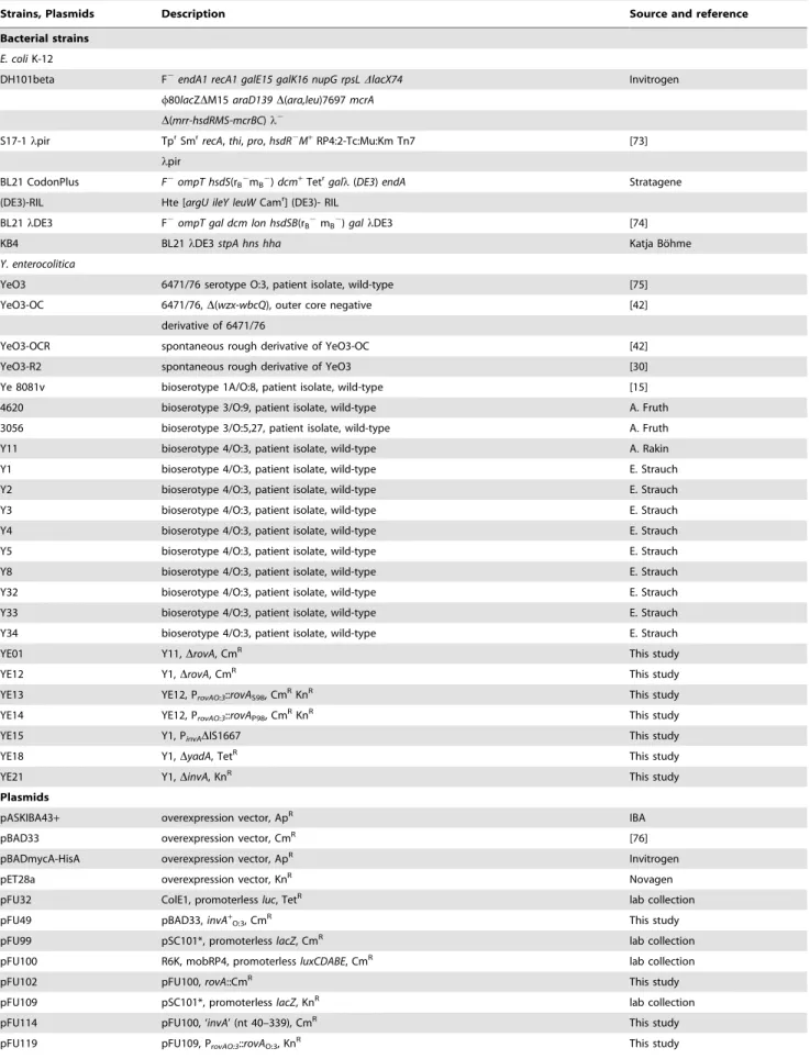
Table 1.Bacterial strains and plasmids.
Strains, Plasmids Description Source and reference
Bacterial strains
E. coliK-12
DH101beta F2endA1 recA1 galE15 galK16 nupG rpsLDlacX74 Invitrogen
w80lacZDM15araD139D(ara,leu)7697mcrA
D(mrr-hsdRMS-mcrBC)l2
S17-1lpir TprSmr
recA,thi,pro,hsdR2M+RP4:2-Tc:Mu:Km Tn7 [73]
lpir
BL21 CodonPlus F2ompT hsdS(r
B2mB2)dcm+Tetrgall(DE3)endA Stratagene
(DE3)-RIL Hte [argU ileY leuWCamr] (DE3)- RIL
BL21lDE3 F2
ompT gal dcm lon hsdSB(rB2mB2)gallDE3 [74]
KB4 BL21lDE3stpA hns hha Katja Bo¨hme
Y. enterocolitica
YeO3 6471/76 serotype O:3, patient isolate, wild-type [75]
YeO3-OC 6471/76,D(wzx-wbcQ), outer core negative [42]
derivative of 6471/76
YeO3-OCR spontaneous rough derivative of YeO3-OC [42]
YeO3-R2 spontaneous rough derivative of YeO3 [30]
Ye 8081v bioserotype 1A/O:8, patient isolate, wild-type [15]
4620 bioserotype 3/O:9, patient isolate, wild-type A. Fruth
3056 bioserotype 3/O:5,27, patient isolate, wild-type A. Fruth
Y11 bioserotype 4/O:3, patient isolate, wild-type A. Rakin
Y1 bioserotype 4/O:3, patient isolate, wild-type E. Strauch
Y2 bioserotype 4/O:3, patient isolate, wild-type E. Strauch
Y3 bioserotype 4/O:3, patient isolate, wild-type E. Strauch
Y4 bioserotype 4/O:3, patient isolate, wild-type E. Strauch
Y5 bioserotype 4/O:3, patient isolate, wild-type E. Strauch
Y8 bioserotype 4/O:3, patient isolate, wild-type E. Strauch
Y32 bioserotype 4/O:3, patient isolate, wild-type E. Strauch
Y33 bioserotype 4/O:3, patient isolate, wild-type E. Strauch
Y34 bioserotype 4/O:3, patient isolate, wild-type E. Strauch
YE01 Y11,DrovA, CmR This study
YE12 Y1,DrovA, CmR This study
YE13 YE12, ProvAO:3::rovAS98, CmRKnR This study
YE14 YE12, ProvAO:3::rovAP98, CmRKnR This study
YE15 Y1, PinvADIS1667 This study
YE18 Y1,DyadA, TetR This study
YE21 Y1,DinvA, KnR This study
Plasmids
pASKIBA43+ overexpression vector, ApR IBA
pBAD33 overexpression vector, CmR [76]
pBADmycA-HisA overexpression vector, ApR Invitrogen
pET28a overexpression vector, KnR Novagen
pFU32 ColE1, promoterlessluc, TetR lab collection
pFU49 pBAD33,invA+
O:3, CmR This study
pFU99 pSC101*, promoterlesslacZ, CmR lab collection
pFU100 R6K, mobRP4, promoterlessluxCDABE, CmR lab collection
pFU102 pFU100,rovA::CmR This study
pFU109 pSC101*, promoterlesslacZ, KnR lab collection
pFU114 pFU100, ‘invA’ (nt 40–339), CmR This study
respectively, fused to theluxCDABEoperon. For their construction PCR fragments were amplified with primer pairs II570/II543 and II571/II543, respectively, and integrated into theBamHI/SalI sites of pFU175. The ProvA::lacZfusions plasmids pFU129 and pFU130
were constructed by insertion of rovA promoter fragments, amplified with primer pair II260/I277 from genomic DNA of YeO:8 strain 8081v or YeO:3 strain Y11, into the BamHI/SalI sites of pFU109.
Following plasmids were engineered for the construction ofrovA,
invA and yadA mutant strains of Y. enterocolitica. For rovA
mutagenesis, plasmid pFU167 was constructed by amplification ofsacBwith primers II421/II422 from pJE4 and integration into the AvrII/NotI sites of pFU102. pFU102 was constructed by insertion of three PCR-derived fragments into theSpeI/NotI sites of pFU100: (i) a SpeI/SacI fragment containing 179 bp of therovA
regulatory region amplified with primers II171/II172, (ii) aSacI/
AatII fragment encoding the chloramphenicol resistance gene of pZA31 luc, and (iii) a AatII/NotI containing 119 bp of the downstream region of rovA amplified with primers II173/II174. For the invA mutagenesis, plasmid pFU213 was constructed by insertion of the SacI/AatII fragment encoding the kanamycin resistance gene of pZS*24MCS into pFU114. The plasmid
pFU114 was constructed by insertion of an ‘invA’ fragment into theSalI/NotI sites of pFU100. The PCR fragment contained base pairs 40–339 of theinvA coding region, amplified from genomic DNA of YeO:3 Y11 with primer pairs II211/II212 which introduce stop codons at the 59 and 39 end in the open reading frame of the ‘invA’ fragment. For yadA mutagenesis, plasmid pFU187 was used. This plasmid was engineered by the insertion of three PCR fragments into theSpeI/NotI sites of pFU167: (i) aSpeI/
SacI fragment which contained 150 bp of the yadA regulatory region amplified from genomic DNA of YeO:3 Y11 with primer pair II513/II514 (ii) aSacI/AatII fragment of pFU32 encoding the tetracycline resistance gene, (iii) and a AatII/NotI fragment containing the last 81 bp of theyadAgene plus 69 bp of theyadA
downstream region amplified from genomic DNA of YeO:3 Y11 with primer pair II515/II516.
For deletion of the IS1667 element in the invAO:3 regulatory region, we used pFU190. Plasmid pFU190 was created by the insertion of three fragments. Two fragments starting from position
21887 to21627 and2249 to21 of theinvA promoter region were amplified with primer pairs II519/II522 and II523/II524, respectively, and ligated at their blunt ends. The resultingXhoI/
NotI fragment was ligated with the SpeI/XhoI fragment from
Strains, Plasmids Description Source and reference
pFU129 pFU109, ProvAO:8::lacZ, KnR This study
pFU130 pFU109, ProvAO:3::lacZ, KnR This study
pFU138 pFU119, ProvAO:3::rovAO:8, KnR This study
pFU156 pET28a,rovA+
O:8, KnR This study
pFU157 pET28a,rovA+
O:3, KnR This study
pFU167 pFU102,rovA::CmR,sacB This study
pFU170 pSC101*, PinvO:8::luxCDABE, KnR This study
pFU171 pSC101*, PinvO:3::luxCDABE, KnR This study
pFU172 pSC101*, PinvO:3DIS::luxCDABE, KnR This study
pFU175 pSC101*, promoterlessluxCDABE, KnR lab collection
pFU182 pBAD33,invA+
O:8, CmR This study
pFU184 pFU119, ProvAO:3::rovAO:3, KnR This study
pFU185 pFU138, ProvAO:3::rovAS98P, KnR This study
pFU187 pFU167,yadA::TetR This study
pFU188 pBADmycA-HisA,yadA+
O:3, ApR This study
pFU190 pFU167 PinvAO:3(21887 to21627/2249 to21), TetR This study
pFU194 pFU175,21169 bpinvAupstream region, KnR This study
pFU195 pFU175,2448 bpinvAupstream region, KnR This study
pFU196 pFU175,2248 bpinvAupstream region, KnR This study
pFU197 pFU175,2146 bpinvAupstream region, KnR This study
pFU198 pFU175,272 bpinvAupstream region, KnR This study
pFU199 pASKIBA43+,hns+
O:3, ApR This study
pFU201 pSC101*, PinvO:3(2248 to2146)-luxCDABE, KnR This study
pFU202 pSC101*, PinvO:3(2448 to2146)-luxCDABE, KnR This study
pFU213 pFU114, ‘invA’ (nt 40–339), KnR This study
pFU220 pBAD33,yadA+
O:3, CmR This study
pJE4 R6K,sacB+
, KnR J. Eitel
pZA31luc expression vector, p15A, PLtetO-1, CmR [77]
pZS*24MCS expression vector, pSC101*, Plac/ara-1, KnR [77]