Specificity of Two Commercially-Available NS1 ELISA
Assays for Dengue Diagnosis
Maria G. Guzman1*, Thomas Jaenisch2, Roger Gaczkowski2, Vo Thi Ty Hang3, Shamala Devi Sekaran4, Axel Kroeger5, Susana Vazquez1, Didye Ruiz1, Eric Martinez1, Juan C. Mercado6, Angel Balmaseda6, Eva Harris7, Efren Dimano8, Prisca Susan A. Leano8, Sutee Yoksan9, Elci Villegas10, Herminia Benduzu10, Iris Villalobos11, Jeremy Farrar3, Cameron P. Simmons3
1Virology Department, Pan American Health Organization/World Health Organization Collaborating Center for the Study of Dengue and Its Vector, Instituto de Medicina Tropical ‘‘Pedro Kouri’’, Habana, Cuba,2Section Clinical Tropical Medicine, Department of Infectious Diseases, University Hospital of Heidelberg, Heidelberg, Germany, 3Oxford University, Clinical Research Unit, Ho Chi Minh City, Vietnam,4Department of Medical Microbiology, Faculty of Medicine, University of Malaya, Kuala Lumpur, Malaysia,5TDR-World Health Organization, Geneva, Switzerland,6CNDR, Managua, Nicaragua,7Division of Infectious Diseases and Vaccinology, School of Public Health, University of California, Berkeley, California, United States of America,8San Lazaro Hospital, Manila, Philippines,9Mahidol University, Bangkok, Thailand,10Instituto Experimental ‘‘Jose W Torrealba’’ Nu´cleo Universitario ‘‘Rafael Rangel’’, Universidad de los Andes Trujillo, Bogota´, Venezuela,11Hospital Central de Maracay, Maracay, Venezuela
Abstract
Background:Early diagnosis of dengue can assist patient triage and management and prevent unnecessary treatments and interventions. Commercially available assays that detect the dengue virus protein NS1 in the plasma/serum of patients offers the possibility of early and rapid diagnosis.
Methodology/Principal Findings:The sensitivity and specificity of the Pan-E Dengue Early ELISA and the PlateliaTMDengue NS1 Ag assays were compared against a reference diagnosis in 1385 patients in 6 countries in Asia and the Americas. Platelia was more sensitive (66%) than Pan-E (52%) in confirmed dengue cases. Sensitivity varied by geographic region, with both assays generally being more sensitive in patients from SE Asia than the Americas. Both kits were more sensitive for specimens collected within the first few days of illness onset relative to later time points. Pan-E and Platelia were both 100% specific in febrile patients without evidence of acute dengue. In patients with other confirmed diagnoses and healthy blood donors, Platelia was more specific (100%) than Pan-E (90%). For Platelia, when either the NS1 test or the IgM test on the acute sample was positive, the sensitivity versus the reference result was 82% in samples collected in the first four days of fever. NS1 sensitivity was not associated to disease severity (DF or DHF) in the Platelia test, whereas a trend for higher sensitivity in DHF cases was seen in the Pan-E test (however combined with lower overall sensitivity).
Conclusions/Significance:Collectively, this multi-country study suggests that the best performing NS1 assay (Platelia) had moderate sensitivity (median 64%, range 34–76%) and high specificity (100%) for the diagnosis of dengue. The poor sensitivity of the evaluated assays in some geographical regions suggests further assessments are needed. The combination of NS1 and IgM detection in samples collected in the first few days of fever increased the overall dengue diagnostic sensitivity.
Citation:Guzman MG, Jaenisch T, Gaczkowski R, Ty Hang VT, Sekaran SD, et al. (2010) Multi-Country Evaluation of the Sensitivity and Specificity of Two Commercially-Available NS1 ELISA Assays for Dengue Diagnosis. PLoS Negl Trop Dis 4(8): e811. doi:10.1371/journal.pntd.0000811
Editor:Ann M. Powers, Centers for Disease Control and Prevention, United States of America ReceivedNovember 23, 2009;AcceptedAugust 4, 2010;PublishedAugust 31, 2010
Copyright:ß2010 Guzman et al. This is an open-access article distributed under the terms of the Creative Commons Attribution License, which permits unrestricted use, distribution, and reproduction in any medium, provided the original author and source are credited.
Funding:This research was supported by the European Commission grant No. 517708, 6th Framework Programme. The funders had no role in study design, data collection and analysis, decision to publish, or preparation of the manuscript.
Competing Interests:The authors have declared that no competing interests exist. * E-mail: [email protected]
Introduction
Dengue is the most important mosquito-borne viral disease of humans and an enormous public health burden in affected countries. An estimated 50–100 million dengue cases occur annually, including 250,000–500,000 cases of severe illness and around 25,000 deaths. Approximately 2.5 billion people live in dengue endemic countries and the illness is reported in Southeast Asia, Western Pacific, the Americas, Africa and Mediterranean regions [1–3].
Dengue viruses (DENVs), of which there are four serotypes, cause a variable spectrum of disease that ranges from an undifferentiated fever to dengue fever (DF) through to more severe syndromes called dengue haemorrhagic fever (DHF) and dengue shock syndrome (DSS). DHF/DSS is a vasculopathy characterized by capillary leakage and haematological dysregulation. There are no licensed vaccines or specific antiviral therapies for dengue, and patient management relies on good supportive care.
management. In principle, early diagnosis could also facilitate timely public health interventions, e.g. vector control targeted at the households of index cases. Existing approaches to dengue diagnosis rely primarily on detection of DENV-reactive IgM; in more specialised settings this is augmented with detection of DENV RNA using home made RT-PCR or rarely, virus isolation [4,5]. Whilst generally robust, a limitation of IgM-based diagnostic approaches is poor sensitivity in the first few days of illness and in some settings, serological cross-reactivity with other Flaviviruses [4,5]. Recently, the diagnostic accuracy of commercial diagnostic assays that detect the DENV NS1 protein in plasma/serum samples have been described [6–13]. NS1 is a 55kDa glycoprotein secreted by DENV infected cells ‘‘in vitro’’ and ‘‘in vivo’’. Whilst
the role of NS1 in DENV biology is not well understood, high plasma NS1 concentrations early in illness have been associated with more severe disease [14,15]. The targeting of NS1 in diagnostic assays potentially offers the opportunity for an early, specific diagnosis of DENV infection since it can be detected prior to the appearance of measurable DENV-reactive IgM [8]. Whilst NS1 is a promising diagnostic target, the assessment of currently available NS1 assays across a breadth of patient populations, viral serotypes and lineages is important in evaluating where and when these assays [16] may fit into the laboratory diagnosis of dengue. At the end of 2006, the Dengue Scientific Working Group under the leadership of the World Health Organization Special Pro-gramme for Research and Training in Tropical Diseases (WHO/ TDR) established priorities for dengue research aimed at improving dengue treatment, prevention and control. The evaluation of new diagnostics were included among these priorities [17,18]. To this end, the purpose of the current study was to assess the sensitivity and specificity of two commercial NS1 assays in six countries.
Materials and Methods
The DENCO study
The DENCO project was a multi-centre prospective observa-tional study of dengue in Southeast Asia (Malaysia, Thailand, The Philippines and Vietnam) and the Americas (Nicaragua and Venezuela). The study sites at which patients were enrolled were: Department of Paediatrics, Faculty of Medicine, University of Malaya, Kuala Lumpur, Malaysia; Queen Sirikit National Institute of Child Health, Bangkok, Thailand; San Lazaro Hospital, Manila, The Philippines; Hospital for Tropical Disease, Ho Chi Minh City, Viet Nam, Children’s Hospital#1, Ho Chi Minh City, Viet Nam; Children’s Hospital#2, Ho Chi Minh City, Viet Nam; Children’s Hospital Manuel Jesus de Rivera, Managua, Nicaragua; Research Centre Jose W. Torrealba, University des Andes, Trujillo and Hospital Central, Maracay, Venezuela.
Author Summary
Dengue is the most important mosquito-borne viral disease of humans and an enormous public health burden in affected countries. Early, sensitive and specific diagnosis of dengue is needed for appropriate patient management as well as for early epidemic detection. Commercially available assays that detect the dengue virus protein NS1 in the plasma/serum of patients offer the possibility of early and rapid diagnosis. Here we evaluated two commercially available ELISA kits for NS1 detection (Pan-E Dengue (Pan-Early (Pan-ELISA and the PlateliaTMDengue NS1 Ag).
Results were compared against a reference diagnosis in 1385 patients in 6 countries in Asia and the Americas. Collectively, this multi-country study suggests that the best performing NS1 assay (Platelia) had moderate sensitivity (median 64%, range 34–76%) and high specific-ity (100%) for the diagnosis of dengue. The combination of NS1 and IgM detection in samples collected in the first few days of fever increased the overall dengue diagnostic sensitivity.
Figure 1. Flow chart summarising multi-country enrolment of dengue patients and basis for the final dataset.
Patient enrolment
Following written informed consent by the study participant, or a parent/guardian in the case of children, patients above 6 months of age with clinically suspected dengue and fever for less than 7 days were enrolled in the study. At 5 centres out-patients were recruited as well as in-patients. Patients were followed daily by trained study physicians using standardised case report forms (CRFs) describing clinical, laboratory, diagnostic and management information in detail. Ethical approval was obtained from the Ethics Review Committee of WHO and each institution involved. All patients in these studies were assessed daily by a study physician and had serial haematocrit and platelet estimations performed, as well as appropriate sampling for diagnostic serology and virology. Two plasma or sera samples were collected from each patient, one at day
of the enrolment and the second 7–14 days after fever onset. Dengue diagnosis was confirmed by either of the following methods: virus isolation in Aedes albopictus cell line (C6/36), by RT/PCR detection as previously described and IgM (MAC-ELISA), IgG (GAC-ELISA or Inhibition ELISA Method, EIM) and total antibody seroconversion (by Hemagglutination Inhibition assay) following the standard procedures at each study site [19–30]. The Hemagglutination Inhibition assay was standardized following WHO criteria and WHO recommended cut-off values were utilized [29]. As previously described, RT/PCR methods used here have sensitivity figures from 90 to 100% [20–23]. Other investigations and clinical management were at the discretion of the attending physicians. After discharge each patient was classified using the former WHO criteria for DF, DHF and DSS [30]. From November
Table 1.List of laboratories performing diagnostic testing for the patients enrolled in the DENCO study.
Location / Country Hospital Laboratory
Bangkok / Thailand Queen Sirikit National Institute of Child Health
AFRIMS, Bangkok* – all tests
Manila / Philippines San Lazaro Hospital AFRIMS certified laboratory at San Lazaro Hospital* for serology
PCR performed at Department of Virology, Nagasaki University, Nagasaki, Japan; serotyping done at San Lazaro Hospital, Manila, biomolecular laboratory (SACCL) Ho Chi Minh City / Vietnam Children’s Hospital No. 1 Oxford University Clinical Research Unit, HCMC** – all tests
Children’s Hospital No. 2 Hospital for Tropical Diseases Kuala Lumpur / Malaysia University of Malaya Medical
Centre, University of Malaya
Department of Medical Microbiology, Faculty of Medicine, University Malaya** - all tests
Managua / Nicaragua Hospital Infantil Manuel de Jesus Rivera La Mascota
Centro Nacional de Diagno´stico y Referencia, Ministry of Health, Managua – all tests
IgM, IgG and NS1 repeated at IPK Cuba** Maracay & Trujillo/ Venezuela Hospital Central de Maracay
Hospital de Trujillo
Trujillo Hospital Laboratory – all tests
IgM, IgG, viral isolation repeated and NS1 done at IPK Cuba**
AFRIMS - Armed Forces Research Institute of Medical Sciences, IPK - Instituto de Medicina Tropical ‘‘Pedro Kouri’’. *AFRIMS laboratory network in Asia.
**Member of the Tropical Disease Research –Pediatric Dengue Vaccine Initiative network of proficiency tested laboratories for dengue diagnostic evaluation. doi:10.1371/journal.pntd.0000811.t001
Table 2.Laboratory criteria employed at country level for dengue laboratory classification as confirmed dengue case.*
Country Confirmed dengue case (one of the following)**
Patients without evidence of recent acute dengue (all countries)
All countries RT-PCR positive or virus isolation positive Having paired plasma or serum specimens (collected$3 days apart) with the last sample collected$7 days after illness onset and RT-PCR negative and virus culture negative (at least one of the two being done on the acute sample), and serologically negative in locally used IgM and IgG assays
Thailand, The Philippines (according to AFRIMS protocol)
IgM.= 40 units (acute or convalescent sample or both) and IgG titer increase to above 100 units (paired samples) Twofold IgG titer increase (paired samples) with a titer.= 100 units in the convalescent sample Malaysia, Nicaragua, Venezuela, Vietnam IgM seroconversion (paired samples)
IgG seroconversion (paired samples) or fourfold or greater increase in titer (paired samples)
*For each test validated local protocols were used at each site. Serology results are based on IgM and IgG Capture ELISA of acute and convalescent specimens except where indicated.
**Four laboratories employed the RT/PCR protocol described by Lanciotti, et al., 1992 [20], one employed the protocols by Kong et al., 2006, J Virol Methods and Yong et al., 2007 Singaporean Med J [22,23],and the other the protocol by Laue et al., . J Clin Microbiol 1999 [21]. All laboratories employed MAC-ELISA. One laboratory employed Inhibition ELISA Method for IgG study while other four used GAC-ELISA. HI: hemagglutination inhibition assay was done in one laboratory (WHO recommendations were followed) [30].
2007 to January 2008, we prospectively tested acute plasma (or serum) samples from children and adults enrolled in these studies.
Characteristics of the study population
Between August 2006 and May 2007 a total of 2259 patients were recruited to the DENCO study at the 11 participating hospitals. NS1 detection was attempted using at least one of the two NS1 tests in 1821 patients. From amongst the 1821 patients, there were 1385 with laboratory-confirmed dengue and 45 with no laboratory evidence of acute or recent dengue. A further 391 had either indeterminate laboratory results or suggestive serology; results from these cases were not included in the analysis. The flow-chart in Figure 1 summarises the numbers and geography of enrolment and the classification of patients according to the results of reference diagnostic tests including demographic information.
Laboratory investigations
Serological and virological dengue diagnostics were performed in each participating country according to local protocols, with support provided by WHO designated laboratories as necessary (for participating laboratories see Table 1) [19–30]. The definitions employed at each site for ‘‘confirmed dengue case’’ are described in Table 2. For NS1 sensitivity analysis, patients with laboratory confirmation of dengue by serological or virological means were the reference population. For an assessment of NS1 specificity, patients in whom there was no evidence of acute or recent dengue (defined as serologically and virologically negative and in whom there were a minimum of 2 plasma or serum samples tested with the second collected$7 days after fever onset and.2 days after the first sample) were studied. As an additional assessment of specificity, two sera panels (one prepared at the Institute of Tropical Medicine ‘‘Pedro Kouri’’ in Cuba and the other at the Mahidol University, Bangkok, Thailand) from healthy individuals and from non-dengue patients were employed.
NS1 detection kits
Pan-E Dengue Early ELISA from Panbio (Brisbane, Australia), (Kit Pan-E) and Platelia Dengue NS1 AG from Bio-Rad (Marnes-la-Coquette, France), (kit Platelia) were evaluated. Both kits are based on a sandwich format microplate enzyme immunoassay for the detection of DENV NS1 employing a peroxidase-labelled murine monoclonal antibodies as probes. Samples were tested for NS1 detection following the manufacturer’s recommendations. Sera were classified as NS1 positive, negative and equivocal according to the manufacturer’s instructions. For the purposes of analysis, equivocal samples were excluded from the analysis.
Data management and analysis
Data were double-entered and checked at two established data-entry facilities in Guatemala (Center for Health Studies, Universidad del Valle de Guatemala) and Thailand (WHO/ TDR Clinical Data Management Collaborating Center, Faculty of Allied Health Sciences, Thammasat University, Thailand) and the two datasets were subsequently merged. Data analysis was performed at the Section of Clinical Tropical Medicine at the University of Heidelberg, Germany, using STATA versions 9.2 and 10, (STATA corporation, College Park, Texas).
Results
Overall sensitivity of NS1 tests versus reference diagnosis of confirmed dengue
The diagnostic sensitivity of kits Pan-E and Platelia assays was evaluated in 854 and 1284 serum samples respectively (Figure 1) from patients with a laboratory confirmed dengue diagnosis. Kit Pan-E it could not be performed in all available samples for logistical reasons relating to assay availability at some sites. The sensitivity of the kit Pan-E ranged from 24% in The Philippines to 72% in Vietnam (overall sensitivity rate of 52%). The sensitivity of the kit Platelia ranged from 34% in Nicaragua to 76% in Thailand (overall sensitivity rate of 66%) (Figure 2A).
NS1 sensitivity in relation to RT/PCR results
Compared to RT/PCR results, sensitivity of kit Pan-E ranged from 29–79% (overall sensitivity rate of 67%; 95% CI 63–71%) and the sensitivity of kit Platelia from 36–88% (overall sensitivity rate of 77%; 95% CI 74–79%) (Figure 2B).
Sensitivity of NS1 tests by day of illness
The sensitivity of both kits Pan-E and Platelia was influenced by the patient’s duration of illness prior to test sample collection.. In Asian patients, kits Pan-E and Platelia were more sensitive in test samples collected early in the disease phase than at later time points (Figure 3A). The analysis was limited to days with more than 40 observations total which is why for Latin America only a narrow range of days can be shown (Figure 3B) and due to small sample size and large confidence intervals no trend is visible. A higher sensitivity of both NS1 detection assays were observed in Asian patients than in Latin-American patients at the first four days of illness (Figure 3B).
NS1 sensitivity in relation to viral serotype
The sensitivity of each NS1 assay was considered in the context of the infecting serotype. Table 3 shows the sensitivity of kit Pan-E and Platelia assays according to DENV serotype as determined by RT-PCR or virus isolation. In our mainly hospital-based patient samples from 2006/2007 DENV-1 was most prevalent in Asia and DENV-2 most prevalent in Latin America (Table 4). For each of the four DENV serotypes kit Platelia had a greater sensitivity except for DENV-2, where the sensitivity was the same in both kits. In kit Platelia, sensitivity for DENV-2 was statistically significantly lower than for the other three serotypes pooled (DENV-2: 63%; 95% CI 57–69% versus 84%; 95% CI 82–88% for DENV-1, 3 and 4). The greater prevalence of DENV-2 in Latin American patients compared with Asian patients may help explain the lower sensitivity of both kit Pan-E and Platelia assays in Latin America (Figure 3B).
NS1 sensitivity in relation to IgM status
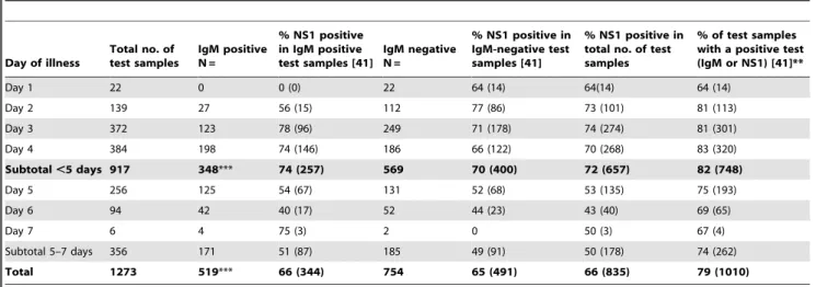
Detection of DENV-reactive IgM by MAC ELISA is the most commonly used approach to making a presumptive diagnosis of acute or recent dengue in endemic countries. Table 5 summarises NS1 sensitivity (kit Platelia assay only) in the context of IgM status and day of illness in confirmed dengue patients. The average sensitivity of NS1 testing in the first 7 days of sample collection was 65% (95%CI 62–69%) in acute samples where the IgM result was negative and 66% (95%CI 62–70%) when the acute test sample
Figure 2. Sensitivity of kits Pan-E and Platelia.Shown are the sensitivities (695% CI) of kits Pan-E and Platelia assays from six Asian and Latin-American countries in 1385 patients with a laboratory confirmed diagnosis of dengue (A) and sensitivities in the subgroup of 933 patients confirmed by PCR or viral isolation (B).
was IgM positive. Sensitivity figures increased to 74% and 70% if only samples collected in the first four days of illness were considered. Taking an algorithmic approach, when either the NS1 test or the IgM test on the acute sample was positive, the sensitivity for a presumptive (IgM) or definitive (NS1) diagnosis versus the reference result was 74% (95%CI 69–78) in samples collected at days 5 to 7. These figure increased to 82% (95%CI 79–84) in samples collected in the first four days of fever. These results suggested a combination of either IgM testing or NS1 testing (with kit Platelia) was sufficient to allow a presumptive (IgM) or definitive (NS1) diagnosis on an average of 82% of dengue cases enrolled in this study when acute early samples are tested. A similar analysis was performed with data obtained in the evaluation of kit Pan-E. Sensitivity figures of 66% (95%CI 60– 72) in samples collected at days 5 to 7 and 71% in samples collected in the first four days of fever (95%CI 67–75) were obtained (Table S1).
NS1 sensitivity according disease severity
The sensitivity of each NS1 assay was considered in the context of disease severity and geographical region (Table 6). Cases were classified according the former WHO criteria for DF and DHF/ DSS [27]. Sensitivity of kit Pan-E ranged from 29% (95%CI 12– 46) in DF to 60% (95%CI 39–82) in DHF cases from Latin-American countries and from 50% (95%CI 43–57) in DF to 62% (95%CI 57–67) in DHF/DSS cases from Asia (overall sensitivity 47% in DF and 62% in DHF/DSS cases). The sensitivity of kit Platelia ranged from 41% (95%CI 28–55) in DF and 68% (95%CI 47–89) in DHF/DSS cases from Latin-American countries and 70% (95%CI 66–75) in DF and 68% (95%CI 64–72) in DHF/ DSS cases from Asia (overall sensitivity of 68% for both DF and DHF/DSS total cases). Kit Pan-E showed higher figures of NS1 positive tests in severe cases, which are borderline statistically significant for Asia. Kit Platelia with overall higher sensitivity figures did not show a statistically significant association with disease severity.
Overall specificity of NS1 tests versus reference diagnosis
The diagnostic specificity of kits Pan-E and Platelia assays was evaluated in 36 and 45 samples respectively from patients with no
virological or serological laboratory evidence of acute or recent dengue. Both kits were negative in all these samples, which translates into a specificity of 100%.
NS1 specificity in healthy blood donors and patients with other confirmed diagnoses
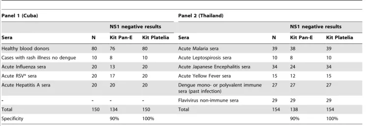
Since the number of patients with no evidence of acute or recent dengue was relatively small (n = 45) in this study, efforts were made to assess the specificity of dengue NS1 assays in patients with other confirmed infectious diseases whose transmission geographically overlaps with dengue, in healthy blood donors, and in blood donors with a serological history of DENV exposure. For the specificity analysis, a total of 304 sera were tested at two study sites (Cuba and Thailand). The specificity of kit Platelia was 100% in both sites whilst the kit Pan-E was 89% (Table 7). The lower specificity of kit Pan-E was in part due to false positive results in patients with Japanese encephalitis, Yellow Fever and acute Influenza.
Discussion
Dengue is increasing in incidence globally and therefore accurate and efficient diagnostic tests are more important than ever for clinical care, surveillance support, pathogenesis studies and vaccine research. Diagnosis is also important for case confirmation, to differentiate dengue from other diseases such as leptospirosis, rubella, and other flavivirus infections, and for the clinical management and evaluation of patients with severe disease [16,31]. The multicentre study described here assessed the diagnostic accuracy of two commercially available NS1 diagnostic tests. Two main findings were observed here: a) NS1 detection was overall only modestly sensitive for dengue diagnosis, with sensitivity highest in patients who presented early in their illness and b) a combined NS1 and IgM detection increased the overall sensitivity of dengue diagnostic.
Figure 3. NS1 sensitivity by day of illness.(A) Shown is the sensitivity (695% CI) of kits Pan-E and Platelia by day of illness in four Asian countries (N = 728 -kit Pan-E; N = 1152 -kit Platelia) amongst patients with a laboratory confirmed diagnosis of dengue where the acute sera were collected between day 2 and day 6 of illness. (B) Shown is the sensitivity (695% CI) of kits Pan-E and Platelia in the first four days of illness in two Latin American countries (N = 93 -kit Pan-E; N = 90 -kit Platelia) amongst patients with a laboratory confirmed diagnosis of dengue where the acute sera were collected between day 2 and day 4 of illness. Data is presented for those days of illness with.= 40 observations respectively. doi:10.1371/journal.pntd.0000811.g003
Table 3.NS1-sensitivity in the context of DENV serotype.
Serotype Kit Pan-E Kit Platelia
*N = 506
% Sensitivity
(95%CI) N = 862
% Sensitivity (95%CI)
DENV-1 223 79 (74–84) 415 87 (83–90)
DENV-2 169 62 (54–69) 257 63 (57–69)
DENV-3 87 60 (49–70) 142 82 (76–88)
DENV-4 27 52 (32–72) 48 79 (67–91)
*Number of DENV-positive samples by virus isolation or RT-PCR and serotype determined.
doi:10.1371/journal.pntd.0000811.t003
Table 4.Geographical and serotype stratification of the study population.
Country DENV-1a DENV-2 DENV-3 DENV-4 Total
Latin America
Nicaragua 6% (2) 94% (32) 0 0 34
Venezuela 35% (19) 16% (9) 36% (20) 13% (7) 55 Mean 24% (21) 46% (41) 22% (20) 8% (7) 89 Asia
Malaysia 62% (48) 9% (7) 17% (13) 13% (10) 78 Thailand 56% (86) 10% (15) 17% (26) 17% (26) 153 Philippines 0 10% (3) 87% (26) 3% (1) 30 Vietnam 47% (268) 35% (198) 10% (59) 1% (5) 568 Mean 48% (411) 27% (227) 15% (126) 5% (45) 829
aPercentages and absolute numbers (in brackets) of identified DENV serotypes
by country.
The global dengue research agenda includes evaluating the validity, role and accessibility of available and new diagnostics of importance to reducing disease severity and case fatality [32]. Recognizing the importance of early diagnosis and taking advantage of the platform of the multicentre DENCO project, two commercial available NS1 detection ELISA kits (Pan-E Early Dengue, Panbio Ltd and PlateliaTMDengue NS1 Ag, Bio-Rad ), named here as kits Pan-E and Platelia, were evaluated in terms of sensitivity and specificity. Overall and within country sensitivity figures were higher for kit Platelia than kit Pan-E. With the exception of Nicaragua and The Philippines, sensitivity figures of kit Platelia varied from 64% to 76% while the sensitivity of kit Pan-E varied from 36% to 72%. Depending on the diagnostic method used for comparison, different figures of sensitivity of NS1 detection have been reported by others [12,33,34]. Kumarasamy et al., obtained an overall sensitivity of 93% using PlateliaTM Den-gue NS1 Ag oscillating from 68% (in samples where the virus was isolated) to 90% in paired sera serologically confirmed as dengue [11,35].
In the present study, relatively higher levels of sensitivity were observed in samples collected in the first four days of fever when
samples from Asian patients were studied (interpretation limited for Latin America because of small sample size per day of illness).. Sensitivity was also higher in Asian patients compared with patients from Nicaragua and Venezuela. The small number of samples from Nicaraguan and Venezuelan patients (including a lower proportion of DHF/DSS cases) as well as the serotypes circulating could partially explain these observations (a high proportion of serotype 2 was found in Nicaraguan samples). The influence of duration of illness at the time of sample collection has been highlighted by others [6,8,10]. Figures of 93–100% sensitivity were obtained in samples collected at days 3 to 5 of fever [8] while others have reported figures higher than 85% in samples from day 1 to 3 in the Platelia assay [6,11].
NS1 protein has been detected concomitant with viremia and coincident with the febrile stage [8].
In the present study, the highest sensitivity was obtained in RT-PCR positive samples. Sensitivity of kit Platelia in RT-RT-PCR positive samples was 71% to 88% in Asian countries and 66% in Venezuela, but much lower in Nicaraguan samples (36%). Samples from this country were retested in a different laboratory by both NS1 detection kits but similar sensitivity results were observed (data not showed). The basis for low sensitivity in Nicaraguan samples remains unclear and will require further studies – but may partly be explained by the high proportion of serotype 2 in Nicaragua, which in both assays was associated with lower sensitivity. Indeed, as 94% (N = 32) of the serotypes recovered from Nicaragua were serotype 2, we cannot determine an estimate of sensitivity for the remaining 6% (N = 2).
Sensitivity varied by infecting serotype for each kit. The sensitivity of kit Pan-E was highest for DENV-1 infection (77%) and significantly lower for DENV-2 (60%), DENV-3 (57%) and DENV-4 (52%). The sensitivity of kit Platelia was also highest for DENV-1 infection (83%) and lowest for DENV-2 (60%). Consistent with DENV-1 infection being associated with high levels of NS1 detection, Xu et al., 2006, reported a sensitivity of 82% in an ‘‘in house’’ ELISA for the detection of NS1 protein of DENV-1 [36]. Similar results for the same serotype were reported by Alcon et al., 2002 [8]. The basis for different sensitivities for different serotypes requires further investigation. Potentially, this reflects different levels of avidity of the test mAbs for the relevant epitope(s) in NS1 from different serotypes, and potentially,
Table 5.NS1 detection (kit Platelia assay only) in relation to IgM status and day of illness.*
Day of illness
Total no. of test samples
IgM positive N =
% NS1 positive in IgM positive test samples [41]
IgM negative N =
% NS1 positive in IgM-negative test samples [41]
% NS1 positive in total no. of test samples
% of test samples with a positive test (IgM or NS1) [41]**
Day 1 22 0 0 (0) 22 64 (14) 64(14) 64 (14)
Day 2 139 27 56 (15) 112 77 (86) 73 (101) 81 (113)
Day 3 372 123 78 (96) 249 71 (178) 74 (274) 81 (301)
Day 4 384 198 74 (146) 186 66 (122) 70 (268) 83 (320)
Subtotal,5 days 917 348*** 74 (257) 569 70 (400) 72 (657) 82 (748)
Day 5 256 125 54 (67) 131 52 (68) 53 (135) 75 (193)
Day 6 94 42 40 (17) 52 44 (23) 43 (40) 69 (65)
Day 7 6 4 75 (3) 2 0 50 (3) 67 (4)
Subtotal 5–7 days 356 171 51 (87) 185 49 (91) 50 (178) 74 (262)
Total 1273 519*** 66 (344) 754 65 (491) 66 (835) 79 (1010)
*Samples from 1273 patients with a confirmed dengue diagnosis between day of illness 1 to 7.
**Percentage of positives IgM samples plus positive NS1 samples of the IgM negative samples in the total tested samples. ***Percentages of IgM positive in total samples collected in the first four days (38%), days 5–7 (48%) and total (41%). doi:10.1371/journal.pntd.0000811.t005
Table 6.Sensitivity of Kit Platelia and Pan-E by geographic region and disease severity.*
LAC** SEA** Total
DF*** DHF/DSS DF DHF/DSS DF DHF/DSS
Platelia 32/13 41% (28–55) 22/15 68% (47–89) 369/260 70% (66–75) 628/427 68% (64–72) 401/273 68% (63–73) 650/442 68% (64–72) Pan-E 31/9 29% (12–46) 23/14 60% (39–82) 228/114 50% (43–57) 396/245 62% (57–67) 259/123 47% (41–54) 419/259 62% (57–66)
*As indicated by the former WHO classification into DF and DHF/DSS for patients with NS1 test result and clinical classification available (N = 1051 for Platelia and 678 for Pan-E).
**LAC (Latin-American countries), SEA (Asian countries). ***N/positive NS1; % ; (95% CI).
different lineages from the same serotype. Also, this could potentially be related to the different sensitivities of the reference RT/PCR methods employed for dengue diagnosis. Alternatively, this might reflect different overall magnitudes of virus burden in patients with different serotypes. A relationship between NS1 detection and viraemia levels has been established previously [12,15]. Since high early viraemia levels have also been linked to increased disease severity, it is plausible that NS1 tests are more sensitive in the first few days of illness in patients at risk of developing severe complications in their illness compared to patients with a more benign disease evolution. However, in our study, no association between NS1 detection and disease severity (indicated by classification of DF or DHF/DSS) was observed. Furthermore a regression analysis on NS1 positivity for DHF/DSS vs. DF (or severe vs. mild) and adjusted for serotype and for country was done and there was no effect seen (data not shown). The specificity of NS1 tests could not accurately be estimated in the DENCO patient population as only a small number of cases had no serological or virological evidence of acute or recent dengue. Nonetheless, in patients who met our criteria for ‘‘not dengue’’, the specificity of both NS1 test kits was very high (100%). To provide further insights into specificity, two sera panels from patients with other confirmed diagnosis and healthy individuals were tested. Kit Platelia showed the higher specificity (100%). Similar specificity values has been previously reported by others [10,11,15,37]. The inclusion into the evaluating panel of samples from patients with acute Yellow fever and Japanese encephalitis virus infections suggest that no cross reaction among flaviviruses is observed with kit Platelia, however a larger number of samples collected from acute flavivirus infected patients need to be studied. The dengue serotype, duration of illness prior to sample collection, and the presence of immunocomplexes (NS1-IgG) in previous dengue immune individuals could explain the low sensitivity observed in the Nicaraguan and The Philippines samples [38]. In the case of Nicaragua, DENV-2 was present in the 94% of the samples where the virus was identified by virus isolation and RT/PCR suggesting that this was the predominant serotype. The generally poor sensitivity for DENV-2 (60%) observed for both assays suggests this partially explains the low sensitivity in Nicaraguan samples [12]. In The Philippines, a conjunction of factors such as to the duration of illness prior to sampling and the high level of individuals with a secondary infection could partially explain the low sensitivity since high sensitivity was observed in RT-PCR positive samples (83%).
One of the limitations of our study is that it is heavily biased towards Asian patients and viruses, with 93% of the total samples coming from this region. The strengths of our study were that it was multicentre, prospective and encompassed a broad range of DENV serotypes and clinical presentations.
It is important to mention that no proficiency panel study on positive or negative samples was performed prior to evaluating the tested samples allowing us to have more comparable reference methods among participant laboratories. However protocols employed at each site, have been extensively evaluated previously [19–30]. In addition, the laboratories participants (including some WHO collaborating centres) are the reference centres for dengue diagnosis and laboratory surveillance in their respective countries and have participated in previous regional and international proficiency testing ([39,40]
This study confirms and extends the findings of others in relation to the use of NS1 detections assays for the early diagnosis of dengue [6–12]. Although we could not study NS1 sensitivity and specificity in primary and secondary cases, in a small subset of samples classified as primary or secondary cases, a higher percentage of diagnose (90% over 80.6%) was obtained in the former (Vazquez S, manuscript in preparation).
In summary, we found the kit Platelia to be more sensitive and specific than kit Pan-E, with the sensitivity of both assays highest in the first few days of illness. Furthermore, we found that NS1 testing combined with IgM testing on the same test sample could yield a presumptive (IgM) or definitive (NS1) diagnose in as many as 82% of confirmed dengue cases using samples collected in the first four days of fever. As IgM detection is widely used for making a presumptive dengue diagnosis and in epidemiological surveil-lance, the use of a combined diagnostic algorithm including NS1 and IgM detection in samples collected in the first days of fever could provide clinically useful information to assist patient triage, management and outbreak response.
Supporting Information
Checklist S1 STARD checklist.
Found at: doi:10.1371/journal.pntd.0000811.s001 (0.13 MB DOC)
Table S1 NS1 detection (kit Pan-E assay only) in relation to IgM status and day of illness.
Found at: doi:10.1371/journal.pntd.0000811.s002 (0.05 MB DOC)
Table 7.NS1 results as determined by Kit Pan-E and Platelia assays in control sera panels.
Panel 1 (Cuba) Panel 2 (Thailand)
NS1 negative results NS1 negative results
Sera N Kit Pan-E Kit Platelia Sera N Kit Pan-E Kit Platelia
Healthy blood donors 80 76 80 Acute Malaria sera 39 38 39
Cases with rash illness no dengue 10 8 10 Acute Leptospirosis sera 10 8 10
Acute Influenza sera 20 13 20 Acute Japanese Encephalitis sera 34 24 34
Acute RSV* sera 20 17 20 Acute Yellow Fever sera 15 12 15
Acute Hepatitis A sera 20 20 20 Dengue mono- or polyvalent immune sera (past infection)
27 27 27
- - - - Flavivirus non-immune sera 29 29 29
Total 150 134 150 Total 154 138 154
Specificity 90% 100% 90% 100%
Author Contributions
Conceived and designed the experiments: MGG TJ AK EM EV IV JF CPS. Performed the experiments: VTTH SDS SV DR JCM AB ED PSAL SY HB. Analyzed the data: MGG TJ RG SDS AK AB EH ED SY EV JF
CPS. Contributed reagents/materials/analysis tools: MGG TJ RG VTTH SV AB EH ED SY EV HB IV CPS. Wrote the paper: MGG TJ RG VTTH SDS AK CPS.
References
1. WHO (2008) Dengue and dengue hemorrhagic fever. http://www.who.int/ mediacentre/factsheets/fs117/en/ (accessed 05 June 2008) fact sheet No117, revised May 2008.
2. Guzman MG, Kouri G (2002) Dengue: an update. Lancet Infect Dis 2: 33–42. 3. Halstead SB, Suaya JA, Shepard DS (2007) The burden of dengue infection.
Lancet 369: 1410–1.
4. Guzman MG, Kouri G (2004) Dengue diagnosis, advances and challenges. Int J Infect Dis 8: 69–80.
5. Vorndam AV, Kuno G (1997) Laboratory Diagnosis of dengue virus infections. CAB International. Kuno DGaG. pp 313–333.
6. Dussart P, Labeau B, Lagathu G, Louis P, Nunes MR, et al. (2006) Evaluation of an enzyme immunoassay for detection of dengue virus NS1 antigen in human serum. Clin Vaccine Immunol.
7. Dussart P, Petit L, Labeau B, Bremand L, Leduc A, et al. (2008) Evaluation of Two New Commercial Tests for the Diagnosis of Acute Dengue Virus Infection Using NS1 Antigen Detection in Human Serum. PLoS Negl Trop Dis 2: e280. 8. Alcon S, Talarmin A, Debruyne M, Falconar A, Deubel V, et al. (2002) Enzyme-linked immunosorbent assay specific to Dengue virus type 1 nonstructural protein NS1 reveals circulation of the antigen in the blood during the acute phase of disease in patients experiencing primary or secondary infections. J Clin Microbiol 40: 376–81.
9. Bessoff K, Delorey M, Sun W, Hunsperger E (2008) Comparison of 2 Commercially Available Dengue NS1 Capture Enzyme-Linked Immunosorbant Assays for Diagnosis of Acute DENV Infection with a Single Clinical Sample. Clin Vaccine Immunol.
10. Kumarasamy V, Wahab AH, Chua SK, Hassan Z, Chem YK, et al. (2007) Evaluation of a commercial dengue NS1 antigen-capture ELISA for laboratory diagnosis of acute dengue virus infection. J Virol Methods 140: 75–9. 11. Chuansumrit A, Chaiyaratana W, Pongthanapisith V, Tangnararatchakit K,
Lertwongrath S, et al. (2008) The use of dengue nonstructural protein 1 antigen for the early diagnosis during the febrile stage in patients with dengue infection. Pediatr Infect Dis J 27: 43–8.
12. Hang VT, Nguyet NM, Trung DT, Tricou V, Yoksan S, et al. (2009) Diagnostic Accuracy of NS1 ELISA and Lateral Flow Rapid Tests for Dengue Sensitivity, Specificity and Relationship to Viraemia and Antibody Responses. PLoS Negl Trop Dis 3: e360.
13. Young PR, Hilditch PA, Bletchly C, Halloran W (2000) An antigen capture enzyme-linked immunosorbent assay reveals high levels of the dengue virus protein NS1 in the sera of infected patients. J Clin Microbiol 38: 1053–7. 14. Avirutnan P, Punyadee N, Noisakran S, Komoltri C, Thiemmeca S, et al. (2006)
Vascular leakage in severe dengue virus infections: a potential role for the nonstructural viral protein NS1 and complement. J Infect Dis 193: 1078–88. 15. Libraty DH, Young PR, Pickering D, Endy TP, Kalayanarooj S, et al. (2002)
High circulating levels of the dengue virus nonstructural protein NS1 early in dengue illness correlate with the development of dengue hemorrhagic fever. J Infect Dis 186: 1165–8.
16. Guzman MG, Rosario D, Kouri G (2006) Diagnosis of dengue virus infection. Molecular Biology of the flavivirus Horizon Bioscience. Borowski P. pp 191–223. 17. Farrar J, Focks D, Gubler D, Barrera R, Guzman MG, et al. (2007) Towards a
global dengue research agenda. Trop Med Int Health 12: 695–9.
18. Kroeger A, Nathan M (2006) Dengue: setting the global research agenda. Lancet 368: 2193–2195.
19. Singh KR, Paul SD (1969) Isolation of Dengue viruses in Aedes albopictus cell cultures. Bull World Health Organ 40: 982–3.
20. Lanciotti RS, Calisher CH, Gubler DJ, Chang GJ, Vorndam AV (1992) Rapid detection and typing of dengue viruses from clinical samples by using reverse transcriptase-polymerase chain reaction. J Clin Microbiol 30: 545–51. 21. Laue T, Emmerich P, Schmitz H (1999) Detection of dengue virus RNA in
patients after primary or secondary dengue infection by using the TaqMan automated amplification system. J Clin Microbiol 37: 2543–7.
22. Kong YY, Thay CH, Tin TC, Devi S (2006) Rapid detection, serotyping and quantitation of dengue viruses by TaqMan real-time one-step RT-PCR. J Virol Methods.
23. Yong YK, Thayan R, Chong HT, Tan CT, Sekaran SD (2007) Rapid detection and serotyping of dengue virus by multiplex RT-PCR and real-time SYBR green RT-PCR. Singapore Med J 48: 662–8.
24. Vazquez S, Saenz E, Huelva G, Gonzalez A, Kouri G, et al. (1998) Deteccion de IgM contra el virus del dengue en sangre entera absorbida en papel de filtro. [Detection of IgM against the dengue++virus in whole blood absorbed on filter paper]. Rev Panam Salud Publica 3: 174–8.
25. Lam SK, Devi S, Pang T (1987) Detection of specific IgM in dengue infection. Southeast Asian J Trop Med Public Health 18: 532–8.
26. Vazquez S, Perez AB, Ruiz D, Rodriguez R, Pupo M, et al. (2005) Serological markers during Dengue 3 primary and secondary infections. J Clin Virol 33: 132–137.
27. Innis BL, Nisalak A, Nimmannitya S, Kusalerdchariya S, Chongswasdi V, et al. (1989) An enzyme-linked immunosorbent assay to characterize dengue infections where dengue and Japanese encephalitis co-circulate. Am J Trop Med Hyg 40: 418–27.
28. Burke DS, Nisalak A, Ussery MA (1982) Antibody capture immunoassay detection of japanese encephalitis virus immunoglobulin m and g antibodies in cerebrospinal fluid. J Clin Microbiol 16: 1034–42.
29. Clark DH, Casals J (1958) Techniques for hemagglutination inhibition with arthropodborne viruses. Am J Trop Hyg 7: 561–573.
30. WHO (1997) Dengue Hemorrhagic Fever. Diagnosis, treatment, prevention and control. Geneva. pp 1–84.
31. Buchy F, Yoksan S, Peeling RW, Hunsperger E Laboratory tests for the diagnosis of dengue virus infection.TDR/Scientific Working Group.TDR/ SWG/08 Geneva, Switzerland, 1–5 October. pp 74–85.
32. TDR/WHO (2006) Report on Dengue.TDR/Scientific Working Group.TDR/ SWG/08 Geneva, Switzerland, 1–5 October.
33. Sekaran SD, Ew CL, Subramaniam G, Kanthesh BM (2009) Sensitivity of dengue virus NS-1 detection in primary and secondary infections. African Journal of Microbiology Research 2: 105–110.
34. Sekaran SD, Ew CL, Kanthesh BM, Appanna R, Subramaniam G (2007) Evaluation of dengue NS1 capture ELISA assay for the rapid detection of dengue. J Infect Developing Countries 1: 182–188.
35. Kumarasamy V, Chua SK, Hassan Z, Wahab AH, Chem YK, et al. (2007) Evaluating the sensitivity of a commercial dengue NS1 antigen-capture ELISA for early diagnosis of acute dengue virus infection. Singapore Med J 48: 669–73. 36. Xu H, Di B, Pan YX, Qiu LW, Wang YD, et al. (2006) Serotype 1-specific monoclonal antibody-based antigen capture immunoassay for detection of circulating nonstructural protein NS1: Implications for early diagnosis and serotyping of dengue virus infections. J Clin Microbiol 44: 2872–8.
37. Blacksell SD, Mammen MP, Jr., Thongpaseuth S, Gibbons RV, Jarman RG, et al. (2008) Evaluation of the Panbio dengue virus nonstructural 1 antigen detection and immunoglobulin M antibody enzyme-linked immunosorbent assays for the diagnosis of acute dengue infections in Laos. Diagn Microbiol Infect Dis 60: 43–9.
38. Koraka P, Burghoorn-Maas CP, Falconar A, Setiati TE, Djamiatun K, et al. (2003) Detection of Immune-Complex-Dissociated Nonstructural-1 Antigen in Patients with Acute Dengue Virus Infections. J Clin Microbiol 41: 4154–4159. 39. Hunsperger EA, Yoksan S, Buchy P, Nguyen VC, Sekaran SD, et al. (2009) Evaluation of commercially available anti-dengue virus immunoglobulin M tests. Emerg Infect Dis 15: 436–40.