Percutaneous endoscopic
versus
surgical gastrostomy
in patients with benign and malignant diseases:
a systematic review and meta-analysis
Jose´ Gonc¸alves Pereira Bravo*, Edson Ide, Andre Kondo, Diogo Turiani Hourneaux de Moura, Eduardo Turiani Hourneaux de Moura, Paulo Sakai, Wanderley Marques Bernardo,
Eduardo Guimara˜es Hourneaux de Moura
Hospital das Clı´nicas da Faculdade de Medicina da Universidade de Sa˜o Paulo (HCFMUSP), Departamento de Gastroenterologia, Unidade de Endoscopia Gastrointestinal, Sa˜o Paulo/SP, Brazil.
To compare the complications and mortality related to gastrostomy procedures performed using surgical and percutaneous endoscopic gastrostomy techniques, this review covered seven studies. Five of these were retrospective and two were randomized prospective studies. In total, 406 patients were involved, 232 of whom had undergone percutaneous endoscopic gastrostomy and 174 of whom had undergone surgical gastrostomy. The analysis was performed using Review Manager. Risk differences were computed using a fixed-effects model and forest and funnel plots. Data on risk differences and 95% confidence intervals were obtained using the Mantel-Haenszel test. There was no difference in major complications in retrospective (95% CI (-0.11 to 0.10)) or randomized (95% CI (-0.07 to 0.05)) studies. Regarding minor complications, no difference was found in retrospective studies (95% CI (-00.17 to 0.09)), whereas a difference was observed in randomized studies (95% CI (-0.25 to -0.02)). Separate analyses of retrospective and randomized studies revealed no differences between the methods in relation to mortality and major complications. Moreover, low levels of minor complications were observed among endoscopic procedures in randomized studies, with no difference observed compared with retrospective studies.
KEYWORDS: Gastrostomy; Mortality; Complication; Surgical Gastrostomy; Percutaneous Endoscopic Gastrostomy.
Bravo JG, Ide E, Kondo A, de Moura DT, de Moura ET, Sakai P, et al. Percutaneous endoscopicversussurgical gastrostomy in patients with benign and malignant diseases: a systematic review and meta-analysis. Clinics. 2016;71(3):169-178
Received for publication onOctober 21, 2015;First review completed onNovember 13, 2015;Accepted for publication onNovember 30,
2015
E-mail: [email protected]
*Corresponding author
’ INTRODUCTION
The use of gastrostomy has expanded over the past decade, and new techniques have been developed that have made the procedure simpler and less risky (1). Gastrostomy is specifically a technique that allows direct access to the stomach to provide food to disabled patients for several reasons. Most commonly, this condition occurs in patients with neurological diseases, impairment following a stroke or obstructive head and neck tumors (1,2).
The absence of systematic reviews and meta-analyses directly comparing endoscopic gastrostomy and surgical gastrostomy (SG) techniques for all pathologies, whether benign or malignant,
in the literature was one of the main reasons for conducting the present comparative review of surgical and endoscopic methods for all pathologies that may result in gastrostomy, taking into account major and minor complications. Two related reviews were found in the literature, but both of these considered specific pathologies. The first, carried out by Grant et al. (9), investigated gastrostomy complications in patients with head and neck tumors and compared radiological methods with endoscopy, not considering the surgical method. The other, by Burkitt et al. (2), similarly compared complications in patients with head and neck tumors but did not address other pathologies and only compared radiological techniques with endoscopy, given that endoscopic gastrostomy has become the technique of choice for carrying out the procedure, with SG only used in cases where the endoscopic procedure is not viable. Most studies associate SG with higher rates of complications and mortality (4,7).
Several techniques for gastrostomy tube insertion have been described. These techniques include percutaneous
DOI:10.6061/clinics/2016(03)09
Copyright&2016CLINICS–This is an Open Access article distributed under the terms of the Creative Commons License (http://creativecommons.org/licenses/by/ 4.0/) which permits unrestricted use, distribution, and reproduction in any medium or format, provided the original work is properly cited.
endoscopic gastrostomy (PEG) and SG (open SG (OSG) or surgical laparoscopic gastrostomy (SLG)) (2). SG was initially suggested in 1837 by Egeberg, a Norwegian surgeon and the first successful gastrostomy was carried out nearly 40 years later, in 1876, by Verneuil in Paris, France (1-3).
The SLG method also avoids the need for a laparotomy but still requires general anesthesia. Although not an absolute contraindication, prior upper abdominal surgery may make the SLG method difficult and risky (8). The SLG method additionally offers better exposure of the stomach than the open technique, in which the incision is usually quite small. However, PEG has nearly entirely displaced SG in clinical practice because the PEG procedure can be carried out more easily and without general anesthesia, which is beneficial for the usually elderly, high-risk patient population. In addition, PEG avoids the mortality and morbidity associated with laparotomy. Despite the minor invasiveness of endoscopic placement of percutaneous feeding tubes, complications remain an important problem (2,3).
There is widespread acceptance of PEG as the insertion technique of choice owing to its simplicity and effectiveness, but certain patients are not candidates for an endoscopic approach (10). The present review provides greater evidence in the current context as to which of these procedures is associated with major complications and mortality.
’ METHODS
Protocol and registration
The present review was conducted in accordance with the PRISMA (Preferred Reporting Items for Systematic reviews and Meta-Analyses) recommendations (7) and it has been registered in the PROSPERO international database (www.crd.york.ac. uk/prospero/) under number CRD42015016493 (20).
Eligibility criteria
a) Types of studies: randomized controlled trials and retro-spective studies.
b) Type of participant: patients undergoing gastrostomy. c) Types of interventions: PEG (intervention) and SG
(comparison).
d) Types of outcome measures: the main outcome parameters were minor and major complications and mortality directly related to the procedure.
There is no literature on the exact classification of com-plications related to gastrostomy and authors have classified these complications in various ways. The methods used here are those found most often in published studies. Complica-tions may be secondary to endoscopic procedures or directly related to gastrostomy, such as cardiopulmonary complications, hypoxemia, phlebitis, bacteremia, perforation and bleeding. Minor complications are treated conservatively. Major complica-tions may require hospitalization (9,2,22), blood transfusions, or endoscopic or surgical therapy. The period in which complica-tions occur may be early (until 15 days) or late (after 15 days) (10). For the present review, both major and minor complications were considered, whether early or late (2,12,22).
Major complications. Bowel perforation, Gastrointestinal hemorrhage, Gastrocutaneous fistula, Intra-abdominal abscess,
Peristomal abscess, Peritonitis requiring surgery, Loss of cath-eter tract, Aspiration pneumonia, Sepsis, Buried bumper syn-drome, Early inadvertent removal of tube.
Minor complications.Dislodged tubes, Inadvertent removal of tubes, Tube malfunction, Other tube problems conservatively managed, Peristomal leaks, Peristomal infection, Mild skin necrosis, Wound granulation, Minor wound bleeding, Wound hematoma, Post-procedure ileus, Symptomatic pneumoperito-neum, Subcutaneous emphysema, Regurgitation, Unsuccessful procedure.
Information sources
The electronic databases searched were MEDLINE (via PubMed), Embase, Scopus, LILACS, the Cochrane Library (via BVS), and CINAHL (via EBSCO), from inception until February 2015.
Search
The descriptors used for the study were as follows: ((Gastrostomies OR Gastrostomie OR Gastrostomy OR Percutaneous endoscopic gastrostomy)) AND random*.
Study selection
The process of including or excluding studies according to PRISMA is presented as a flow chart. The eligibility assessment and selection of records shown were per-formed independently in a standardized manner by two reviewers. Disagreements between the reviewers were resolved by consensus and the search was conducted in all languages, with no limit regarding time.
Studies comparing patients undergoing endoscopic gastro-stomy and SG were included regardless of cause and enrolled patients aged over 18 years and with a minimum period of one month of follow-up. Abstracts, letters, editorials, expert opinions, case reports and reviews were excluded. Studies that did not consider the desired outcomes or that compared other techni-ques were also not included.
Data collection process and items
Data were extracted independently by two reviewers (JGPB, BWM) using forms (checklists) that are standard for cohort studies (8) and randomized clinical trials (7). The data were drawn from all studies comparing endoscopic gastrostomy and SG using the following as main variables: endoscopic gastro-stomy, gastrogastro-stomy, follow-up, early and late complications and minor and major complications. Complications not related to the procedure, mortality not related to the procedure, were excluded.
Risk of bias
indicated or if the study based its findings on key secondary endpoints, the study was rejected.
Planned methods of analysis
The analysis was performed using Review Manager (RevMan) 5.3 (21) from the Cochrane Informatics & Knowledge Management Department website. Risk differences for dichot-omous variables were computed using a fixed-effects model and the respective forest and funnel plots were obtained. Data on risk differences and the 95% confidence intervals (CIs) for each out-come were calculated using the Mantel-Haenszel test. Incon-sistency (heterogeneity) was qualified and reported using the Chi-squared (Chi2) and Higgins methods and was termed I2. Data on the absolute risk reduction (ARR) or increase (ARI) and the number needed to treat (NNT) or harm (NNH) were obtained for validity and applicability using Critically Appraised Topics (CAT) software (19).
’ RESULTS
Initially, 2,042 studies were retrieved. A total of 2,024 studies were excluded for various reasons after reading: 720 presented no direct comparison of the techniques under study, 104 were narrative reviews and another 1,200 had no direct relation to the review. For evaluation and eligibility, the full text of the remaining eighteen articles was read and eleven studies were further excluded for various reasons: two studies compared endoscopic gastrostomy with gastrojejunostomy, six were comparative reviews of radiological and endoscopic techniques and three were case series. Selected a total of seven studies of the remaining studies, two were prospective rando-mized studies and five were retrospective cohort studies. The retrospective and randomized studies were evaluated inde-pendently. Few randomized studies compared one technique with the other. All strategies and the selection procedure are represented in the diagram below (Figure 1).
Study characteristics
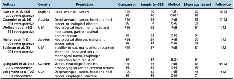
Seven studies were included for review, including two rando-mized controlled trials and five retrospective cohort studies. The total population was 406 individuals, with 232 having under-gone endoscopic gastrostomy and 172 having underunder-gone SG. The main indications were neurological; traumatic; tumors of the head and neck; and other situations, such as stenosis or eso-phageal atresia (Table 1). All studies used the Gauderer-Ponsky or ‘‘pull’’endoscopic gastrostomy technique described in 1980 (17,24, 27). Certain studies did not mention whether the patients received antibiotic prophylaxis. The main outcomes studied were procedure-related complications, divided into major, minor and mortality complications directly related to the procedure.
Risk of bias within studies
For the prospective randomized studies, the Jadad score (11), ranging from 0 to 5, was used and only the studies with a Jadad score X3 were selected. For observational studies, the Newcastle-Ottawa rating scale (12) was used and only studies with a score X6, of a maximum total of 9 points, were selected. Publication bias is related to what is likely to be published among what is available to be published (Tables 4 and 5, Supplementary file).
Results of individual studies
Of the 406 patients in total, 232 had undergone PEG and 174 had undergone SG. Among these patients, 27 major
complications were observed, 16 of which were related to the endoscopic procedure and 11 of which were related to the surgical procedure. Minor complications occurred in 57 patients with SG and in 56 with PEG. Moreover, mortality related to the procedures was higher in the group with SG (five cases) compared with the group with PEG (one case).
All complications in retrospective studies
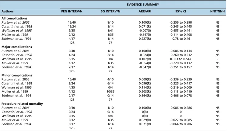
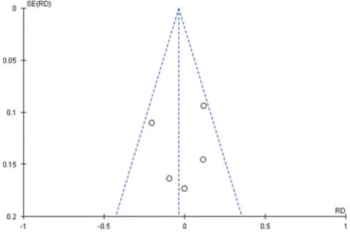
In the retrospective studies, with a sample of 205 indi-viduals, complications occurred in 125 for PEG and in 77 for SG (23,4,6,16,29). There was no significant difference favor-ing either group.
Major complications in retrospective studies
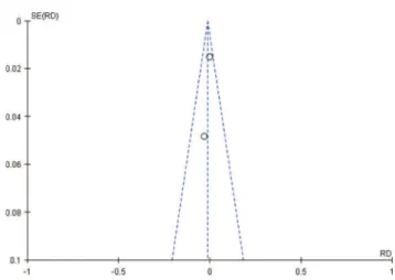
Of a total of 18 complications, 12 were for PEG and 6 were for SG. There was no significant difference between the four studies. Only one study (29) showed a significant difference (95% CI (0.333 to 0.547), ARR=10.7%, NNT=9, i.e., it would be necessary to treat nine patients for one to have a higher number of complications) (Table 2). Analysis of the retrospective studies (23,4,6,16,29) showed that the most frequent major complica-tions were peritonitis requiring surgical intervention, aspiration and sepsis, with certain cases resulting in death.
Minor complications in retrospective studies
Of 59 minor complications reported in the retrospec-tive studies, 31 were for PEG and 28 were for SG; this difference was not significant (Table 2). The most frequent complications were small tube leaks, stoma leakage, displacement of the tube and superficial cellulitis. There was no death related to minor complications in most studies (Table 2) (23,4,6,16,29).
Mortality related to the procedure in retrospective studies
Three deaths related to the procedure occurred, all of which were following SG. The leading causes of death were peritonitis and aspiration pneumonia. The risk difference analysis did not show a statistically significant trend favoring any group. Mortality occurred in only three retro-spective studies (Table 2) (23,6,16).
All complications in randomized studies
Among the 201 patients in total, there were 104 complica-tions for PEG and 97 for SG. In all, 29 complicacomplica-tions were related to PEG and 42 were related to SG. There was no significant difference between the groups (Table 3).
Major complications in randomized studies
Four complications occurred in the PEG group and 5 occurred in the SG group. There was no significant diff-erence between PEG and SG. The most common major complications were pneumonia and peritonitis (Table 3).
Minor complications in randomized studies
Procedure-related mortality in randomized studies
Three deaths were related to the procedures, with one in the PEG group and two related to SG in the study by Ljungdahl et al. (14). In contrast, in the study by Stiegmann et al. (26), there were no deaths related to the procedure. The main cause of death was aspiration pneumonia. However, there was no significant difference between PEG and SG (Table 3).
Summary of results (meta-analyses) Risk of bias across studies and additional analyses
Risk of bias across studies and additional analyses. The data on effect estimates and CIs for each study are illustrated graphically below. The numerical group-specific summary information, effect sizes, CIs and percentage weights are also
Records identified through database searching Medline
(n = 410 )
Additional records identified through other sources
EMBASE = 220, CINAHL = 705, LILACS = 40, Cochrane = 386, Scopus = 281
Records screened (n = 2042)
Records excluded, with reasons (n = 2024 )
-did not compare the techniques directly (720)
-Narrative review (104)
Other study not related to review (1200)
Full-text articles assessed for eligibility
(n = 18)
Full-text articles excluded, with reasons (n = 11 )
-Articles that compared endoscopic gastrostomy to gastrojejunostomy (2) -Systematic review that compares two RIG and PEG (6) - Case series (3)
Studies included in qualitative synthesis
(n = 7)
Studies included in quantitative synthesis
(meta-analysis) (n = 7)
Studies included: (5) Retrospective (2) Randomized
Adapted from: Moher D, Liberati A, Tetzlaff J, Altman DG, The PRISMA Group (2009). Preferred Reporting Items for Systematic Reviews and Meta-Analyses: The PRISMA Statement (15).
Screening
Eligibility
Included
Identification
Figure 1 -Search strategy and selection of studies.
Table 1-Characteristics of retrospective and randomized studies.
Authors Country Population Comparison Sample (n=523) Method Mean age (years) Follow-up
Rustom et al. (23) 2006 retrospective
England Head and neck tumors PEG 40 Pull* 63 18 M
SG 10 OSG* 65
Cosentini et al. (4) 1998 retrospective
Austria Oropharyngeal cancer, head and neck cancer, neurological disorder
PEG 35 Pull 58 17 M
SG 4 OSG 28
Wollman et al. (30) 1995 retrospective
USA Neurological impairment, head and neck cancer, gastrointestinal decompression
PEG 69 Pull 61 1 M
SG 62 OSG 54
Muller et al. (16) 1999 retrospective
Sweden Neurological disorder, malignant cancer, other
PEG 24 Pull 55 1 M
SG 14 OSG 39
Edelman et al. (6) 1994 retrospective
USA Inability to eat, malnutrition, recurrent aspiration, head and neck or
esophageal tumor, esophageal obstruction from radiation
PEG 17 Pull 81 1 M
SG 14 SLG* 61
Ljungdahl et al. (14) 2006 randomized
Sweden Stroke, neurological disease,
oropharyngeal cancer, cerebral trauma
PEG 35 Pull 69 45 M
SG 35 OSG 65
Stiegmann et al. (26) 1990 randomized
USA Oropharyngeal cancer, head and neck cancer, esophageal stricture
PEG 12 Pull 48 4 M
SG 35 OSG 71
presented in the following tables. Sensitivity analysis was carried out using the heterogeneity test and is represented in the form of forest and funnel plots.
There was no statistically significant difference between PEG and SG (risk difference = -0.04, 95% CI (-0.18 to 0.10), Figure 2a). For major and minor complications in particular, there was no difference between PEG and SG (risk difference = -0.00, 95% CI (-0.11 to 0.10)), Figure 2b and risk difference = -0.04,
95% CI (-0.17 to 0.09), Figure 2c, respectively). Additionally, for mortality related to the procedures (Figure 8), there was no difference between PEG and SG (risk difference = -0.06, 95% CI (-0.15 to 0.03), Figure 2d). Sensitivity analysis for retrospective studies (Figures 4-7).
In the randomized studies related to endoscopic gastrostomy, the procedure was associated with significantly fewer complica-tions (risk difference = -0.15, 95% CI (-0.27 to -0.03)), although Table 2-Statistical summary of complications and mortality for retrospective studies.
EVIDENCE SUMMARY
Authors PEG INTERV/N SG INTERV/N ARR/ARI 95% CI NNT/NNH
All complications
Rustom et al. 2006 12/40 8/10 0.100(R) -0.256 to 0.398 NS
Cosentini et al. 1998 16/24 5/14 0.071(R) -0.245 to 0.445 NS
Wollman et al. 1995 9/35 1/41 -0.007(I) -0.455 to 0.441 NS
Moller et al. 1999 2/12 1/35 -0.147(I) -0.114 to 0.408 NS
Edelman et al. 1994 4/17 1/14 0.227(R) -0.78 to 0.46 NS
128 77
Major complications
Rustom et al. 2006 0/40 1/10 0.100(R) -0.086 to 0.134 NS
Cosentini et al. 1998 4/24 2/14 -0.024(I) -0.260 to 0.212 NS
Wollman et al. 1995 5/35 1/4 0.107(R) 0.333 to 0.547 9
Moller et al. 1999 1/12 1/35 -0.054(I) -0.220 to 0.112 NS
Edelman et al. 1994 2/17 1/14 -0.047(I) -0.251 to 0.157 NS
128 77
Minor complications
Rustom et al. 2006 16/40 4/10 0.000(R) -0.339 to 0.339 NS
Cosentini et al. 1998 8/24 6/14 0.096(R) -0.225 to 0.417 NS
Wollman et al. 1995 4/35 0/4 0.114(R) -0.219 to 0.009 NS
Moller et al. 1999 1/12 10/35 0.203(R) -0.113 to 0.410 NS
Edelman et al. 1994 2/17 0/14 0.164(R) -0.406 to 0.078 NS
128 77
Procedure-related mortality
Rustom et al. 2006 0/40 1/10 0.100(R) -0.086 to 0.286 NS
Cosentini et al. 1998 0/24 0/14 0(R) 0 NS
Wollman et al. 1995 0/35 0/4 0(R) 0 NS
Moller et al. 1999 0/12 1/35 0.029(R) -0.027 to 0.085 NS
Edelman et al. 1994 0/17 1/14 0.071(R) -0.064 to 0.206 NS
128 77
PEG: percutaneous endoscopic gastrostomy; SG: surgical gastrostomy; ARR/ARI: absolute risk reduction or increase – (R): reduction, (I): increase; 95% CI: 95% confidence interval; NNT/NNH: number needed to treat or harm, (-): negative, NS: not statistically significant; Interv: intervention.
Table 3-Statistical summary of complications and mortality for randomized studies.
EVIDENCE SUMMARY
Authors PEG INTERV/N SG INTERV/N ARR/ARI 95% CI NNT/NNH
All complications
Ljungdahl et al. 2006 13/35 27/35 0.400(R) -0.188 to 0.612 NS
Stiegmann et al. 1990 16/69 15/62 -0.010(I) -0.136 to 0.156 NS
104 97
Major complications
Ljungdahl et al. 2006 0/35 2/35 -0.057(I) -0.020 to 0.134 NS
Stiegmann et al. 1990 4/69 3/62 -0.054(I) -0.087 to 0.067 NS
104 97
Minor complications
Ljungdahl et al. 2006 13/35 25/35 0.343(R) 0.124 to 0.562 3
Stiegmann et al. 1990 12/69 12/62 0.020(R) -0.133 to 0.153 NS
104 97
Procedure-related mortality
Ljungdahl et al. 2006 1/35 2/35 0. 028(R) -0.067 to 0.123 NS
Stiegmann et al. 1990 0/69 0/62 0(R) 0 NS
104 97
high heterogeneity (I2=89%) was present (Figure 3a1-a2). The study by Ljungdahl (3) was outside of the funnel, which could indicate important bias in the interpretation (Figure 8a, Supple-mentary file) that PEG is associated with fewer complications. Furthermore, its weight in the analysis was 34% lower compared with the value in the study by Stiegmann (14) or at 65.1%, which led to a new analysis of sensitivity and exclusion of the study by Ljungdahl (3) (Figure 8b, Supplementary file). No significant difference was found (risk difference = -0.01, 95% CI (-0.22 to 0.20)) between PEG and SG (Figure 3a1-a2).
Regarding major complications in the randomized studies, the forest plot demonstrated no significant difference between PEG and SG (risk difference = -0.01, 95% CI (-0.07 to 0.05), Figure 3b). In sensitivity analysis funnel plot of major complica-tions in randomized studies. All studies are inside the funnel plot (Figure 9). Regarding minor complications in the rando-mized studies, the forest plot showed that PEG was associated with significantly fewer complications compared with SG (risk difference = -0.13, 95% CI (-0.25 to -0.02)). There was great heterogeneity between studies, which may have been due to
the varying characteristics of the studies, times of publication and distributions of the populations as well as other population differences (Figure 3c). In the analysis of sensitivity, two studies were found to lie inside the funnel plot, demonstrating true heterogeneity (Figure 10, Supplementary file). Regarding mor-tality related to the procedures in the randomized studies, there was no significant difference between PEG and SG (risk difference = -0.01, 95% CI (-0.05 to 0.03), Figure 3d).
’ DISCUSSION
The aim of the present systematic review and meta-analysis was to compare the complications and mortality directly related to PEG and SG. Seven studies were included, namely, two prospective randomized studies and five retrospective studies and these were evaluated differently, given that there are few published randomized studies comparing the two techniques. The review demonstrates, via separate analyses of the randomized trials, that endoscopic gastrostomy has a low
Figure 2 -Summary of analysis of retrospective studies. (A) analysis--all complications in retrospective studies. (B)
Figure 3 -Summary of analysis of randomized studies. (A1-A2) analysis of all complications for randomized trials. (B) analysis of major complications in randomized studies. (C) analysis of minor complications in randomized studies. (D) meta-analysis of mortality related to procedures in randomized studies.
Figure 4 - Funnel plot of all complications in retrospective
studies. All studies are inside the funnel plot.
Figure 5 -Funnel plot of major complications in retrospective
rate of minor complications compared with SG. In contrast, the retrospective studies exhibited no significant differences.
Endoscopic gastrostomy is used as the method of choice in nearly all centers worldwide, replacing SG (24,25,30). Many
studies indicate that SG is associated with more complications and higher mortality, mainly because it is a more invasive procedure with a longer recovery period. In addition, this method is more expensive and involves operating room reservations and an anesthesia team in 100% of cases and in certain cases, patients need intensive care. Although more practical, SG is also associated with complications and mortality. In particular, several studies have reported many complications and considerable cases of mortality linked to this procedure and there have additionally been many unreported cases of complications and mortality directly associated with the procedure (12,13). Many of these com-plications and cases of mortality involve seriously ill patients, the bedridden, or the elderly, with multiple comorbidities during hospitalization.
Despite high heterogeneity in the randomized studies (14,26), in the sensitivity analysis, it was found that there was true heterogeneity and inconsistency (outliers outside of the funnel). There were no significant differences between the two techniques regarding major complications or mortality related to the procedure in either the retrospective or randomized studies. Analysis of overall complications in the randomized trials revealed that SG had a higher rate of complications, but when sensitivity was analyzed and discrepancies (outliers) were removed, there was no differ-ence between the two techniques.
Another problem is the lack of adequate standardization in the literature regarding the definitions of major and minor complications, which has also been a key factor in increasing bias. Both SG and endoscopic gastrostomy are associated with many complications, large and small. Specifically, there are many reports of complications that led to the death of patients as a result of associated diseases. For example, Grant et al. (9) assessed complications of PEG in patients with head and neck tumors; a total of 253 gastrostomy cases were observed, with 1% of deaths, 3.3% of minor complications and 28.9% of major complications related to PEG. Addition-ally, many studies do not report whether they used antibiotics for prophylaxis, which reduces complications and mortality from infections. In a study by Lipp et al. (1), 1,100 patients in ten randomized clinical trials were evaluated and the use of antibiotic prophylaxis was found to reduce complications related to infections (1). In the present review and meta-analysis, the main major complica-tions reported were pneumonia aspiration, sepsis, and peritonitis, and the main minor complications were wound
Figure 6 - Funnel plot of major complications in retrospective
studies. All studies are inside the funnel plot.
Figure 7 - Funnel plot of mortality related to procedures in
retrospective studies. All studies are inside the funnel plot.
Figure 8 -Funnel plot of all complications in randomized studies. The study by Ljungdahl (3) was outside of the funnel, which could
infection, probe displacement, and leakage at the site of the puncture.
Limitations
Few randomized studies are available in the literature There is a lack of recent studies comparing the two
techniques using the means available today
There is a lack of uniformity in surgical techniques in studies
Certain studies do not mention whether they used antibiotic prophylaxis or not
Many retrospective studies have a small population There is a lack of standardization regarding major and
minor complications
This review indicates that PEG and SG are equivalent methods based on the evidence and that, furthermore, PEG is associated with fewer comorbidities.
Separate analysis of retrospective and randomized studies revealed no differences between the methods in relation to mortality and major complications, with low levels of minor complications for endoscopic procedures in randomized
studies and no difference observed when compared with retrospective studies.
’ ACKNOWLEDGMENTS
This study was supported entirely by the Gastrointestinal Endoscopy Unit, Gastroenterology Department, University of São Paulo Medical School (Center of Excellence 2015-2020 World Endoscopy Organization).
’ AUTHOR CONTRIBUTIONS
Bravo JG was responsible for the study design, data collection, data analysis, data interpretation, literature search, generation of figures, manuscript writing. Moura ET was responsible for the study design, data collection, data analysis, data interpretation, literature search. Kondo A, Moura DT were responsible for the study design and manuscript writing. Ide E, Sakai P were responsible for the study design and manuscript writing. Bernardo WM was responsible for the study design, data collection, data analysis, data interpretation, literature search. Moura EG was res-ponsible for the study design, data collection, data analysis, data inter-pretation, literature search, generation offigures, manuscript writing.
’ REFERENCES
1. Lipp A, Lusardi G. A systematic review of prophylactic antimicrobials in PEG placement. J Clin Nurs. 2009;18(7):938-48.
2. Burkitt P, Carter LM, Smith AB, Kanatas A. Outcomes of percutaneous endoscopic gastrostomy and radiologically inserted gastrostomy in patients with head and neck cancer: a systematic review. Br J Oral Maxillofac Surg. 2011;49(7):516-20.
3. Bankhead RR, Fisher CA, Rolandelli RH. Gastrostomy tube placement outcomes: comparison of surgical, endoscopic, and laparoscopic meth-ods. Nutr Clin Pract. 2005;20(6):607-12.
4. Cosentini EP, Sautner T, Gnant M, Winkelbauer F, Teleky B, Jakesz R. Outcomes of surgical, percutaneous endoscopic, and percutaneous radio-logic gastrostomies. Arch Surg. 1998;133(10):1076-83.
5. Centre for Evidence-Based Medicine, Headington, Oxford. Critically Appraised Topics (CAT). Available at: http://www.cebm.net/catmaker-ebm-calculators/. (ACCESS DATE 8/3/2015).
6. Edelman DS, Arroyo PJ, Unger SW. Laparoscopic gastrostomy versus percu-taneous endoscopic gastrostomy. A comparison. Surg Endosc. 1994;8(1):47-9. 7. Eisem GM, Baron TH, Dominitz JA, et al. ASGE Standards of Practice Commitee. Complication of upper GI endoscopy. Gastrointest Endosc. 2002; 55:784-93.
8. Finocchiaro, Concetta et al. Percutaneous endoscopic gastrostomy: A long-term follow-up. Nutrition, Volume 13, Issue 6, 520-3.
9. Grant DG, Bradley PT, Pothier DD, Bailey D, Caldera S, Baldwin DL, et al. Complications following gastrostomy tube insertion in patients with head and neck cancer: a prospective multi-institution study, systematic review and meta-analysis. Clin Otolaryngol. 2009;34(2):103-12.
Figure 9 - Funnel plot of major complications in randomized
studies. All studies are inside the funnel plot.
Figure 10 - Funnel plot of minor complications in randomized
studies. The heterogeneity between studies was high (Figure 11, Chi2=6.31, DF=1 (p=0.01), I2=84%), which may have been due to the varying characteristics of the studies, times of publication and distributions of the populations as well as other population differences. However, two studies were found to lie inside the funnel plot, demonstrating true heterogeneity.
Figure 11 - Funnel plot of mortality related to procedures in
10. Hoffer EK, Cosgrove JM, Levin DQ, Herskovits MM, Sclafani SJ. Radi-ologic gastrojejunostomy and percutaneous endoscopic gastrostomy: a prospective, randomized comparison. J Vasc Interv Radiol. 1999;10(4): 413-20.
11. Jadad AR, Moore RA, Carroll D, Jenkinson C, Reynolds DJ, Gavaghan DJ, et al. Assessing the quality of reports of randomized clinical trials: is blinding necessary? Control Clin Trials. 1996;17(1):1-12.
12. Jones M, Santanello SA, Falcone RE. Percutaneous endoscopic vs surgical gastrostomy. JPEN J Parenter Enteral Nutr. 1990;14(5):533-4.
13. Leeds JS, McAlindon ME, Grant J, Robson HE, Lee FK, Sanders DS. Survival analysis after gastrostomy: a single-centre, observational study comparing radiological and endoscopic insertion. Euro J Gastroenterol Hepatol. 2010; 22(5):591-6.
14. Ljungdahl M, Sundbom M. Complication rate lower after percutaneous endoscopic gastrostomy than after surgical gastrostomy: a prospective, randomized trial. Surg Endosc. 2006;20(8):1248-51.
15. Liberati A, Altman DG, Tetzlaff J, Mulrow C, Gøtzsche PC, Ioannidis JP, et al. The PRISMA statement for reporting. Available at: http://www. prisma-statement.org./statement.htm. (ACCESS DATE 2/02/2015). 16. Möller P, Lindberg CG, Zilling T. Gastrostomy by various techniques:
evaluation of indications, outcome, and complications. Scand J Gastro-enterol. 1999;34(10):1050-4.
17. Neeff M, Crowder VL, McIvor NP, Chaplin JM, Morton RP. Comparison of the use of endoscopic and radiologic gastrostomy in a single head and neck cancer unit. ANZ J Surg. 2003;73(8):590-3.
18. Ottawa Hospital Research Institute. The Newcastle-Ottawa Scale (NOS) for assessing the quality of nonrandomized studies in meta-analysis. (ACCESS DATE 15/1/2015).
19. OCEBM Levels of Evidence Working Group. The Oxford Levels
of Evidence 200. Oxford Centre for Evidence-Based Medicine. Available
at: http://www.cebm.net/index.aspx?o=5653. (ACCESS DATE 20/1/ 2015).
20. Prospero Centre for Reviews and Dissemination, University of York. Guidance notes for registering a systematic review with PROSPERO. Available at: http://www.crd.york.ac.uk/PROSPERO/. (ACCESS DATE 1/03/2015). 21. Review Manager (RevMan) [Computer program]. Version 5.3.
Copenha-gen: The Nordic Cochrane Centre, The Cochrane Collaboration, 2014. (ACCESS DATE 20/3/2015).
22. Rahnemai-Azar AA, Naghshizadian R, Kurtz A, Farkas DT. Percutaneous endoscopic gastrostomy: indications, technique, complications and man-agement. World J Gastroenterol. 2014;20(24):7739-51.
23. Rustom IK, Jebreel A, Tayyab M, England RJ, Stafford ND. Percutaneous endoscopic, radiological and surgical gastrostomy tubes: a comparison study in head and neck cancer patients. J Laryngol Otol. 2006;120(6):463-6. 24. Sacks D, Mthelenny TE, Cardella JF, and Lewis CA. Society of Interven-tional Radiology clinical practice guidelines. J Vasc Interv Radiol. 2003; 14(9 Pt 2):S199-202.
25. Scott JS, de la Torre RA, Unger SW. Comparison of operative versus percutaneous endoscopic gastrostomy tube placement in the elderly. Am Surg. 1991;57(5):338-40.
26. Stiegmann GV, Goff JS, Silas D, Pearlman N, Sun J, Norton L. Endoscopic versus operative gastrostomy: final results of a prospective randomized trial. Gastrointest Endosc. 1990;36(1):1-5.
27. SIGN Scottish Intercollegiate Guidelines Network, Healthcare Improve-ment Scotland, Edinburgh. Available at: http://www.sign.ac.uk/metho-dology/checklists.html. (ACCESS DATE 1/2/2015).
28. Schapiro GD, Edmundowicz SA Complications of percutaneous endoscopic gastrostomy. Gastrointest Endosc Clin N Am. 1996;6(2):409-2210.
29. Wo’lman B, D’Agostino HB, Walus-Wigle JR, Easter DW, Beale A.
Radi-ologic, endoscopic, and surgical gastrostomy: an institutional evaluation. Radiology. 1995;197(3):699-704.