Review / Revisão
Importance of detecting FLT3 and NPM1 gene mutations in acute myeloid
leukemia - World Health Organization Classification 2008
AAcute myeloid leukemia is a group of malignancies characterized by uncontrolled proliferation of hematopoietic cells resulting from mutations that occur at different stages in the differentiation of myeloid precursor cells. In 2008, the World Health Organization (WHO-2008) published a new classification for cancers of the hematopoietic and lymphoid system. According to this classification, FLT3 and NPM1 gene mutations should be investigated for a more precise diagnosis and prognostic stratification of acute myeloid leukemia patients. It is well known that the presence of FLT3 gene mutations is considered an unfavorable prognostic factor and type-A NPM1 gene mutations are considered to be favorable. In developed countries, an analysis of FLT3 and NPM1 mutations is considered important for therapeutic decisions in acute myeloid leukemia patients. Hence, an analysis of FLT3 (internal tandem duplication -ITD- and D835 point mutation) and NPM1 gene mutations is extremely important as molecular markers for diagnosis, prognosis and monitoring of minimal residual disease in acute myeloid leukemia patients.
Keywords: Leukemia, myeloid, acute/genetics; Mutation; World Health Organization;
Hematologic neoplasms/classification; Leukemia/classification Marley Aparecida Licínio1
Maria Cláudia Santos da Silva2
Introduction
Leukemias are a group of malignant neoplasms characterized by uncontrolled proliferation of hematopoietic cells in bone marrow and/or lymphoid tissues, which eventually reach the peripheral circulation and can infiltrate other organic systems.(1) The uncontrolled proliferation of leukemic cells
results from a clonal expansion of a single stem cell that has undergone a series of genetic alterations that accumulate in
Universidade Federal de Santa Catarina - UFSC – Florianópolis (SC), Brazil
1Post Graduation Program in Pharmacy, Universidade Federal de Santa Catarina – UFSC, Florianópolis (SC), Brazil
2Clinical Analysis Department, Health Sciences Center (CCS), Universidade Federal de Santa Catarina – UFSC, Florianópolis (SC), Brazil
Financial support: Marley Aparecida Licínio receives a grant from CAPES
Conflict of interest: none
Submitted: 26/11/2009 Accepted: 18/3/2010
Correspondence: Santos-Silva M.C
Departamento de Análises Clínicas, Centro de Ciências da Saúde, Universidade Federal de Santa Catarina Hospital Universitário – HU/Universidade Federal de Santa Catarina – UFSC
Campus Universitário – Trindade 88040-970 - Florianópolis (SC), Brazil Phone: +55 48 37218146
Fax: +55 48 37219542 [email protected]
the single cell clone which has a proliferative advantage over other cells and prevents their differentiation. Due to this proliferation, leukemic cells inhibit the production of normal blood cells such as leukocytes, erythrocytes and platelets. Moreover, due to the functionality of the leukemic cells, affected individuals, besides suffering from anemia and bleeding disorders, are more susceptible to infections.(2)
Licínio MA et al. Rev Bras Hematol Hemoter. 2010;32(6):476-481
which characterizes this as a heterogeneous disease from the biological and morphological points of view. As a heterogeneous group of diseases, leukemias differ in etiology, pathogenesis, prognosis and response to treatment.(3,4) The
fact that leukemia is a genetic disease makes the identification of changes in blast cells essential to identify subgroups of patients with distinct clinical features in order to guide treatment, stratify prognosis and monitor therapeutic response.(5)
Establishing an accurate diagnosis and clues as to prognosis based on the analysis of clinical, biological, genetic and molecular factors is one of the reasons for the therapeutic success in patients with acute leukemia.(6) With recent
technological advances, new methodologies to diagnose acute leukemia able to identify important prognostic factors have emerged. Thus, in 2008, the World Health Organization (WHO) established new criteria for the diagnosis of acute leukemia (WHO-2008) which include the cell origin and lineage, the maturation stage and type of cytogenetic or molecular abnormalities involved in the pathogenesis of the disease.(1)
Classification of acute myeloid leukemia (WHO-2008)
The WHO-2008 classification established seven subcategories for acute myeloid leukemia (AML): AML associated with recurrent genetic abnormalities, AML with myelodysplasia-related changes, myeloid neoplasms associated with treatment, AML not classified in previous items, myeloid sarcoma, myeloid-related proliferation with Down syndrome and neoplasia of plasmacytoid dendritic cells.(1)
Although some studies show that the cytogenetic criteria are important for the stratification of prognosis as favorable, intermediate and unfavorable(7,8) others show that
only the evaluation of the karyotype is unsatisfactory for a correct stratification of AML patients.(9,10) Moreover, it is
known that just chromosomal changes are insufficient for the development of acute leukemia(11) and that about 45% of
AML patients have normal karyotypes.(9,12) Thus, it is
increasingly clear that the changes that disrupt genes involved in the regulation of intracellular signal transduction of cell death and proliferation act in collaboration with factors resulting from chromosomal changes. Thus, the investigation of possible changes is crucial for prognostic stratification, which serves as a tool to predict the risk of relapse, resistance to therapy and disease-free survival.(10)
Gilliland & Speck (13) suggest the following classification
for the mutations found in AML patients: those that confer proliferative advantages and/or survival of hematopoietic progenitors but do not affect differentiation (class I mutations) and those that affect transcription or components of the transcriptional complex and impair hematopoietic
differentiation (class II mutations). Thus, FLT3 gene mutations cKIT and N-RAS belong to the class I mutations(13-15) NPM1(14-16) and C/EBPa gene mutations and
AML1/ETO and abnormalities in CBFB/MYH11, PML/ RARA and MLL are class II mutations.(13-15)
The classification proposed by the WHO(1) in 2008,
suggests a new subtype, interim in nature, in the AML subcategory associated with recurrent genetic abnormalities: AML with NPM1 gene mutations. According to the WHO, mutations of the FLT3 gene are associated to more than one subcategory, and, therefore, were not classified as a distinct subtype, despite having clinical importance.(1) Consequently, several research groups
emphasize that one should investigate FLT3 gene(1,14,17,18)
and NPM1 gene mutations,(1,14,17-19) in all AML patients as
they have clinical relevance that is crucial for correct prognostic stratification.
It is known that FLT3 gene mutations are associated with higher propensity for recurrence and shorter overall disease-free survival and thus prognosis is unfavorable
(14,20,21) while the presence of NPM1 gene mutations confer a
favorable prognosis with greater disease-free survival for AML patients.(14,22-24)
Importance of detecting NPM1 gene mutations for the classification of acute myeloid leukemia (WHO-2008)
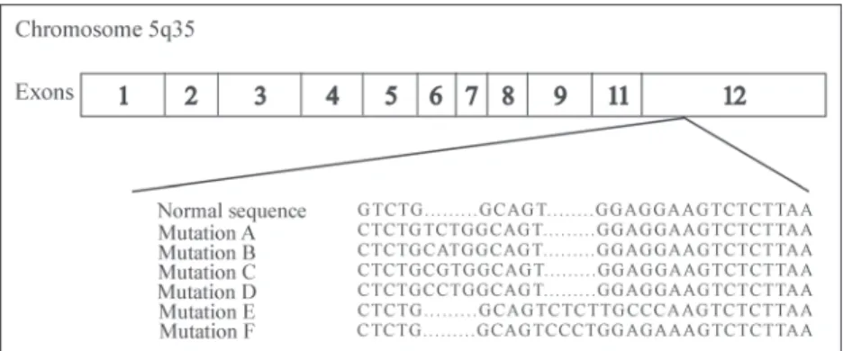
The gene responsible for the synthesis of nucleofosmine (NPM), also known as B23, NO38 or numatrin, was mapped on chromosome 5q35 in humans. This gene contains 12 exons that encode three isoforms of NPM: NPM1 (B23.1), NPM1.2 (B23.2) and NPM1.3 (B23.3). The NPM1 isoform is the most prevalent and has a C-terminal domain and an N-terminal region.(25) The isoform NPM1.2 is a
truncated isoform found at very low levels in tissues and isoform NPM1.3 has not been described much in the literature. (26)
NPM1 is a nucleolar phosphoprotein which moves between the nucleus and cytoplasm during the cell cycle and interacts with several proteins. Because of this behavior, NPM1 is a multifunctional protein, and is involved in the processing of ribosomal RNA, centriole duplication, response to stress stimuli such as UV irradiation and hypoxia, maintenance of genomic stability by controlling cell ploidy, participation in processes of DNA repair and transcription regulation through the modulation of events of condensation and decondensation of chromatin.(19,23,27) Moreover, NPM1
binds to p53 and regulates retinoblastoma (pRb)(28) the
p19ARF(29) and HDM2 proteins.(30)
F according to the insertion or deletion of four base pairs in exon 12 of the C-terminal region (Figure 1), which makes a new nuclear export signal occur.(26,31) NPM1 gene mutations,
apart from being identified by the mutations found in the gene, can also be investigated by phenotypic changes investigating abnormalities in the NPM1 protein.(1,31)
Many studies showed that NPM1 gene mutations were relevant only for patients with normal karyotypes.(17,24,31,32)
On the other hand, Haferlach et al.(33) recently reported that,
although AML patients with abnormal karyotypes represent a minority of individuals with NPM1 gene mutations, its definition, in respect to the biological, pathological and clinical characteristics, is very important. This study of Haferlach et al.(33) was conducted with 631 AML patients
who had NPM1 gene mutations; 14.7% of patients had abnormal karyotypes. However no differences were found in AML patients who had NPM1 mutations (normal or abnormal karyotypes) in respect to overall survival. This result confirms the concept that AML with NPM1 gene mutations should be clinically treated as a subtype, regardless of karyotype.(33)
In the few studies with chromosomal abnormalities associated with NPM1 gene mutations in AML patients, (22,23)
the combination of biological and clinical importance has not been thoroughly investigated. Hence, during the preparation of the WHO-2008 classification, a point of debate was that the name of the subtype "AML with NPM1 gene mutations" was provisional and not definitive.
Falini et al.(26) reported that around 75% - 80% of AML
patients with NPM1 gene mutations had type-A mutations. A recent study reported that individuals with type-A NPM1 mutations have a favorable prognosis, as was already known for NPM1 mutations in general, but also that NPM1 non-type-A mutations are of unfavorable prognosis.(34)
Prognostic value of FLT3 gene mutations in acute myeloid leukemia
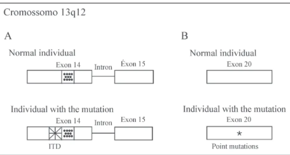
The gene encoding the FLT3 protein is located on chromosome 13q12 [Fms-like tyrosine kinase-3, also known
as fetal liver kinase-2 (FLK-2)] was first cloned in 1991 by Rosnet et al.(35) and by
Matthews et al.. (36) Under physiological conditions, transcription of the FLT3 gene encodes a monomeric protein consisting of an extracellular domain, a transmembrane region, a juxtamembrane region and two intracellular tyrosine kinase domains. In the presence of a ligand, dimerization of monomers occurs followed by phosphorylation of effector substrates of intracellular signal transduction pathways. The main pathways affected by FLT3 activation are PI3K (phosphatidilinositol 3-kinase) of the RAS/ERK/MAPK (mitogen-activated protein kinase) and STAT5 (signal transducers and activators of Transcription 5) pathways, resulting in increased proliferation and inhibition of apoptosis.(37)
When the FLT3 gene undergoes a mutation, a modified final product is produced, i.e. an FLT3 receptor with structural changes is generated. Thus, the tyrosine kinase domain is permanently activated, independent of ligands, which leads to uncontrolled proliferation of myeloid cells.(38) When this
occurs, the leukemia has a poor prognosis and requires appropriate treatment to contemplate this variable.(18) The
changes identified in the FLT3 gene are of two types: internal tandem duplications (ITD) or point mutations.(38) The ITD in
the FLT3 gene is the second most common mutation found in AML patients.(39) This change occurs in exons 14 or 15
(previously described as exons 11 or 12) (Figure 2-A) and affects 20% -35% of all AML patients and 5% - 10% of acute lymphoblastic leukemias.(20)
The incidence of ITD depends on the age of AML patients. In over 60-year-old individuals, the frequency of FLT3-ITD mutations is 30%-35%, while in younger AML patients, these molecular aberrations are detected in 20%-25%. These results are explained by a higher incidence of chromosomal aberrations as well as by a higher rate of secondary AML (e.g. secondary to myelodysplastic syndrome), associated with an unfavorable outcome in elderly AML patients.(37,41)
Nakao et al.(42) first described the ITD in the
juxtamembrane region of one FLT3 allele in AML patients. The molecular model of the functioning of the altered protein has been described by in vitro studies that demonstrated uncontrolled dimerization of the juxtamembrane region caused by ITD, which loses its autoinhibitory ability and allows dimerization of the receptors independent of the ligand leading to autophosphorylation and autonomous growth of mutant cells.(43) With the presence of ITD,
autophosphorylation of the FLT3 receptor occurs, leading to permanent activation of this receptor, resulting in the activation of cell signaling pathways such as ERK and STAT5.(44)
Licínio MA et al. Rev Bras Hematol Hemoter. 2010;32(6):476-481
The clinical significance of FLT3 mutations in AML was established from the correlation between the presence of ITD associated with leukocytosis, high percentages of blasts and unfavorable response to therapy.(20) Some groups
have also detected the presence of FLT3-ITD type mutations concomitant with other changes, for example, t(15;17), indicating a favorable prognosis, whereas the presence of t(6,9) is associated with unfavorable prognosis.(21,45) However,
the prognostic value of ITD in the FLT3 gene, associated with other changes, has not yet been well established, since there are published reports about ITD associated with high levels of relapse, with remission levels similar to a reference group without these mutations, or even associated with favorable clinical response.(46,47)
FLT3 gene point mutations involve the activation loop of the second kinase domain, with the most common mutation resulting from the substitution of an aspartic acid residue at position 835 of exon 20 (formerly exon 17) (Figure 2-B) for a tyrosine residue (D835).(48) The activation loop is a common
component of tyrosine-kinase receptors and its function is to block access of ATP and substrate to the kinase domain when the receptor is inactive. With a mutation in this region, the receptor is autoactivated and thus as in the presence of ITD, control of the signaling cascade promoted by FLT3 is lost, leading to cell proliferation.(49)
The D835 type mutation, also known as FLT3-TKD, was described in approximately 5%-10% of AML patients and has been correlated to an unfavorable prognosis.(48,50,51) In the presence of two changes in FLT3,
the low incidence (1.7%) reported makes an assessment of the clinical value difficult, however, the poor prognosis seems to prevail and in vitro studies suggest the development of resistance to conventional and specific therapies for the receptor.(46)
In general, changes in FLT3, identified as a prognostic factor, have already been incorporated to determine risk and
intensification of therapy in recently updated protocols in developed countries.(18) While
there is disagreement as to the aggressiveness of the disease, all studies reveal high white blood cell counts and lower remission rates with this mutation. Some studies have also assessed the possibility of using changes in FLT3 as tumor markers to identify minimal residual disease. The fact is that gains and losses of mutations have been observed during the course of treatment, which restricts the value of FLT3 as a marker indicative of relapse.(52,53)
As already described in this article, AML patients with NPM1 gene mutations have a favorable prognosis, but if the patient has concomitant ITD for the FLT3 gene, the prognosis is considered unfavorable.(26,31,33,34,39,54)
Final considerations
In the literature, some phase I and II,(55) and phase III
clinical trials(56) investigated the benefits of the use of FLT3
inhibitors in patients with FLT3 gene mutations. These results show the importance of the investigation of FLT3 gene mutations for stratification of prognosis in AML, because the presence of these mutations will affect the choice of treatment.(18) In addition, new studies involving the
investigation of NPM1 gene mutations are extremely important because they can clarify the characterization of the subtype "AML with NPM1 gene mutations."
Resumo
As leucemias mieloides agudas (LMA) constituem um grupo de neoplasias malignas caracterizadas pela proliferação descontrolada de células hematopoéticas, decorrente de mutações que podem ocorrer em diferentes fases da diferenciação de células precursoras mieloides. Em 2008, a Organização Mundial da Saúde (OMS-2008) publicou uma nova classificação para neoplasias do sistema hematopoético e linfoide. De acordo com essa classificação, para um diagnóstico mais preciso e estratificação de prognóstico de pacientes com leucemias mieloides agudas, devem-se pesquisar mutações nos genes FLT3 e NPM1. Sabe-se que a presença de mutações no gene FLT3 é de prognóstico desfavorável e que as mutações no gene NPM1 do tipo A são de prognóstico favorável. Assim, nos países desenvolvidos, a análise das mutações no gene FLT3 e NPM1 tem sido considerada como um fator de prognóstico importante na decisão terapêutica em pacientes com diagnóstico de leucemias mieloides agudas. Considerando essas informações, é de extrema importância a análise das mutações no gene FLT3 (duplicação interna em tandem - DIT - e mutação pontual D835) e no gene NPM1 como marcadores moleculares para o diagnóstico, o prognóstico e a Figure 2. Diagram of mutations in the FLT3 gene
monitoração de doença residual mínima em pacientes com leucemias mieloides agudas.
Descritores: Leucemia mieloide aguda/genética; Genes neoplásicos; Mutação; Organização Mundial da Saúde; Neoplasias hemato-lógicas/classificação; Leucemia/classificação
References
1. Swerdlow SH, Campo E, Harris NL, Jaffe ES, Pileri SA, Stein H, et al. WHO Classification of tumours of haematopoietic and lymphoid tissues. Lyon: IARC Press; 2008.
2. Estey EH. Therapeutic options for acute myelogenous leukemia. Cancer. 2001;92(5):1059-73.
3. Pui CH, Evans WE. Acute lymphoblastic leukemia. N Engl J Med. 1998;339(9):605-15.
4. Bain BJ. Diagnóstico em Leucemias. 2a ed. Rio de Janeiro: Revinter; 2003.
5. Rubnitz JE, Pui CH. Molecular diagnostics in the treatment of leukemia. Curr Opin Hematol. 1999;6(4):229-35.
6. Greaves M. Molecular genetics, natural history and the demise of childhood leukaemia. Eur J Cancer. 1999;35(2):173-85. Review.
7. Slovak ML, Kopecky KJ, Cassileth PA, Harrington DH, Theil KS, Mohamed A, et al. Karyotypic analysis predicts outcome of preremission and postremission therapy in adult acute myeloid leukemia: a Southwest Oncology Group/Eastern Cooperative Oncology Group Study. Blood. 2000;96(13):4075-83.
8. Byrd JC, Mrózek K, Dodge Rk, Carroll AJ, Edwards CG, Arthur DC, et al. Pretreatment cytogenetic abnormalities are predictive of induction success, cumulative incidence of relapse, and overall survival in adult patients with "de novo" acute myeloid leukemia: results from Cancer and Leukemia Group B (CALGB 8461). Blood. 2002;100(13):4325-36.
9. Baldus CD, Mrózek K, Marcucci G, Bloomfield CD. Clinical outcome of de novo acute myeloid leukaemia patients with normal cytogenetics is affected by molecular genetic alterations: a concise review. Br J Haematol. 2007;137(5):387-400.
10. Huang Q, Chen W, Gaal KK, Slovak ML, Stein A, Weiss LM. A rapid, one step assay for simultaneous detection of FLT3/ITD and NPM1 mutations in AML with normal cytogenetics. Br J Haematol. 2008;142(3):489-92.
11. Schnittger S, Schoch C, Dugas M, Kern W, Staib P, Wuchter C, et al. Analysis of FLT3 length mutations in 1003 patients with acute myeloid leukemia: correlation to cytogenetics, FAB subtype, and prognosis in the AMLCG study and usefulness as a marker for the detection of minimal residual disease. Blood. 2002;100(1):59-66.
12. Mrózek K, Heerema NA, Bloomfield CD. Cytogenetics in acute leukemia. Blood Rev. 2004;18(2):115-36.
13. Speck NA, Gilliland DG. Core-binding factors in haematopoiesis and leukaemia.Nat Rev Cancer. 2002;2(7):502-13.
14. Schlenk RF, Döhner K, Krauter J, Fröhling S, Corbacioglu A, Bullinger L, et al. Mutations and Treatment Outcome in Cytogenetically Normal Acute Myeloid Leukemia. N Engl J Med. 2008;358 (18):1909-18. Comment in: N Engl J Med. 2008; 359(6):652; author reply 652-3, N Engl J Med. 2008;359(6):652; author reply 652-3, N Engl J Med. 2008;359(6):651; author reply 652-3, N Engl J Med. 2008;359(6):651-2; author reply 652-3, N Engl J Med. 2008;358(18):1960-2.
15. Ishikawa Y, Kiyoi H, Tsujimura A, Miyawaki S, Miyazaki Y, Kuriyama K, et al. Comprehensive analysis of cooperative gene
mutations between class I and class II in de novo acute myeloid leukemia. Eur J Haematol. 2009;83(2):90-8.
16. Andersen MT, Andersen MK, Christiansen DH, Pedersen-Bjergaard J. NPM1 mutations in therapy-related acute myeloid leukemia with uncharacteristic features. Leukemia. 2008 ;22(5):951-5.
17. Mrózek K, Marcucci G, Paschka P, Whitman SP, Bloomfield CD. Clinical relevance of mutations and gene-expression changes in adult acute myeloid leukemia with normal cytogenetics: are we ready for a prognostically prioritized molecular classification? Blood. 2007;109(2):431-48.
18. National Comprehensive Cancer Network (NCCN). Practice Guidelines in Oncology: Acute Myeloid Leukemia . Washington: NCCN; c2008. Vol.1.
19. Grisendi S, Mecucci C, Falini B, Pandolfi PP. Nucleophosmin and cancer. Nat Rev Cancer. 2006;6(7):493-505.
20. Kottaridis PD, Gale RE, Frew ME, Harrison G, Langabeer SE, Belton AA, et al. The presence of a FLT3 internal tandem duplication in patients with acute myeloid leukemia (AML) adds important prognostic information to cytogenetic risk group and response to the first cycle of chemotherapy: analysis of 854 patients from the United Kingdom Medical Research Council AML 10 and 12 trials. Blood. 2001;98(6):1752-9.
21. Thiede C, Steudel C, Mohr B, Schaich M, Schäkel U, Platzbecker U, et al. Analysis of FLT3-activating mutations in 979 patients with acute myelogenous leukemia: association with FAB subtypes and identification of subgroups with poor prognosis. Blood. 2002;99(12):4326-35.
22. Verhaak RG, Goudswaard CS, van Putten W, Bijl MA, Sanders MA, Hugens W, et al. Mutations in nucleophosmin (NPM1) in acute myeloid leukemia (AML): association with other gene abnormalities and previously established gene expression signatures and their favorable prognostic significance. Blood. 2005;106 (12):3747-54.
23. Thiede C, Koch S, Creutzig E, Steudel C, Illmer T, Schaich M, et al. Prevalence and prognostic impact of NPM1 mutations in 1485 adult patients with acute myeloid leukemia (AML). Blood. 2006;107(10):4011-20.
24. Gale RE, Green C, Allen C, Mead AJ, Burnett AK, Hills RK, et al. The impact of FLT3 internal tandem duplication mutant level, number, size, and interaction with NPM1 mutations in a large cohort of Young adult patients with acute myeloid leukemia. Blood. 2008;111(5):2776-84.
25. Namboodiri VM, Akey IV, Schmidt-Zachmann MS, Head JF, Akey CW. The Structure and Function of Xenopus NO38-Core, a Histone Chaperone in the Nucleolus. Structure. 2004;12(12):2149-60. 26. Falini B, Nicoletti I, Martelli MF, Mecucci C. Acute myeloid leukemia
carrying cytoplasmic/mutated nucleophosmin (NPMc + AML): biologic and clinical features. Blood. 2007;109(3):874-85. 27. Lim MJ, Wang XW. Nucleophosmin and human cancer. Cancer
Detect Prev. 2006;30(6):481-90.
28. Takemura M, Ohoka F, Perpelescu M, Ogawa M, Matsushita H, Takaba T, et al. Phosphorylation-Dependent Migration of Retinoblastoma Protein into the Nucleolus Triggered by Binding to Nucleophosmin/B23. Exp Cell Res. 2002;276(2):233-41.
29. Bertwistle D, Sugimoto M, Sherr C. Physical and Functional Interactions of the Arf Tumor Suppressor Protein with Nucleophosmin/B23. Mol Cell Biol. 2004;24(3):985-96. 30. Kurki S, Peltonen K, Latonen L, Kiviharju TM, Ojala PM, Meek
D, et al. Nucleolar protein NPM interacts with HDM2 and protects tumor suppressor protein p53 from HDM2-mediated degradation. Cancer Cell. 2004;5(5):465-75.
Licínio MA et al. Rev Bras Hematol Hemoter. 2010;32(6):476-481
with a Normal Karyotype. N Engl J Med. 2005;352(3):254-66. Erratum in:N Engl J Med. 2005;352(7):740. Comment in:N Engl J Med. 2005 Jan 20;352(3):291-2,N Engl J Med. 2005; 352 (17): 1819-20; author reply 1819-20.
32. Akagi T, Ogawa S, Dugas M, Kawamata N, Yamamoto G, Nannya Y, et al. Frequent genomic abnormalities in acute myeloid leukemia/ myelodysplastic syndrome with normal karyotype. Haematologica. 2009;94(2):213-23.
33. Haferlach C, Mecucci C, Schnittger S, Kohlmann A, Mancini M, Cuneo A, et al. AML with mutated NPM1 carrying a normal or aberrant karyotype show overlapping biological, pathological, immunophenotypic, and prognostic features. Blood. 2009;114 (14):3024-32.
34. Koh Y, Park J, Bae EK, Ahn KS, Kim I, Bang SM, et al. Non-A type nucleophosmin 1 gene mutation predicts poor clinical outcome in de novo adult acute myeloid leukemia: differential clinical importance of NPM1 mutation according to subtype. Int J Hematol. 2009;90(1):1-5.
35. Rosnet O, Matteï MG, Marchetto S, Birnbaum D. Isolation and chromosomal localization of a novel FMS-like tyrosine kinase gene. Genomics. Genomics. 1991;9(2):380-5.
36. Matthews W, Jordan CT, Wiegand GW, Pardoll D, Lemischka IR. A receptor tyrosine kinase specific to hematopoietic stem and progenitor cell-enriched populations. Cell. 1991;28(7): 1 1 4 3 - 5 2 .
37. Stirewalt DL, Kopecky KJ, Meshinchi S, Appelbaum FR, Slovak ML, Willman CL, et al. FLT3, RAS, and TP53 mutations in elderly patients with acute myeloid leukemia. Blood. 2001; 97 (11):3589-95.
38. Reindl C, Bagrintseva K, Vempati S, Schnittger S, Ellwart JW, Wenig K, et al. Point mutations in the juxtamembrane domain of FLT3 define a new class of activating mutations in AML. Blood. 2006;107(9):3700-7.
39. Scholl S, Fricke HJ, Sayer HG, Höffken K. Clinical implications of molecular genetic aberrations in acute myeloid leukemia. J Cancer Res Clin Oncol. 2009;135(4):491-505.
40. Stirewalt D, Radich JP. The role of FLT3 in haematopoietic malignancies. Nat Rev Cancer. 2003;3(9):650-65.
41. Andersson A, Johansson B, Lassen C, Mitelman F, Billström R, Fioretos T. Clinical impact of internal tandem duplications and activating point mutations in FLT3 in acute myeloid leukemia in elderly patients. Eur J Haematol. 2004;72(5):307-13.
42. Nakao M, Yokota S, Iwai T, Kaneko H, Horiike S, Kashima K, et al. Internal tandem duplication of the FLT3 gene found in acute myeloideukemia. Eur J Haematol. 2004;72(5):307-13.
43. Kiyoi H, Towatari M, Yokota S, Hamaguchi M, Ohno R, Saito H, Naoe T. Internal tandem duplication of the FLT3 gene is a novel modality of elongation mutation which causes constitutive activation of the product. Leukemia. 1998;12(9):1333-7. 44. Griffith J, Black J, Faerman C, Swenson L, Wynn M, Lu F, et al.
The Structural basis for autoinhibition of FLT3 by the juxtamembrane domain. Mol Cell. 2004;13(2):169-78. 45. Libura M, Asnafi V, Tu A, Delabesse E, Tigaud I, Cymbalista F, et al.
FLT3 and MLL intragenic abnormalities in AML reflect a common category of genotoxic stress. Blood. 2003;102(6):2198-204. 46. Lacayo NJ, Meshinchi S, Kinnunen P, Yu R, Wang Y, Stuber CM,
et al. Gene expression profiles at diagnosis in de novo childhood AML patients identify FLT3 mutations with good clinical outcomes. Blood. 2004;104(9):2646-54.
47. Gale RE, Hills R, Pizzey AR, Kottaridis PD, Swirsky D, Gilkes AF, et al. Relationship between FLT3 mutation status, biologic characteristics, and response to targeted therapy in acute promyelocytic leukemia. Blood. 2005;106(12):3768-76.
48. Yamamoto Y, Kiyoi H, Nakano Y, Suzuki R, Kodera Y, Miyawaki S, et al. Activating mutation of D835 within the activation loop of FLT3 in human hematologic malignancies. Blood. 2001;97(8):2434-9.
49. Griffin JD. Point mutations in the FLT3 gene in AML. Blood. 2001;97(8):2193A-2193.
50. Choudhary C, Schwäble J, Brandts C, Tickenbrock L, Sargin, B, Kindler T, et al. AML-associated FLT3 kinase domain mutations show signal transduction differences compared with FLT3 ITD mutations. Blood. 2005;106(1):265-73.
51. Grundler R, Miething C, Thiede C, Peschel C, Duyster J. FLT3-ITD and tyrosine kinase domain mutants induce 2 distinct phenotypes in a murine bone marrow transplantation model. Blood. 2005;105(12):4792-9.
52. Kottaridis PD, Gale RE, Langabeer SE, Frew ME, Bowen DT, Linch DC. Studies of FLT3 mutations in paired presentation and relapse samples from patients with acute myeloid leukemia: implications for the role of FLT3 mutations in leukemogenesis, minimal residual disease detection and possible therapy with FLT3 inhibitors. Blood. 2002;100(7):2393-8.
53. Heidel F, Solem FK, Breitenbuecher F, Lipka DB, Kasper S, Thiede MH, et al. Clinical resistance to the kinase inhibitor PKC412 in acute myeloid leukemia by mutation of Asn-676 in the FLT3 tyrosine kinase domain. Blood. 2006 Jan 1;107(1): 2 9 3 - 3 0 0 .
54. Schnittger S, Schoch C, Kern W, Mecucci C, Tschulik C, Martelli MF, et al. Nucleophosmin gene mutations are predictors of favorable prognosis in acute myelogenous leukemia with a normal karyotype. Blood. 2005;106(12):3733-9.
55. Shah M, Agarwal B. Recent advances in management of acute myeloid leukemia (AML). Indian J Pediatr. 2008;75(8):831-7.Ohtake S. Acute myeloid leukemia. Gan To Kagaku Ryoho. 2007;34(13):2175-9.