Arq Neuropsiquiatr 2006;64(3-A):664-667
1N e u ro s u rg e ry, Fundação Benjamin Guimarães/Hospital da Baleia, Belo Horizonte MG, Brasil;2N e u ro s u rg e ry, To ronto We s t e rn
Hospital - University of Toronto, Toronto, M5T 2S8 Canada.
Received 14 September 2005, received in final form 23 January 2006. Accepted 4 April 2006. Dr. Leodante B. da Costa - Rua Três Corações 13 / 302 - 30480-110 Belo Horizonte MG - Brasil
SYMPTOMATIC NON-ATHEROSCLEROTIC
BILATERAL EXTRACRANIAL VERTEBRAL
ARTERY OCCLUSION TREATED WITH
EXTRACRANIAL TO INTRACRANIAL BYPASS
Case report
Leodante B. da Costa
1,2, Michael Tymianski
2ABSTRACT - Posterior fossa ischemia is not a very frequent situation. It is responsible for about 25% of all ischemic strokes, and the vast majority of the cases are related to atherosclerotic stenosis of the vertebral and/or basilar arteries. Acute ischemia can also occur in the setting of vertebral art e ry dissection, traumat-ic or spontaneous. Recently, blunt trauma has been increasingly recognized as a cause for craniocerv i c a l a rt e ry injury. The management options for both traumatic and athero s c l e rotic lesions of the posterior fos-sa are still under debate. We present a case of a delayed onset of hemodynamic ischemic symptoms due to bilateral vertebral art e ry occlusion probably related to remote trauma to the head and neck in a 55-year-old- man treated successfully with extracranial to intracranial bypass.
KEY WORDS: EC-IC bypass, ischemic stroke, surgical revascularization.
Oclusão sintomática não-aterosclerótica da artéria vertebral extracraniana tratada com bypass: relato de caso
RESUMO - Acidentes vasculares cerebrais (AVC) isquêmicos no sistema vert e b ro-basilar não são fre q u e n t e s . R e p resentam cerca de 25% dos AVCs isquêmicos, e a maioria é relacionada com atero s c l e rose das art é r i a s v e rtebrais e/ou basilar. Isquemia aguda pode também ser resultado de dissecções da artéria vert e b r a l , traumáticas ou espontâneas. Recentemente, traumatismos fechados têm sido cada vez mais re c o n h e c i d o s como causa de lesão das artérias craniocervicais, podendo ou não resultar em sintomas isquêmicos. O trata-mento para estas lesões, sejam traumáticas ou ateroscleróticas, ainda é motivo de debate. Relatamos o caso de um homem de 55 anos com sintomas isquêmicos, hemodinâmicos, tardios, devido a oclusão bilate-ral das artérias vertebrais, provavelmente relacionada a lesão traumática das artérias vertebrais, tratada com sucesso com bypass extra-intracraniano.
PALAVRAS-CHAVE: bypass, isquemia cerebral, revascularização intracraniana.
Posterior fossa ischemia is not a very frequent sit-uation. It is responsible for about 25% of all ischemic s t rokes, the vast majority related to athero s c l e ro t i c stenosis of the vertebral and/or basilar arteries. Blunt trauma to the head and neck is being recognized as a cause of carotid and vertebral art e ry injury, most c o m m o n l y, carotid dissections. Since its institution in the 50s, anticoagulation has been the main stem of medical management of vert e b ro-basilar insuff i c i e n-c y. The use of surgin-cal means to treat intran-cranial is-chemic symptoms has been disfavored since the
EC-IC bypass study, although the surgical treatment of v e rt e b robasilar insufficiency can be considered in cas-es where there is no rcas-esponse to maximal medical the-rapy and no endovascular options. We present a case w h e resurgical treatment of posterior fossa ischemia resulted in complete resolution of symptoms.
CASE
Arq Neuropsiquiatr 2006;64(3-A) 665
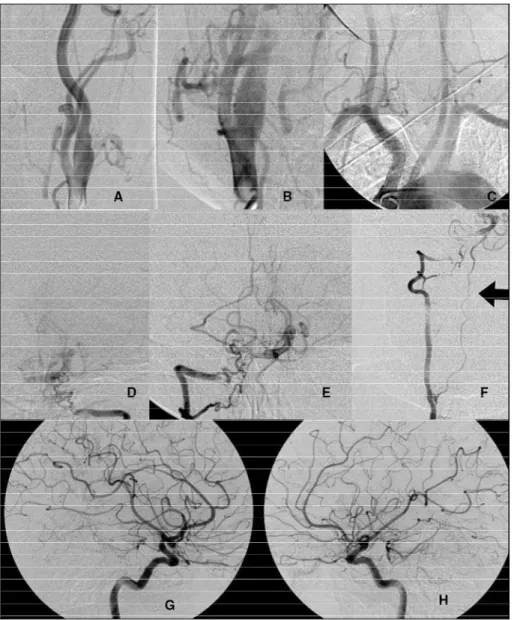
trauma with loss of consciousness 12 years ago. For the last one and a half years he had been presenting sudden epi-sodes of double vision, unsteadiness and vertigo, and occa-sionally left-sided weakness and numbness. He had multi-ple episodes a day, and noticed that they improved faster with some physical exercise. He was mildly hypert e n s i v e , but could not tolerate antihypertensive medications, which a p p e a red to aggravate his symptoms. He was also using antiplatelets, but still having symptoms fre q u e n t l y. Neuro l o-gical examination was unremarkable. Physical examination was also normal, except for mild hypertension. A magnet-ic resonance imaging study (MRI) was done, and suggest-ed bilateral extracranial vertebral occlusion, which was
con-f i rmed by an angiogram. The angiogram also showed that the posterior fossa circulation was supplied only by collat-eral circulation through very small posterior communicat-ing arteries and some collateral flow from extracranial art e r-ies. Of note, an enlarged anterior spinal art e ry was seen being responsible for most of the flow in the lower basilar a rt e rysegment (Fig 1). The patient was then submitted to a bilateral occipital to PICA bypasses, in order to incre a s e the blood flow to the posterior fossa (Fig 2). Postoperative angiogram done two days after the pro c e d u re showed good blood flow through the bypasses (Fig 3). In his last vi-sit, two months after the pro c e d u re, he had not had any episode of the previous ischemic symptoms.
666 Arq Neuropsiquiatr 2006;64(3-A)
DISCUSSION
Posterior fossa ischemia represents roughly 25% of all ischemic strokes. The most common cause is a t h e ro s c l e rotic stenosis of the vertebral and/or basi-lar arteries. The anatomy of the posterior fossa cir-culation is such that most of the times the occlusion of one of the vertebral arteries is well tolerated. In the absence of congenital anomalies, the collateral circulation is usually sufficient to keep an adequate blood flow. Trauma, soft tissue or osseous compres-sions are also cited as cause of compression and steno-sis of the vertebral artery in the neck, although less frequently1-4.
Blunt trauma to the head and neck is being re c o g-nized as a cause of carotid and vertebral art e ry inju-ry, most commonly, carotid dissections. The real inci-dence of vertebral art e ry injury after blunt trauma is unknown as also is its clinical significance. Series re-p o rt an incidence of 30 to 60% of carotid and/or ver-tebral art e ry injury associated with some patterns of blunt head and neck trauma. Some patterns of
cer-vical spine fracture are being related to high risk of vessel injury, with some authors suggesting that al-most every patient with a cervical spine fracture should be screened for craniocervical vessel injury5 - 1 0.
The prognosis of posterior fossa stenosis is not well determined, although an increased incidence of s t roke is suggested1. In a series from the Cleveland
C l i n i c1 1, including also asymptomatic patients with
a t h e ro s c l e rotic stenosis, an increased stroke rate of 5.25% per year, most in the anterior circulation was o b s e rved. The presence of intracranial vertebral art e ry stenosis, which is often associated with basilar art e ry stenosis, seems to increase the risk of posterior fos-sa stroke to 7% per annum, if compared to extracra-nial stenosis alone. The inclusion of patients with as-sociated carotid disease and asymptomatic patients may have biased the results, however. Traumatic le-sions are associated with early of delayed ischemic symptoms, and aggressive medical management with full anticoagulation is suggested. Better pro g n o s i s (lower stroke rates) for the patients with lesions
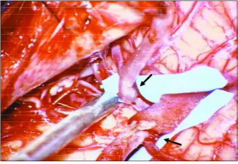
diag-Fig 2. Intraoperative picture showing the bilateral occipital to PICA bypasses (arro w s ) .
Arq Neuropsiquiatr 2006;64(3-A) 667
nosed and treated before the onset of ischemic symp-toms has been demonstrated in some case series5 , 6 , 8 , 9.
Hemodynamic insufficiency seems to be the most common mechanism of symptoms associated with vertebro-basilar stenosis. In vertebral stenosis cases, the involvement of both arteries or some anomaly (eg: ending in a PICA art e ry, hypoplastic vertebral ar-t e ry) ar-thaar-t compromises ar-the conar-tralaar-teral blood flow need to be present for symptoms to occur2,3,10-12.
Since its institution in the 50s, anticoagulation has been the main stem of medical management of ver-t e b ro-basilar insuff i c i e n c y. Anver-tiplaver-telever-ts may also be used if embolism is a consideration. Endovascular techniques are being more and more commonly used in the treatment of intracranial vascular insufficien-c y, and it has it is now the first insufficien-choiinsufficien-ce in insufficien-cases of s y m p-tomatic proximal vertebral artery occlusion11-14.
S u rgical treatment of vert e b robasilar insuff i c i e cy can be considered in cases where there is no re sp o n-se to maximal medical therapy and no endovascular options. In the past, both extracranial and intracra-nial vertebral endart e rectomies were done1 5 - 1 8. The
extracranial vertebral endart e rectomy is being re-placed by the more convenient and less invasive en-dovascular angioplasty and stenting. The long term duration of this treatment is still to be determ i n e d . The complexity and high risk of the intracranial vert e-bral art e ry endart e rectomy prompt the search for new options for the treatment of intracranial vert e-b roe-basilar insuff i c i e n c y, and angioplasty with or with-out stenting is rising as an option. The use of EC-IC bypass for the treatment of intracranial vascular insuf-ficiency was nearly abandoned after the publication of the results of the EC-IC bypass trial1 9. Extracranial
to intracranial circulation bypasses are an intuitive-ly very attractive technique to increase the blood flow to a given area of the brain, and in spite of the results of the EC-IC trial, it seems to have an indica-tion in selected cases15,20-22.
We believe that our patient had bilateral extracra-nial vertebral art e ry occlusion due to traumatic injury of the arteries. He is otherwise healthy, with no risk factors for athero s c l e rotic disease except for mild hy-p e rtension, and the angiograhy-phic ahy-phy-pearance of the stenosis does not suggest atherosclerotic lesions. Its late presentation, for which we do not have an expla-nation, makes his case unique. The clear hemodyna-mic nature of his symptoms indicated to us that a p ro c e d u reto improve the blood flow in the posteri-or fossa was indicated. The need fposteri-or better blood supply could also be inferred from the extremely
en-l a rged anterior spinaen-l art e ry. Endovascuen-lar tre a t m e n t was not considered an option in his case, and bilat-eral occipital to PICA bypasses were perf o rmed, with complete resolution of the symptoms. In spite of not being commonly used nowadays, we believe that re-vascularization pro c e d u res still have an import a n t role in the management of patients with cere b ro v a s-cular insuff i c i e n c y, especially in carefully selected sub-groups.
REFERENCES
1. P rognosis of patients with symptomatic vertebral or basilar artery steno-sis. The Warfarin-Aspirin Symptomatic Intracranial Disease (WASID) Study Group. Stroke 1998;29:1389-1392.
2. Cloud GC, Markus HS. Diagnosis and management of vertebral artery stenosis. QJM 2003;96:27-34.
3. Moufarrij NA, Little JR, Furlan AJ, Williams G, Marzewski DJ. Ve r t e b r a l artery stenosis: long-term follow-up. Stroke 1984;15:260-263. 4. Voetsch B, DeWitt LD, Pessin MS, Caplan LR. Basilar artery occlusive
disease in the New England Medical Center Posterior Circ u l a t i o n Registry. Arch Neurol 2004;61:496-504.
5. B i ffl WL, Moore EE, Elliott JP, et al. The devastating potential of blunt vertebral arterial injuries. Ann Surg 2000;231:672-681.
6. B i ffl WL, Ray CE Jr., Moore EE, et al. Tre a t m e n t - related outcomes fro m blunt cere b rovascular injuries: importance of routine follow-up arte-riography. Ann Surg 2002;235:699-707.
7. C o t h ren CC, Moore EE, Biffl WL, et al. Cervical spine fracture patterns predictive of blunt vertebral artery injury. J Trauma 2003;55:811-813. 8. Kerwin AJ, Bynoe RP, Murray J, et al. Liberalized screening for blunt
carotid and vertebral artery injuries is justified. J Trauma 2001;51:308-314.
9. Miller PR, Fabian TC, Bee TK, et al. Blunt cere b rovascular injuries: diag-nosis and treatment. J Trauma 2001;51:279-285.
10. Miller PR, Fabian TC, Croce MA, et al. Prospective screening for blunt c e re b rovascular injuries: analysis of diagnostic modalities and out-comes. Ann Surg 2002;236:386-393.
11. Robertson JT. Current management of vertebral basilar occlusive dis-ease. Clin Neurosurg 1983;31:165-187.
12. Oliveira-Filho J, Pedreira BB, Jesus PA P, et al. Pharmacologically-induced hypertension in a patient with vertebro-basilar territory ischemia associated with bilateral vertebral stenosis. A rq Neuro p s i q u i a t r 2002;60:498-501.
13. D rescher P, Katzen BT. Percutaneous treatment of symptomatic verte-bral artery stenosis with coronary stents. Catheter Cardiovasc Interv 2001;52:373-377.
14. Misra M, Alp MS, Hier D, Ausman JI. Multidisciplinary treatment of posterior circulation ischemia. Neurol Res 2004;26:67-73.
15. Ausman JI, Diaz FG, Dujovny M. Posterior circulation re v a s c u l a r i z a-tion. Clin Neurosurg 1986;33:331-343.
16. Ausman JI, Diaz FG, Sadasivan B, Dujovny M. Intracranial vertebral endarterectomy. Neurosurgery 1990;26:465-471.
17. Hopkins LN, Martin NA, Hadley MN, Spetzler RF, Budny J, Carter LP. Ve r t e b robasilar insuff i c i e n c y. Part 2. Micro s u rgical treatment of intracra-nial vertebrobasilar disease. J Neurosurg 1987;66:662-674.
18. Spetzler RF, Hadley MN, Martin NA, Hopkins LN, Carter LP, Budny J. Ve r t e b robasilar insuff i c i e n c y. Part 1: Micro s u rgical treatment of extracranial vertebrobasilar disease. J Neurosurg 1987;66:648-661. 19. F a i l u re of extracranial-intracranial arterial bypass to reduce the risk of
ischemic stroke. Results of an international randomized trial. The EC/IC Bypass Study Group. N Engl J Med 1985;313:1191-1200.
20. Ausman JI, Caplan LR, Diaz FG. Surgically created posterior circ u l a-tion vascular shunts. Clin Neurosurg 1986;33:327-330.
21. Ausman JI, Diaz FG, Vacca DF, Sadasivan B. Superficial temporal and occipital artery bypass pedicles to superior, anterior inferior, and pos-terior inferior cerebellar arteries for vertebrobasilar insuff i c i e n c y. J Neurosurg 1990;72:554-558.