Arq. NeuroPsiquiatr. vol.57 número3A
Texto
Imagem


Documentos relacionados
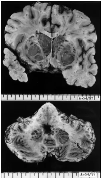
A contrast- enhanced C T scan showed retractile, hypodense, circumscribed lesions, which involves the cortex and white- matter, in both the temporal opercular regions, extending to
The present work aimed to study the cytotoxic activity of NK cells and T cell subsets in peripheral blood of 13 patients with primary tumors in central nervous system (CNS). As
In Benton’s analysis from patients with impairment in the visuo-spatial orientation (they were not cases of myotonic dystrophy), there were cases with full rotation of the figure
Sleep patterns in ages ranging from 2 to 10 year olds studied by us in the isolated rural Black community of Furnas do Dionísio have quite distinct characteristics.. Cosleeping
The cranial CT scan showed twenty- eight rounded hypodense lesions located at cerebral and cerebellar hemispheres, measuring between 6 and 18 mm (average, 13 mm), with
The cervical radiographs showed a lytic expansible lesion involving the body and especial- ly the posterior arch of C2, which presented multiple luid-luid levels at the CT scan
Computed tomography (CT) showed a bulk tumor in the right temporal lobe, with ir- regular contrast enhancement (Fig 1).. A radical resection of the neoplasia was performed, and
às instituições de ensino superior (IES) na sua capacidade de desenvolverem, mas também de aplicarem políticas promotoras de interculturalidade; Objetivos Conhecer as
