The role of Tumor Necrosis
Factor -Alpha (TNF-
α
) in bone
resorption present in middle
ear cholesteatoma
Summary
Rodrigo Faller Vitale 1, Fernando de Andrade
Quintanilha Ribeiro 2
1 Preceptor of Otorhinolaryngology – Otorhinus Clinic and Santa Marcelina Hospital. 2 Adjunct Professor of Otorhinolaryngology – School of Medical Sciences - Santa Casa de São Paulo.
Mailing Address: R. Zacarias de Goes 1296 Pq. Colonial 04610-003 São Paulo SP.
Paper submitted to the ABORL-CCF SGP (Management Publications System) on June 30th, 2005 and accepted for publication on July 2nd, 2006. cod. 477.
C
holesteatoma may cause bone erosion, with high morbidity and mortality rates. Tumor Necrosis Factor -Alpha (TNF-a) is one of the main cytokines involved in this process. Our goal was to evaluate the role of TNF-a in Bone Resorption and its effect on cholesteatoma. Material and Methods: analysis and critical literature review. Results: Different studies have demonstrated that TNF-a is capable of causing bone erosion. It may stimulate the differentiation and maturation of osteoclasts or it may act on the bone matrix, exposing it to the action of the osteoclasts. It is possible to inhibit TNF-a, reducing its effects and prevent bone loss in illnesses such as rheumatoid arthritis,and there has been no specific investigation regarding cholesteatomas. All studies agree on the importance of TNF-a in the bone resorption process present in cholesteatomas, and on the degree of destruction observed; however, there is no consensus as to its location. These differences are probably due to receptor site. Conclusion: TNF-a, present in cholesteatomas, promotes bone resorption, along with other cytokines (RANKL and IL-1) related to complications.Keywords: cholesteatoma, tumor necrosis factor, ear middle, bone resorption.
REVIEW ARTICLE Rev Bras Otorrinolaringol
INTRODUCTION
Acquired middle ear cholesteatoma was first des-cribed by Curveilhier in 1829. It is characterized by the invasion of keratinized stratified squamous epithelium into the tympanic cavity. This epithelium is different from that usually found in the middle ear.1
Cholesteatoma may cause bone destruction and result in intratemporal and intracranial complications,2 which have high morbidity and mortality rates. This jus-tifies our study of the bone destruction mechanisms in cholesteatoma.
There are numerous theories to explain the des-tructive properties (erosion) of cholesteatomas. Pressure due to accumulated keratin and other waste was initially proposed as the cause of destruction. A biochemical theory eventually was postulated, in which enzymes and cytokines released by cholesteatomas would cause bone lysis and destruction.3
Cytokines are a group of low molecular weight proteins for communication between cells.4 Cytokines are released as a result of a variety of stimuli, and interact with their receptors to regulate cell function.5 They are closely related to inflammation. The tumor necrosis factor alpha (TNF-α) was discovered by Carswell et al.6 in 1975; it is considered one of the main cytokines related to inflam-mation and immune processes, and operates in various parts of the body. TNF-α is secreted by macrophages, lymphocytes and monocytes mostly as a result of the pre-sence of bacterial lipopolysaccharides (LPS). TNF-α and other cytokines cause bone destruction and remodelling7 in cholesteatomas.
The aim of this paper is to analyze the importance of tumor necrosis factor alpha (TNF-α) in bone resorption and its action in acquired middle ear cholesteatoma based on a review and an analysis of literature.
REVIEW OF LITERATURE
Properties of TNF-α
TNF-α is produced by activated macrophages, lym-phocytes or monocytes.8 The main stimulus for its produc-tion are lipopolysaccharides that are part of gram-negative bacterial membranes.9 After production and release, TNF-α links to specific receptors called TNF receptors I and II (TNF-R) to produce biological effects. TNF receptors (par-ticularly TNF-RII) may also initiate apoptosis. The mecha-nism that defines the dominant effect, however, has not been fully clarified.5 Thus, the main physiological effect of TNF-α is to promote the immunological and inflammatory response by recruiting neutrophils and monocytes to an infection site followed by their activation. TNF-α therefore has a number of effects in the body.5
on endothelial cells to promote vasodilation and stimula-tes these cells to secrete a group of leukocyte-chemotaxis cytokines named chemokines. Resulting local inflammation combats infections.5 In the hypothalamus TNF-α is an endogenous pyrogen that causes fever. In the liver,
TNF-α stimulates the production of acute phase inflammatory proteins and fibrinogen.5
TNF-α leads to bone destruction by acting directly on osteoclast differentiation and maturation, and by indi-rectly exposing the bone matrix.10 It does so together with interleukin 1 (IL-1) and RANKL (ligandins for activating NF-κB receptors), which are also abundant in inflamma-tion sites associated with bone destrucinflamma-tion. Together, these substances can recruit, differentiate, and activate osteoclasts.11 There is, therefore, synergy between TNF-α and RANKL that increases osteoclast function by a coope-ration mechanism,8,12 to which may be added interleukin 1 and 6 (IL-1 and IL-6).13,14
TNF-α may be inhibited by antagonist solutions containing a soluble portion that block its receptors. As-suma et al.12 (1998) observed a roughly 60% reduction of bone loss by using these antagonists. TNF-α may also be inhibited by anti-TNF-α antibodies.15
Action of TNF-α in acquired middle ear cholesteatoma
The first papers that investigated the role of TNF-α in cholesteatoma were published in the beginning of the 1990s.16,17 Since then many authors have studied the action of TNF- on bone resorption in cholesteatoma.18
Yan and Huang11 (1991) analyzed five experimen-tally induced cholesteatomas in animals and five surgical fragments. They found that human and experimental cho-lesteatomas had varying degrees of inflammation, including mononuclear infiltrates (lymphocytes and macrophages) and signs of vascular proliferation. Immunohistochemistry demonstrated TNF-α in granulation tissue. The authors noted that adding TNF-α to bone tissue cultures stimu-lated the formation of multinucleated cells (osteoclasts) that produced a gap on the bone surface (Howship’s lacunae). They concluded that TNF-α is present in gra-nulation tissue adjacent to the cholesteatoma and causes bone resorption11.
Marenda and Aufdemorte19 (1995) attempted to find TNF-α and other cytokines in surgically collected choleste-atomas, comparing them with normal skin fragments from the external auditory meatus (EAM). TNF-α was found in suprabasal layers of cholesteatomas and in macrophages and fibroblasts; it was not found in the basal layer or in the EAM.
exposure of the dura mater and the sigmoid sinus).
TNF-αconcentration was significantly higher in more aggres-sive cholesteatomas. Amar et al.20 (1996) concluded that cytokines - TNF-α in particular - are directly involved in bone destruction caused by cholesteatomas, acting as an autocrine growth factor and indirectly stimulating hydro-lase lysozymes.
Chung and Yoon21 (1998) separated the subepithe-lial and epithesubepithe-lial portion of cholesteatomas and cultured these tissues. These authors found that epithelial cells only produced interleukins (cytokines similar in function to TNF-α) when associated with the subepithelial tissue. They concluded that substances from the perimatrix pos-sibly stimulate the cholesteatoma matrix to produce IL1, resulting in bone resorption.
Sastry et al.7 (1999) dosed serum TNF-α levels in twenty normal patients (control group) and in twenty pa-tients that were to undergo surgery for middle ear choleste-atoma. They collected surgical fragments of cholesteatomas and noted the degree of bone destruction. According to their findings, patients undergoing otological surgery had significantly higher preoperative serum TNF-α levels. They also found differences in serum TNF-α levels depending on the degree of bone destruction.
Akimoto et al.22 (2000) studied the role of TNF-α in inflammation in congenital and acquired cholesteatomas. They noted that TNF-α and IL-1 levels are higher in these conditions compared to the skin of the EAM.
Yong-Soo et al.13 (2001) also attempted to identify the importance of TNF-α in middle ear inflammation in rats with secretory otitis media induced by injecting Psudomo-nas aeuroginosas endotoxins. They noted the possibility of inhibiting inflammation. This was done dividing the rats into two groups. Saline solution containing a TNF-α antagonist (sTNF RI) was injected in rats belonging to the first group; rats in the second group received no injection. The authors found that the concomitant injection of sTNF RI inhibited inflammation. According to these authors, a TNF-α antagonist could be an important measure in the treatment of secretory otitis media, avoiding complications and cholesteatomas.
Yetiser et al.23 (2002) compared TNF-α and IL-1 levels in sixteen cholesteatoma-free chronic otitis media patients and twenty chronic otitis media patients with cholesteatomas. They found significantly higher levels of IL-1 and TNF-α in the second group, and concluded that bone destruction was mediated by these cytokines.
Alves and Ribeiro18 (2004) reviewed the role of cytokines in acquired middle ear cholesteatoma. They found a lack of consensus in literature about the localiza-tion of TNF-α in cholesteatoma layers. According to these authors TNF-α acts as a mediator in bone destruction.
DISCUSSION
Cytokines are proteins formed within cells that, when released, interact with transmembrane cell receptors to produce their biological effect.4 Depending on the site of action, they may have an autocrine action (when acting within their mother cell), a paracrine action (when acting on neighboring cells), or an endocrine action (acting at a distance). Each cytokine has multiple actions (pleiotropic effect), operating in synergy or antagonism, such as the synergism between TNF-α and interleukin 1 (IL-1), the main cytokines released in inflammation.4,5
TNF-α is produced by activated macrophages or by lymphocytes and monocytes. After linking with its receptors, TNF-α stimulates transcription and production of the IκB kinase enzyme, which in turn produces the nuclear factor κB (NF-κB). When activated, NF-κb acts on the cell nucleus inducing the production of a variety of proteins involved in inflammation and immunological processes responsible for the biological action of TNF-α.8 (Figure 1).
Figure 1. Action of TNF-α on a cell. After linking to its receptor, TNF-α
activates NF-κB, which produces and releases a variety of substances involved in inflammation and the immunological response.
we reviewed agree that TNF-α is one of the main factors responsible for bone resorption and destruction seen in diseases that course together with chronic inflammation.
Osteoclasts are the main cells involved in bone re-sorption. They originate from monocytes and macrophages and act directly on the bone matrix, causing bone erosion and remodeling. TNF-α, IL-6, IL-1 α or β, interleukin 11 (IL-11), and the macrophage colony stimulating factor (M-CSF) are all responsible for osteoclast differentiation and regulation. These factors, therefore, act directly on bone resorption.8,10,12
Other factors, however, have an indirect action on osteoclasts or on bone-lining cells, by exposing the bone matrix to osteoclasts. These include the parathyroid hormone, prostaglandins, estrogen, IL-11, IL-1 α or β, and TNF- α (which acts both directly and indirectly).10,12
Osteoblasts release a substance - RANKL - that sti-mulates differentiation of macrophages into osteoclasts. There is synergy between TNF-α and RANKL in that both cooperate to increase the action of osteoclasts.11 Authors that have studied TNF-α action mechanisms believe that it promotes bone resorption by two pathways (direct and indirect),8,10,12 interacting with other cytokines (RANKL and IL-1) in this process.11,13
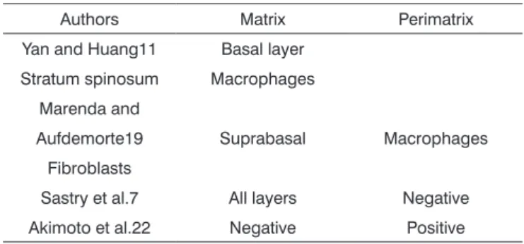
All of the authors we reviewed agree that TNF-α is present in cholesteatomas. There is no consensus, ho-wever, about its localization. Alves and Ribeiro18 (2004) summarized the differences in opinion. (Table 1)
the cholesteatoma epithelium (matrix). Sufficient studies to prove the location of TNF-α receptors in cholesteatoma and the factors that enhance its expression are lacking in literature.
The quantity of TNF-α, and not only the number of receptors, also seems to be related to bone destruction. Various authors have attempted to relate TNF-α levels with intraoperative findings.7,20,23 Cholesteatomas with increased bone destruction had a higher local amount of TNF-α,7,20,23 a finding seen both in acquired and congenital cholesteatomas.22 There is, however, only one published paper comparing congenital and acquired cholesteatomas, so this last findings needs to be further substantiated. The plasmatic concentration of TNF-α in cholesteatoma patients with variable degrees of bone destruction is proportionally higher compared to normal patients. A correlation may be established between systemic TNF-α levels and its local behavior.7 In future it may become possible to predict the aggressiveness of cholesteatoma through a blood test, predicting the associated degree of bone destruction and the possibility of complications.
Based on these papers, we may conclude that TNF-α is associated with bone destruction and defines its severity.7,20,23 TNF-α is increased both systemically and locally in aggressive cholesteatomas. None of the authors, however, investigated why TNF-α levels are increased. The difference in TNF-α levels may be related to various other factors such as, for instance, duration of the disease, the intensity of local infection, or the presence and distribution of TNF-α receptors in the cholesteatoma matrix. There are, however, insufficient published papers for a more detailed analysis of this hypothesis.
TNF-α is one of the main factors responsible for bone resorption and resulting complications in a variety of diseases. The possibility of interrupting the inflamma-tion seen in such chronic diseases was raised following the discovery of natural inhibitors against TNF-α, which could reduce morbidity and mortality.24-26 The first large-scale clinical trial of these inhibitors in systemic diseases demonstrated the efficacy of this treatment in reducing bone resorption.27 Studies have focused on inhibiting bone destruction by blocking the action of TNF-α using TNF-α antagonists or anti-TNF-α antibodies.
In periodontitis injection of a solution containing a TNF-α antagonist (s NF RI) inhibits osteoclast formation, reduces inflammatory cell recruitment and reduces bone loss by 60%.12,28 Thus, TNF-α antagonists can reduce bone destruction.
Anti-TNF-α antibodies can also reduce and pos-tpone bone destruction in rheumatoid arthritis.27 Bone resorption may, therefore, be inhibited using two different pathways. There are, however, no such studies applied to cholesteatoma. Based on these findings, there may be
Table 1. Location of TNF-α in cholesteatoma, according to various authors.
Authors Matrix Perimatrix Yan and Huang11 Basal layer
Stratum spinosum Macrophages Marenda and
Aufdemorte19 Suprabasal Macrophages Fibroblasts
Sastry et al.7 All layers Negative Akimoto et al.22 Negative Positive
Source: Based on work by Alves and Ribeiro18 (2004)
(perima-control bone destruction, which would reduce the mor-bidity and mortality of cholesteatoma.
CONCLUSION
TNF-α is found in cholesteatomas, promoting bone resorption by different routes. It acts on osteoclast differen-tiation and maturation (direct pathway) and also exposes the bone matrix (indirect pathway). TNF-α, however, is not the only factor involved in bone resorption; it acts in association with interleukin 1 and RANKL. Furthermore, local levels of TNF-α are related with the degree of bone destruction in this disease.
REFERENCES
1. Ribeiro FAQ, Pereira CSB. Otite média crônica colesteatomatosa em: Tratado de otorrinolaringologia. 1ª ed. São Paulo: Editora Roca; 2003, pp.93-102.
2. Lessa HA; Freitas EB; Cruz OLM. Complicações das otites médias em: Tratado de otorrinolaringologia. 1ª ed. São Paulo: Editora Roca; 2003, pp 43-49.
3. Pereira CSB. Análise de estudos da expressão das citoqueratinas no colesteatoma adquirido. São Paulo; 1997 (dissertação - mestrado - Faculdade de Ciências Médicas da Santa Casa de São Paulo). 4. Hamblin AS. Cytokines and cytokine receptors. 2nd ed. New York:
Oxford University Press; 1993.
5. Abbas AK, Lichtman AH, Pober JS. Citocinas. In: Imunologia celular e molecular. 2ª ed. Rio de Janeiro: Revinter 1998, pp.253-276. 6. Carswell EA, Old LJ, Kassel RL, Green S, Fiore N, Williamson B. An
endotoxin-induced serum factor that causes necrosis of tumor. Proc Natl Acad Sci USA 1975;72:3666.
7. Sastry KVSSR, Sharma SC, Mann SBS, Ganguly NK e Panda NK. Aural cholesteatom: role of tumor necrosis factor-alpha in bone destruction. Am J Otol 1999;20:158-61
8. Bingham CO. The pathogenesis of rheumatoid arthritis: pivotal citokynes involved in bone degradation and inflammation. J Reum 2002;29 supl 65:3-9.
9. Iino K, Toryama M, Ogawa H, Kawakami M. Cholesteatoma debris as an activador of human monocytes. Acta otolaryngol 1990;110:410-15.
10. Kreutzer DL, Yellon RF, Leonard G, Marucha PT, Craven R, Carpenter RJ et al. Characterization of cytokines present in middle ear effusions. Laryngoscope 1991; 101:165-69.
11. Yan SD, Huang CC. The role of tumor necrosis fator-alpha in bone reabsorption of cholesteatoma.Am J otolaryngol 1991;12:83-9. 12. Assuma R, Oates T, Cochram D, Amar S, Graves DT. IL-1 and TNF
antagonists inhibit the inflammatory response and bone loss in ex-perimental periodontitis. J Immunol 1998;160(1):403-9.
13. Yong-Soo P, Sang WY, Young CC, Timothy TKJ. Effect of inhibitor of tumor necrosis factor-alpha on experimental otitis media with effusion. Ann Otol Laryngol 2001;110:917-21.
14. Goldring SR. Bone and joint destruction in rheumatoid arthritis: What is really happening? J Rheumatol Suppl 2002; 65:44-8.
15. Goldring SR, Gravallese EM. Pathogenesis of bone erosions in rheu-matoid arthritis. Curr Opin Rheumato 2000;12(3):195-9,2.
16. Zhang YH, Heulsmann A, Tondravi MM, Mukherjee A, Abu-Amer Y. Tumor necrosis factor-alpha (TNF) stimulates RANKL-induced osteoclastogenesis via coupling of TNF type 1 receptor and RANK signaling pathways. J Biol Chem 2001;276(1):563-8.
17. Steeve KT, Marc P, Sandrine T, Dominique H, Yannick F. IL-6, RANKL, TNF-alpha/IL-1: interrelations in bone resorption pathophysiology. Cytokine & Grown factor reviews 2004;15:49-60.
18. Alves AL, Ribeiro FAQ. O papel das citocinas no colesteatoma adqui-rido da orelha média: revisão da literatura. RBORL 2004; 70(6):813-18.
19. Marenda SA; Aufdemorte TB. Localization of cytokines in choleste-atoma tissue. Otolaryngol Head Neck Surg 1995;112(3):359-68. 20. Amar MS; Wishahi HF; Zakhary MM Clinical and biochemical
stu-dies of bone destruction in cholesteatoma. Amer J Laryngol Otol 1996;110(6):534-9.
21. Chung JW; Yoon TH Different production of interleuk1alpha, in-terleukin-1beta and interleukin-8 from cholesteatomatous and normal epithelium. Acta Otolaryngol 1998:118(3):386-91.
22. Akimoto R, Pawankar R, Yagi T, Baba S. Acquired and congenital cho-lesteatoma: determination of tumor necrosis factor-alpha, intercellular adhesion molecule-1,interleukin-1-alpha and lymphocyte functional antigen-1 in the inflammatory process. ORL 2000;62:257-65. 23. Yetiser S, Satar B, Aydin N. Expression of epidermal growth factor,
tumor necrosis factor-alpha, and interleukin-1alpha in chronic otitis media with or without cholesteatoma. Otol Neurotol 2002;23(5):647-52.
24. Seckinger P, Isaaz S, Dayer JM. Purification and biologic characte-rization of specific tumor necrosis factor α inhibitor. J Biolo Chem 1989;264:11966-73.
25. Olsson I, Lantz M, Nilsson E, Peetre C, Thysell H, Grubb A, e cols. Isolation and characterisation of tumor necrosis factor binding protein from urine. Eur J Hematol 1989;42:270-5.
26. Engelmann H, Aderka D, Rubintein M, Rotman D, Wallach D. A tumor necrosis factor-binding protein purified to homogeneity from human urine protects cells from tumor necrosis toxicity. J Biol Chem 1989; 264:11974-80.
27. Elliot MJ, Maini, RN, Feldmann M, Kalden JR, Antoni C, Smolen JS, e cols. Randomised double-blind comparison of chimeric monoclonal antibody to tumor necrosis factor alpha (cA2) versus placebo in rheumatoid arthritis. Lancet 1994;344:1105-10.