Changes in the Production of IL-10 and TNF-
α
in Skeletal Muscle of
Rats with Heart Failure Secondary to Acute Myocardial Infarction
Renato Delacio Lopes
2,3, Miguel Luiz Batista Júnior
1, José Cesar Rosa
1, Fabio Santos de Lira
1, Eivor Martins Jr
1,
Alex Yamashita Shimura
1, Patrícia Chakur Brum
1, Antonio Herbert Lancha Jr
1, Marília C. L. Seelaender
1,
Antonio Carlos Lopes
2Universidade de São Paulo1, São Paulo; Universidade Federal de São Paulo2; Universidade de Mogi das Cruzes3, Mogi das Cruzes – Brazil; Duke Clinical Research Institute4, Durham, North Caroline – USA
Abstract
Background: Recent studies show that the expression of inflammatory mediators, such as cytokines, is an important factor for the development and progression of heart failure (HF), especially in the presence of left ventricular dysfunction. These changes have been demonstrated both in the plasma and heart muscle and, more recently, in skeletal muscle of rats and in patients with HF.
Objective: To investigate the production and expression of tumor necrosis factor-α (TNF) and interleukin-10 (IL-10) in the soleus and the extensor digitorum longus (EDL) muscles of animals with left ventricular dysfunction after myocardial infarction (MI).
Methods: We used male Wistar rats that underwent ligation of the left coronary artery without reperfusion. Four weeks after this procedure, the animals underwent echocardiography and were divided into the following experimental groups: sham operated (sham) and IM. They remained under observation for a further period of 8 weeks.
Results: The level of the cytokine TNF-α increased by 26.5% (p <0.05), and its gene expression increased 3 times (p <0.01). The level of IL-10 decreased by 38.2% (p <0.05). Both changes occurred only in the soleus muscle, with no change in the EDL. The decrease (36.5%, p <0.05) in the IL-10/TNF-α ratio was due to both increased tissue levels of TNF-α and decreased tissue levels of IL-10.
Conclusion: Our results showed significant changes in the IL-10/TNF-α ratio, which may have an additive role in the assessment of deterioration and progression of left ventricular dysfunction post-MI. Furthermore, our study suggests that these changes seem to be related to the muscle fiber type. (Arq Bras Cardiol 2010; 94(3):293-300)
Key words: Ventricular dysfunction, left; muscle, skeletal; cytokines; inflammation.
Mailing address: Miguel Luiz Batista Júnior •
Av. Lineu Prestes, 1524 - 05508-900 - Butantã - São Paulo, SP - Brazil. E-mail: [email protected]
Manuscript received on April 15th, 2009; revised manuscript received on July
Introduction
Heart failure (HF) is a clinical syndrome of complex pathophysiology that may result from any structural or functional disorder that strikes the heart and consequently undermines the ability of the ventricles to satisfactorily fill and pump blood1,2. The cardinal manifestations of HF are dyspnea
and fatigue, which may limit the ability to perform physical activity (exercise intolerance) and culminate in processes leading to pulmonary and systemic congestion and increased peripheral vascular resistance1,3. The main causes of HF of
cardiac origin are coronary artery disease, high blood pressure, dilated cardiomyopathy and heart valve diseases1,4.
Currently, the deteriorative changes involved in the progression of HF with left ventricular dysfunction do not depend only on hemodynamic parameters, but also on
the processes that culminate in local (heart) and systemic inflammatory activation5-7, which are evidenced by the increase
in both gene and protein expression of pro-inflammatory cytokines, such as: tumor necrosis factor (TNF-α), interleukin 1β (IL-1β), and interleukin 6 (IL-6), among others. These changes have been demonstrated both in plasma and in skeletal and heart muscles of rats and patients with HF. These inflammatory mediators may contribute to the pathophysiology and progression of HF, and also directly influence the onset of clinical manifestations of the HF syndrome, particularly the changes related to the reduction and the functioning of muscle mass8,9.
Therefore, although in recent years the vast majority of studies on HF have focused on pro-inflammatory cytokines, the anti-inflammatory cytokines, especially IL-10, have currently gained prominence and may have an important role in the pathophysiology of HF10,11. In patients with HF, and
of IL-10 and TNF-α (IL-10/TNF-α ratio) has recently been demonstrated as a more accurate indicator of the degree of ventricular dysfunction12.
In fact, in animal models and patients with HF, the distribution of muscle fibers into type I (aerobic), type IIa (aerobic and anaerobic) and type IIb (anaerobic) undergoes changes in its composition. Notably, the skeletal muscle fibers type I are the most affected, because they develop some characteristics similar to the type IIA13, besides a reduction in
capillary density3 and a reduction in the maximum activity of
the enzyme cytochrome c oxidase9, in comparison to the same
muscle fiber type in individuals without pathological changes in skeletal muscle. Similarly, studies in rats with post-MI left ventricular dysfunction have reproduced this behavior14,
showing thereby that the type I fibers in skeletal muscle seem to be the most affected in this condition.
Furthermore, these changes have been correlated to the increase in gene expression and production of cytokines (TNF-α, IL-1β, and IL-6) in the quadriceps muscle of humans15
and animals7. Although the deleterious changes occur
predominantly in type I fibers, recent studies have shown the presence of a chronic inflammatory condition in that tissue. However, to date, no studies have evaluated the production of these cytokines in the different muscle fiber types, a fact that becomes even more significant when we consider that this tissue may be an important systemic “supplier” of pro-inflammatory cytokines in the presence of left ventricular dysfunction. In turn, the analysis of the gene expression and the consequent production of these cytokines may be a more accurate and/or additional indicator of the presence of changed chronic inflammatory markers in ventricular dysfunction.
On this basis, we proposed to study the relationship between the levels of pro-TNF-α cytokines and anti-inflammatory (IL-10) cytokines in skeletal muscle in an experimental model in rats with post-MI left ventricular dysfunction. Still, we used the soleus muscle (fiber type I, predominantly) and the extensor digitorum longus (EDL) muscle (fiber type IIb, predominantly) to compare the profiles of these cytokines in different muscle types.
Methods
Animals
The experimental procedures were in accordance with the ethical principles for animal experimentation adopted by the Brazilian College of Animal Experimentation (Cobea), and the protocol for the use of animals in experimentation (009/2005) was approved by the Animal Experimentation Ethics Committee (Ceea) at a meeting held on February 17th, 2005.
We used male Wistar rats aged 8 weeks. During the experimental period, they were kept in collective cages for 5 mice, in a vivarium with a light/dark cycle of 12/12 hours, with the light phase starting at 7 am, in the Department of Cell Biology and Development of the Institute of Biomedical Sciences of the University of São Paulo. Room temperature
was maintained at 25 ± 2° C. Humidity was maintained at 60
± 5%. All groups received a balanced commercial diet (Nuvilab CR1 - Nutrivital Nutrients Ltda.) and water ad libitum. In order to minimize the acute influence of the prandial status in the experimental results, all animals were fasted overnight before the morning sacrifice.
Induction of acute myocardial infarction (MI) by myocardial injury through ischemia without reperfusion
This surgical procedure was performed using the techniques of the exercise and cellular physiology laboratory (KCM or MAC). The animals were anesthetized with a solution of Ketalar ® (80 mg/kg) + Rompun ® (12 mg/kg). After intubation, the animals received mechanical ventilation with a Harvard ventilator, model 683 (frequency of 90 m/ min, and tidal volume of 2.0 ml). After shaving the chest, a left thoracotomy was performed in the intercostal space of the apex beat, and the heart was quickly exposed. The anterior interventricular branch of the left coronary artery was identified and ligated between the edges of the left auricular appendage and the pulmonary artery with 6-0 prolene sutures. The lungs were hyperinflated, and the chest wall was closed with a previously performed purse-string suture. For the sham operation, we used the same surgical procedure but without the disruption of the pericardium and the ligation of the left anterior descending coronary artery.
After the coronary artery ligation, the animals were confined to the vivarium for 3 more weeks. By the fourth week, an echodopplercardiogram (ECHO) was performed to confirm the presence of infarction, to evaluate its size, and to conduct echocardiographic measurements, including left ventricular ejection fraction.
Transthoracic doppler echocardiography
During 4 and 12 weeks after MI, the animals were anesthetized with a solution of Ketalar ® (50 mg/kg) + Rompun ® (10 mg/kg) intraperitoneally for echocardiography.
The echocardiography was performed in an HP Sonos 5500 (Hewlett Packard, Andover, MA, USA) using a transducer fundamental frequency of 12 MHz, to obtain images at 2 cm depth. After anesthesia and using the same procedure, the animals were placed in the left lateral position, and three electrodes were placed in their legs for the electrocardiogram.
muscles, and apical region), according to earlier patterning characterized by the group of Professor Paul J. F. Tucci16,17,
using the formula: MI (%) = (AHR / EP) x 100.
The cardiac function was evaluated by the ejection fraction (EF). The ventricular diastolic (DV) and systolic (SV) volumes were obtained using the Simpson biplane (apical, four and two chambers) method, and the EF was calculated by the formula EF = (DV-SV/DV) x 100. The diastolic function was assessed using indices derived from the velocity curve of the mitral diastolic flow obtained by the pulsed Doppler technique. We obtained the velocity curve of the mitral diastolic flow from the apical four-chamber view, placing the sample volume close to the ventricular face of the mitral valve. We determined the E wave, the A wave, and the E/A ratio. At the end of the ECHO assessment, the animals in which the MI was greater than 40% of the LV were selected for the group of myocardial infarction (MI). After 12 weeks, the animals in the MI group were reevaluated as to the aforementioned parameters.
Experimental design
After the echocardiography, the animals were divided into two experimental groups: sham operated (sham, n = 13) and myocardial infarction (MI, n=17). The sham group animals underwent all surgical procedures except the coronary artery ligation. After this, the animals were kept for 8 weeks under observation. Following this period, the animals were sacrificed by decapitation without anesthesia.
Collection and homogenization of tissues
Immediately after sacrifice, tissues and organs (liver, lungs, heart, soleus and extensor digitorum longus-EDL muscles) were removed, weighed, frozen in liquid nitrogen, and stored at -80° C for subsequent measurements.
Quantiication of tissue cytokines
For measurement of TNF-α (DY510) and IL-10 (DY522) cytokines, we used the enzyme-linked immunoabsorbent assay (DuoSet ELISA) capture method (R&D System, Inc., Minneapolis, USA). This assay was performed on homogenized samples from the different experimental groups, according to the standards described by the manufacturer. The results are expressed in pg/μg of total protein in the assessed tissues according to the colorimetric method described by Bradford18.
Quantiication of tissue cytokine gene expression
To isolate total RNA from the EDL and the soleus muscles (right hindpaw), samples weighing between 0.2 and 0.5g were homogenized in TRIZOL® Reagent, according to the standards described by the manufacturer19. The RNA
samples were transcribed to DNA in a thermocycler (Techne, Cambridge, UK). To synthesize cDNA, we used 2μg of total RNA from each sample. The samples were incubated with 0.5 μg/ml of oligo DT12-18 (Invitrogen, USA) at 65° C, for 5 minutes, to obtain the first tape of cDNA.
Primers were synthesized by Integrated DNA Technologies (Prodimol Biotecnologia S/A). The sequence was determined
using the software Primer 3, available at http://www.genome. wi.mit.edu. Primers were selected in accordance with the following parameters described by Marone et al20: TNF-α (NM
012675.2); sense: 5`TCT CAA AAC TCG AGT GAC AAG C 3`;
anti-sense: 5`GGT TGT CTT TGA GAT CCA TGC3`; IL-10 (NM 012854.1), sense: 5`GAG AGA AGC TGA AGA CCC TCT G3`;
anti-sense: 5`TCA TTC ATG GCC TTG TAG ACA C3`; and 18S (NM 213557.1), sense: 5`ACCAGTTCGCCATGGATGA3`;
anti-sense: 5`TGCCGGAGCCGTTGTC3`.
The gene expression was quantified by real-time PCR (Higuchi et al21) using a 7500 Fast Real-Time PCR (Applied
Biosystems, USA) machine and a Syber Green kit as fluorescence markers (number 11744-500, Invitrogen, USA). Reactions were performed in 25µl of a mixture containing 1µl of cDNA sample, 0.5 µl of primers (adjusted to the concentration shown below), 10.5 µl of Depec water, and 12.5 µl of Syber green master mix (dNTP - reaction buffer, Taq DNA polymerase Syber green I). The conditions of real-time PCR were: first cycle (single), at 95°C, for 15 minutes to activate the enzyme; 40 cycles with denaturation phases, at 95°C, for 15 seconds; and annealing, at 60°C, for 30 seconds. We used the primer sequence according to the following concentrations:
• soleus muscle: TNF-α and IL-10 (200 nM) and 18S (300 nM).
• EDL - TNF-α: IL-10 (200 nM) and 18S (200 nM).
Statistical analysis
Data analysis was performed using the software SigmaStat (version 3.1, SigmaStat, Systat, Point Richmond, CA). The mean and standard error of the mean (SEM) were calculated for all variables. After the trial period, the values of the different analyses of the results were performed by unpaired Student’s t-test. The level of significance used in all tests was less than 5%, i.e., p <0.05.
Results
General characteristics of the experimental groups
The postoperative mortality was 23.8% (n = 5 animals that evolved to death during the 24 hours after surgery) in the MI group, and 13.3% (n = 2) in the sham group. The total weight of the lungs, liver, soleus and EDL, in absolute value, was not different in any of the groups evaluated (Table 1). The MI group showed a 23.8% (p <0.012) increase in the absolute heart weight when compared to the sham group. The animals with MI showed an increase in the wet weight/dry weight ratio of the lungs (4.7%, p <0.011), of the liver (3.8, p <0.035), and a 27.7% (p <0.002) decrease in the soleus muscle weight relative to the body weight (Table 2).
Left ventricular function in animals with MI
Absolute weight (g) Sham (13) MI (17)
Body 319,3±17,9 346,2±20,1
Heart 0,90±0,02 1,12±0,06
Liver 7,66±0,50 7,52±0,79
Lung 4,22±0,08 4,41±0,14
Soleus 0,136±0,01 0,132±0,01
EDL 0,150±0,01 0,144±0,01
Note: The results are expressed in grams and represent mean ± SEM of 30
animals. * Signiicant difference in comparison with the sham group, p <0.05
(Student t test).
Table 1 - Total body weight and total tissue weight in rats that
underwent sham operation and operation with acute myocardial infarction produced by coronary artery ligation after 12 weeks
Relative weight Sham (13) MI (17)
Heart (mg/g) 2,99±0,16 3,24±0,16
Liver (sec. x umi.) 3,04±0,03 3,15±0,03*
Lung (sec. x umi.) 4,22±0,08 4,41±0,14*
Soleus (mg/g) 0,44±0,02 0,35±0,02*
EDL (mg/g) 0,42±0,01 0,43±0,01
Table 2 - Total relative tissue weight in rats that underwent sham
operation and operation with acute myocardial infarction produced by coronary artery ligation after 12 weeks
Note: The results are expressed as values relative to total body weight, wet weight (umi.) by dry weight (sec.) and represent mean ± SEM of 30 animals. * Significant difference in comparision with the sham group, p
<0.05 (Student t test).
Parameters Sham (13) MI (17)
EF (%) 67,4±0,10 40,4±0,06*
SF (mm/s) 43,0±3,0 19,5±6,5*
PAVED (mm) 7,73±0,32 10,06±0,77*
EDV (mL) 1,69±0,32 3,08±1,48*
MI (%) 39%±0,05
Table 3 - Values of the functional variables of the left ventricle by
Doppler echocardiography after 4 weeks in rats that underwent sham operation and operation with acute myocardial infarction produced by coronary artery ligation (MI)
EF - ejection fraction, SF - shortening fraction, Paved - anterior wall of the left ventricle during diastole, EDV - end diastolic volume and MI - index of myocardial infarction. The results represent mean ± SEM of 30 animals. * Signiicant difference in comparison with the sham group, p <0.05 (Student t test).
IL-10 TNF-alpha
Sham MI
C
ytok
ines
pg/m
ic
rogram
of ti
ss
ue protei
n
250
200
150
100
50
0
Figure 1 - Quantiication of IL-10 and TNF-α in the soleus muscle in rats that underwent sham operation and operation with acute myocardial infarction produced by coronary artery ligation after 12 weeks. The cytokine content was analyzed by ELISA and normalized by the total protein content of the tissues evaluated. The results represent mean ± SEM of 14 animals. * Signiicant difference in comparison with the sham group, p <0.05 (Student t test).
shortening fraction (SF) showed a 40.1% and a 55.7% (p <0.001) reductions, respectively, when these results were compared with those obtained in the sham group. The end diastolic volume (EDV) increased by 82.2% (p <0.01) when compared to that obtained in the sham group. The animals in the MI group had an infarct area of 39 ± 0.05%.
TNF-α levels in skeletal muscle
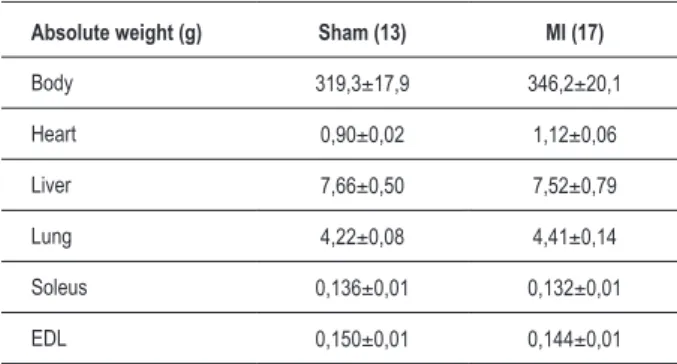
The MI group animals showed a significant 26.5% increase (p <0.05) in the production of TNF-α in the soleus muscle when compared to the sham group (Fig. 1). In EDL muscle, the levels of TNF-α were not detected. The animals in the MI group (Fig. 2A) showed a significant 3-fold increase (p <0.01) in TNF-α gene expression in the soleus muscle, with no change in the EDL muscle (Fig. 2B).
IL-10 levels in skeletal muscle
The MI group animals showed a significant 38.2% decrease (p <0.05) of production of IL-10 in the soleus muscle, when compared to the sham-S group (Fig. 1). In the EDL muscle, there was no change in the levels of this cytokine. The animals in the MI group (Fig. 2A) showed a significant 3.6-fold increase (p <0.05) in IL-10 gene expression in the soleus muscle, with no change in the EDL muscle (Fig. 2B).
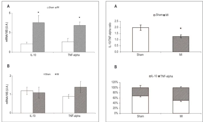
IL-10/TNF-α ratio in skeletal muscle
The MI group animals showed a significant 36.5% decrease (p <0.05) in the IL-10/TNF-α ratio when compared to the sham group. The chart 3B data show the participation of each cytokine in the IL-10/TNF-α ratio.
Discussion
Figure 2 - IL-10 and TNF-α gene expression in the soleus muscle (A) and in the EDL muscle (B) in rats that underwent sham operation and operation with acute myocardial infarction produced by ligation of coronary artery after 12 weeks. The mRNA expression was analyzed by real-time PCR. The results represent mean ± SEM, in duplicates of 14 animals. * Signiicant difference
IL-10 TNF-alpha
5
3
0
m
R
N
A
/18S
(U
.A
.)
Sham IM
A
IL-10
m
R
N
A
/18S
(U
.A
.)
Sham IM
TNF-alpha
2
1
0
B
were accompanied by a reduction in the concentration of anti-inflammatory cytokine (IL-10) levels, also observed only in the soleus muscle, in turn, the IL-10/TNF-α ratio was altered, suggesting the predominance of TNF-α to the detriment of IL-10 in that tissue.
The ligation of the left anterior descending coronary artery was used as an experimental model of MI in animals, by the induction of myocardial ischemia, with or without reperfusion7,22,23. However, data on left ventricular function
are insufficient for the clinical characterization of HF, because this syndrome is defined by the presence of hemodynamic abnormalities, fluid retention (congestion), and physical exercise intolerance24,25. Although our study showed data
referring to the congestive picture, such as the dry weight/ wet weight ratio of liver and lungs, which suggests the presence of pulmonary edema and hepatic congestion, we chose to classify the MI group as animals with severe ventricular post-MI, and not with HF. It is possible that most of the animals in the MI group also had HF clinical changes, but this has not been studied in this paper.
Initially, it was assumed that the production of TNF-α
was performed by monocytes and macrophages exclusively when appropriately stimulated26. Recently, TNF-α gene and
protein expression in skeletal muscle has been characterized in humans27, in a study that showed for the first time the presence
of this cytokine in the absence of infiltrating monocytes and
Figure 3 - Cytokine levels ratio (A) and proile of cytokine levels (B) in the
muscle IL-10/TNF-α ratio. The cytokine content was analyzed by ELISA and normalized by the total protein content of the tissues evaluated. The proportion refers to the percentage of each cytokine in the ratio. The results represent mean ± SEM of 14 animals. * Signiicant difference in comparison with the
Sham MI
Sham MI
2.5
2.0
1.5
1.0
0.5
0.0
IL-10/TN
F-al
pha rati
o
120%
100% 80%
60% 40% 20% 0%
IL-10 TNF-alpha
Sham MI
A
B
macrophages. This indicated that the skeletal muscle fiber itself can produce it, and so it could act in an autocrine and paracrine manner, in sufficient quantity to mediate a series of morphological and functional changes in the tissue3,28.
In our study, rats with left ventricular dysfunction post-MI showed an increased content of pro-inflammatory cytokine TNF-α, which was only evident in the soleus muscle, with no change in the EDL muscle (not detected by ELISA). This condition was evidenced by the increase in TNF-α gene expression, suggesting that the increase in the TNF-α protein content may have been due to increased levels of mRNA. This result is consistent with another study7 that used the same
experimental model and demonstrated an increase in both gene expression and muscle levels of TNF-α. However, these parameters were assessed in the quadriceps muscle, which is mixed with regard to its muscle fiber type.
Therefore, despite the fact that an increase in inflammatory mediators (TNF-α, IL-1 β, IL-6) in skeletal muscle is well characterized both in patients with HF15 and in rats with
heart failure and left ventricular dysfunction post-MI7, both
HF, the most affected fiber, both morphologically and functionally, is the type I fiber3. Thus, we showed, for the
first time, that this condition appears to be specific to muscle tissue with a predominance of type I fibers. As we did not separate the total homogenate in a soluble fraction bound to the cell membrane, we could not infer that the protein assessed was the one which was produced exclusively by the tissue evaluated.
Plasma levels of these cytokines in patients with HF have been related to both prognosis and severity of the disease, a fact which has been shown to be even more evident in patients with more advanced HF (functional class IV, NYHA)6,29.
Therefore, it has been suggested that this increase results from a peripheral inflammatory process, which would ideally be initiated in skeletal muscle, because of reduced blood flow and a consequent increase in reactive oxygen species5. This
hypothesis has gained consistency, as studies conducted both in animals7,12,23,30,31 and humans2,32,33 have shown that the
increase in local inflammatory milieu occurs independently of plasma levels changes, especially in patients in functional classes II and III (NYHA). Therefore, the increased gene and protein expression of these cytokines, even without plasma levels changes, confirms the results obtained by studies that suggest that local inflammation precedes the increase in plasma and may be a more accurate indicator with respect to the progression of HF.
The effects of IL-10 in vivo have been observed in models of inflammation, autoimmunity, tolerance and parasitic infections in animals34. In mice, IL-10 attenuated the deleterious effects
induced by lipopolysaccharide (LPS) and SEB (staphylococcal enterotoxin B). Furthermore, these effects were correlated with decreased TNF-α circulating levels. In macrophages cultured in the presence of LPS, besides the increase in production of proinflammatory cytokines, this condition is also followed by an increase in IL-10, and TNF-α is an important inducer of IL-10 gene expression and production34,35. Therefore, IL-10 is
the main anti-inflammatory cytokine and plays an important role in modulating the production of TNF-α.
In rats with left ventricular dysfunction post-MI, IL-10 tissue levels were reduced only in the soleus muscle. In these animals, the gene expression was increased in the same tissue, showing that perhaps the decrease in its production is not related to changes in mRNA expression, at least in this aspect. This suggests that the effects of this experimental model in the concentration of IL-10 may have been mediated in a post-transcriptional level, like the production of IL-10 post-transcriptional regulatory elements, such as enhancers and/or mechanisms of post-transcriptional control. The last possibility is described in T cell clones, which express IL-10, with no detectable changes in mRNA levels36. Therefore, a combination of post-transcriptional
mechanisms and ubiquitination for the expression of IL-10 may have a crucial role for this cytokine37. Furthermore, although
TNF-α can induce IL-10 gene expression34 and consequently
its protein expression, this effect seems to be dependent on the intensity of the inflammatory condition, and thus the increase in the IL-10 gene expression could have occurred because of a decrease in protein production (mRNA/protein ratio) toward the restoration of a balanced condition.
Therefore, the new fact we present is that there was an increase not only in gene expression, but also in protein content, in the case of TNF-α, and a decrease in IL-10 protein, suggesting, in this condition, a tendency towards an imbalance in their production. This condition may tend to the predominance of a pro-inflammatory milieu at the expense of the anti-inflammatory milieu, especially through an increased production of TNF-α and a decreased production of IL-10. This association was observed by Stumpf et al10 who used the
IL-10/TNF-α ratio to characterize the pro/anti-inflammatory "balance". In this study, patients with HF (NYHA functional class III/IV) showed increased levels of TNF-α and decreased levels of IL-10 when compared with control subjects (without IC) of the same age.
Although the molecular mechanisms are not well characterized, studies using IL-10 as treatment (rhIL-10) in humans and in knocked out mice (IL-10-/-) stressed the
importance of this cytokine in reversing the alterations caused by local inflammation, especially those mediated by TNF-α10. This result is consistent with ours, since the
IL-10/TNF-α ratio decreased because of an increase in TNF-α and, especially, a decrease in IL-10, suggesting an important role of IL - 10 in the local inflammatory "control" in the soleus muscle.
In patients with HF, muscle mass loss or atrophy of begins in the early stages of HF8 development, and this
is more specifically related to intrinsic changes in skeletal muscle3,38 than in those resulting from physical inactivity
(a more determining factor in women). Recently, it has been shown that, in this condition, there is an immune activation, especially by inflammatory mediators, which may contribute to the development of this condition3,7,8,39. In rats
with heart failure and left ventricular dysfunction after MI, this condition has been reproduced both by the decrease in cross-sectional area and by increased production of proinflammatory cytokines (quadriceps muscle)7. Similarly,
our study showed a skeletal muscle weight reduction relative to body weight, a condition that proved to be dependent on the predominant type of muscle fiber, since it was present only in the soleus muscle, with no change in the EDL muscle. These changes directly and indirectly affect skeletal muscle, and recent studies13,14 in rats with heart failure and
left ventricular dysfunction after MI showed that muscle tissues with predominantly type I fibers (e.g., soleus muscle) may be more sensitive to changes that are characteristic of this experimental model and that compromise the blood and oxygen supply, when compared to tissues in which type IIa and IIb fibers are predominant (e.g., EDL muscle). Therefore, the changes found in proinflammatory and anti-inflammatory cytokines have demonstrated the presence of a chronic inflammatory state in the soleus muscle, as assessed by the IL-10/TNF-α ratio, which was paralleled by a reduction in muscle mass, a condition which was only observed in muscle tissue with a predominance of type I fibers (soleus).
References
1. Sociedade Brasileira de Cardiologia. Revisão das II Diretrizes da Sociedade Brasileira de Cardiologia para o diagnóstico e tratamento da insuficiência cardíaca. Arq Bras Cardiol. 2002; 79: 1-30.
2. Aukrust P, Ueland T, Lien E, Bendtzen K, Müller F, Andreassen AK, et al. Cytokine network in congestive heart failure secondary to ischemic or idiopathic dilated cardiomyopathy. Am J Cardiol. 1999; 83 (3): 376-82.
3. Larsen AI, Lindal S, Aukrust P, Toft I, Aarsland T, Dickstein K. Effect of exercise training on skeletal muscle fibre characteristics in men with chronic heart failure: correlation between skeletal muscle alterations, cytokines and exercise capacity. Int J Cardiol. 2002; 83 (1): 25-32.
4. Hunt SA, Abraham WT, Chin MH, Feldman AM, Franas GS, Ganiats TG, et al. ACC/AHA 2005 Guideline update for the diagnosis and management of chronic heart failure in the adult: A report of the American College of Cardiology/American Heart Association task force on practice guidelines (Writing Committee to Update the 2001 Guidelines for the Evaluation and Management of Heart Failure): Developed in collaboration with the American College of Chest Physicians and the International Society for Heart and Lung Transplantation: Endorsed by the Heart Rhythm Society. Circulation. 2005; 112 (12): e154-235.
5. Coats A, Clark A, Piepoli M, Volterrani M, Poole-Wilson P. Symptoms and quality of lyfe in heart failure: the muscle hypothesis. Br Heart J. 1994; 72: S36-9.
6. Anker SD, von Haehling S. Inflammatory mediators in chronic heart failure: an overview. Heart. 2004; 90 (4): 464-70.
7. Schulze P, Gielen S, Adams V, Linke A, Mobius-Winkler S, Erbs S, et al. Muscular levels of proinflammatory cytokines correlate with a reduced expression of insulinlike growth factor-1 in chronic heart failure. Basic Res Cardiol. 2003; 98 (4): 267-74.
8. Toth MJ, Ades PA, Tischler MD, Tracy RP, LeWinter MM. Immune activation is associated with reduced skeletal muscle mass and physical function in chronic heart failure. Int J Cardiol. 2006; 109 (2): 179-87.
9. Gielen S, Adams V, Linke A, Erbs S, Mobius-Winkler S, Schubert A, et al. Exercise training in chronic heart failure: correlation between reduced local inflammation and improved oxidative capacity in the skeletal muscle. Eur J Cardiovasc Prev Rehabil. 2005; 12 (4): 393-400.
10. Stumpf C, Lehner C, Yilmaz A, Daniel WG, Garlichs CD. Decrease of serum levels of the anti-inflammatory cytokine interleukin-10 in patients with advanced chronic heart failure. Clin Sci. 2003; 105 (1): 45-50.
11. Yamaoka M, Yamaguchi S, Okuyama M, Tomoike H. Anti-inflammatory cytokine profile in human heart failure: behavior of interleukin-10 in association with tumor necrosis factor-alpha. Jpn Circ J. 1999; 63 (12): 951-6.
12. Kaur K, Sharma A, Singal P. Significance of changes in TNF-{alpha} and IL-10 levels in the progression of heart failure subsequent to myocardial infarction. Am J Physiol Heart Circ Physiol. 2006; 291 (1): H106-13.
13. Richardson TE, Kindig CA, Musch TI, Poole DC. Effects of chronic heart failure on skeletal muscle capillary hemodynamics at rest and during contractions. J Appl Physiol. 2003; 95 (3): 1055-62.
14. Behnke BJ, Delp MD, McDonough P, Spier SA, Poole DC, Musch TI. Effects of chronic heart failure on microvascular oxygen exchange dynamics in muscles of contrasting fiber type. Cardiovasc Res. 2004; 61 (2): 325-32.
15. Gielen S, Adams V, Mobius-Winkler S, Linke A, Erbs, S, Yu J, et al. Anti-inflammatory effects of exercise training in the skeletal muscle of patients
16. Nozawa E, Kanashiro RM, Murad N, Carvalho AC, Cravo SL, Tucci PJ, et al. Performance of two-dimensional Doppler echocardiography for the assessment of infarct size and left ventricular function in rats. Braz J Med Biol Res. 2006; 39: 687-95.
17. Kanashiro R, Saraiva R, Alberta A, Antonio E, Moisés V, Tucci P. Immediate functional effects of left ventricular reduction: a doppler echocardiographic study in the rat. J Card Fail. 2006; 12 (2): 163-9.
18. Bradford MM. A rapid and sensitive method for the quantitation of microgram quantities of protein utilizing the principle of protein-dye binding. Anal Biochem. 1976; 72: 248-54.
19. Chomczynski P, Sacchi N. Single-step method of RNA isolation by acid guanidinium thiocyanate-phenol-chloroform extraction. Anal Biochem. 1987; 162 (1): 156-9.
20. Marone M, Mozzetti S, De Ritis D, Pierelli L, Scambia G. Semiquantitative RT-PCR analysis to assess the expression levels of multiple transcripts from the same sample. Biol Proced Online. 2001; 3: 19-25.
21. Higuchi R, Dollinger G, Walsh PS, Griffith R. Simultaneous amplification and detection of specific DNA sequences; Biotechnology (NY). 1992; 10 (4): 413-7.
22. Bregagnollo EA, Okoshi K, Matsubara BB, Tucci PJF. End-systolic pressure-diameter relation of the left ventricle during transient and sustained elevations of blood pressure. Arq Bras Cardiol. 2000; 75: 26-32.
23. Ono K, Matsumori A, Shioi T, Furukawa Y, Sasayama S. Cytokine gene expression after myocardial infarction in rat hearts: possible implication in left ventricular remodeling. Circulation. 1998; 98 (2): 149-56.
24. Hill M, Singal P. Right and left myocardial antioxidant responses during heart failure subsequent to myocardial infarction. Circulation. 1997; 96 (7): 2414-20.
25. Armstrong PW, Moe GW. Medical advances in the treatment of congestive heart failure. Circulation. 1993; 88 (6): 2941-52.
26. Beyaert R, Fiers W. Tumor necrosis factor and lymphotoxin. In: Mire-Sluis A, Thorpe R (eds). Cytokines. 2nd ed. California: Academic Press; 1999. p.
335-45.
27. Saghizadeh M, Ong J, Garvey W, Henry R, Kern P. The expression of TNF alpha by human muscle: relationship to insulin resistance. J Clin Invest. 1996; 97 (4): 1111-6.
28. Spate U, Schulze PC. Proinflammatory cytokines and skeletal muscle. Curr Opin Clin Nutr Metab Care. 2004; 7: 265-9.
29. Anker SD, Coats AJS. Cardiac cachexia: a syndrome with impaired survival and immune and neuroendocrine activation. Chest. 1999; 115 (3): 836-47.
30. Batista ML Jr, Santos RV, Cunha LM. Changes in the pro-inflammatory cytokine production and peritoneal macrophage function in rats with chronic heart failure. Cytokine. 2006; 34 (5-6): 284-90.
31. Batista ML Jr, Santos RVT, Oliveira EM, Seelaender MC, Costa Rosa LF. Endurance training restores peritoneal macrophage function in post-MI congestive heart failure rats. J Appl Physiol. 2007; 102 (5): 2033-9.
32. Yndestad A, Kristian Damås J, Øie E, Ueland T, Gullestad L, Aukrust P. Systemic inflammation in heart failure – The whys and wherefores. Heart Fail Rev. 2006; 11 (1): 83-92.
33. Paulus WJ. How are cytokines activated in heart failure? Eur J Heart Fail. 1999;
muscle are evident in animals with left ventricular dysfunction, and thus the soleus muscle may be a site of production of inflammatory mediators in this condition.
In summary, our results suggest an important role of the IL-10/TNF-α ratio, which may have an additive role in
34. Malefyt R. Interleukin-10. In: Mire-Sluis A, Thorpe R (eds.). Cytokines. 2nd
ed. California: Academic Press; 1999. p. 151-61.
35. Moore KW, de Waal Malefyt R, Coffman RL, O’Garra A. Interleukin-10 and the interleukin-10 receptor. Annu Rev Immunol. 2001; 19 (1): 683-765.
36. Naora H, Altin JG, Young IG. TCR-dependent and -independent signaling mechanisms differentially regulate lymphokine gene expression in the murine T helper clone D10.G4.1. J Immunol. 1994; 152 (12): 5691-702.
37. Powell MJ, Thompson SAJ, Tone Y, Waldmann H, Tone M. Posttranscriptional regulation of IL-10 gene expression through sequences in the 3’-untranslated region. J Immunol. 2000; 165 (1): 292-6.
38. Duscha BD, Annex BH, Green HJ, Pippen AM, Kraus WE. Deconditioning fails to explain peripheral skeletal muscle alterations in men with chronic heart failure. J Am Coll Cardiol. 2002; 39 (7): 1170-4.