Arq. Bras. Oftalmol. vol.77 número3
Texto
Imagem
Documentos relacionados
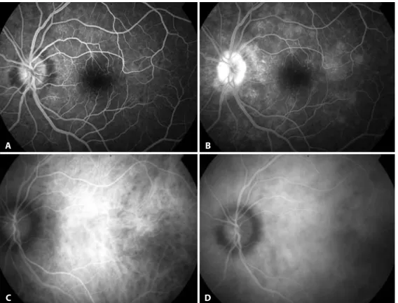
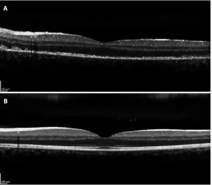
Epiretinal membrane formation associated with idiopathic macular telangiectasia: case report.. 2 6 6 Arq
Arq Bras Oftalmol. B) FAF exhibits hypoautoluo rescence in the areas corresponding to intraretinal hemorrhage and mild perimacular hyperautoluorescence. C) Recent subretinal
There were statistically signiicant diferences in subfoveal, temporal, and nasal choroidal thickness among the groups ( p < 0.05). Figure 2 shows the distribution of
Purpose : To investigate the distribution of axial length, anterior chamber depth, lens thickness, vitreous chamber depth, and central corneal thickness in children at different
Ophthalmic radiation may be used as a curative therapy, as adjuvant treatment following surgical excision, or as palliative therapy for advanced cases of eye- lid and
The SCC measurement on the Schwind Custom Ablation Mana- ger (CAM) platform is based on an algorithm that registers landmarks and patterns on the iris and limbus. These landmarks
Methods : We conducted a cohort study with patients screened in the 2011 Rapid Assessment of Avoidable Blindness (RAAB) who had reported that the cost was the main barrier to
Compared with the NG group, the sclera and choroid of the HG group showed a significant increase in IL-6 expression (p=0.002) (Table 2), characterized by the predominance of
