Arq Neuropsiquiatr 2011;69(4)
721
Letters
DISCUSSION
Cavernoma is a benign tumor and it is considered a dysplasia of the vessels-forming mesoderm4. Cavernous hemangiomas in the vertebral, extradural, intradural ex-tramedullary and inex-tramedullary spaces are responsible for 3 to 16% of spinal vascular anomalies4,5.
Extradural cavernous hemangioma represent 4% of all spinal epidural lesions5. Modern diagnostic imaging techniques are increasing the number of diagnosis and its frequency may be more than previously reported in the medical literature5.
There are four clinical syndromes described: slow and progressive spinal cord syndrome, which is the most common form; acute spinal cord syndrome; back pain; and radiculopathy3.
Imaging diagnostic exams such as spine X-rays, my-elography, CT and MRI are important for evaluating the relationship of the lesion with the surrounding anatomic structures1. Currently, MRI is the modality of choice5.
he treatment for these lesions is total removal of the tumor with microsurgical technique1.
REFERENCES
1. Hatiboglu MA, Iplikcioglu AC, Ozcan D. Epidural spinal cavernous heman-gioma-case Report. Neurol Med Chir (Tokyo) 2006;46:455-458. 2. Goyal A, Singh AK, Gupta V, Tatke M. Spinal epidural cavernous
haemangioma:a case report and review of literature. Spinal Cord 2002;40: 200-202.
3. Zevgaridis D, Buttner A, Weis S, Hamburger C, Reulen HJ. Spinal epidural cavernous hemangiomas. Report of three cases and review of the litera-ture. J Neurosurg 1988;88:903-908.
4. Saringer W, Nobauer I, Haberler C, Ungersbock K. Extraforaminal, thoracic, epidural cavernous hemangioma:case report with analysis of magnetic resonance imaging characteristics and review of the literature. Acta Neu-rochir (Wien) 2001;43:1293-1297.
5. Santoro A, Piccirilli M, Bristot R, Norcia V, Salvati M, Delini R. Extradural spinal cavernous angiomas: report of seven cases. Neurosurg Rev 2005;28: 313-319.
HEMANGIOMA CAVERNOSO EXTRADURAL DA COLUNA TORÁCICA
Neurosurgical Unit, Hospital de Clínicas de Porto Alegre, Porto Alegre RS, Brazil:
1Head of Neurosurgical Unit, Associate Professor of Neurosurgery, FAMED,
UFRGS; 2Resident of Neurosurgery.
Correspondence: Apio Antunes - Unidade de Neurocirurgia / Serviço de Neurologia do HCPA - Rua Ramiro Barcelos 2350 / 2° andar - 90035-903 Porto Alegre RS - Brasil. E-mail: [email protected]
Received 13 February 2011. Received in final form 31 November 2011. Accepted 7 April 2011.
Bilateral traumatic avulsion of abducens nerve
Bruno S.C. Lopes¹, Lazaro L.F. do Amaral¹, Higor G. Bezerra¹, Ricardo M. Rogério¹, Antônio A. Zambon²
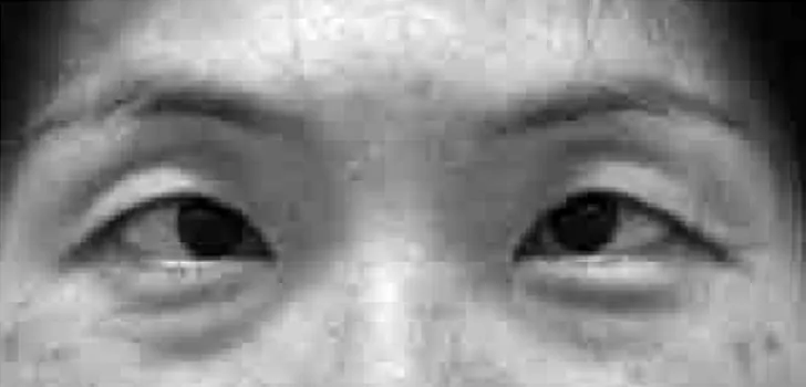
A 45-year-old previously healthy woman sufered a head trauma with neck hyperextension during bike ex-ercise, losing consciousness for about 24 hours. Upon awakening, she presented bilateral lateral gaze palsy and convergent strabismus (Fig 1). No bone fracture was detected on CT studies (not shown). his clinical pic-ture persisted unchanged for over one year and a MRI study done at our service showed bilateral avulsion of the sixth cranial nerve using FIESTA sequence (Fig 2A, B, C and D).
he abducens innervates the lateral rectus muscle, which is responsible for the horizontal lateral move-ment of the ocular globe. It has a long course, begin-ning at its nucleus, on the ventral pons, going through the pre-pontine cistern to its dural entry point on the petroclival region, coursing through Dorello’s canal, be-neath petroesphenoidal ligament, where it is covered by an envelope composed of one dural layer and one ar-achnoidal layer1 to the cavernous sinus, lateral to the in-ternal carotid artery, reaching the superior orbital issure
and orbital apex. his long course makes it more suscep-tible to injuries.
Various diferent diseases can cause sixth nerve palsy, neoplasic and traumatic etiologies being more common in children, while vascular and idiopathic are responsible for the majority of cases in adult population2.
Traumatic injuries of abducens nerve are a well-known consequence of severe head trauma, reported in
Arq Neuropsiquiatr 2011;69(4)
722
Letters
1-2,7% of the cases, with or without associated cervical or skull base fracture3. Usually, the mechanism of injury is contusion/stretching along its course and vertical dis-placement (downward and/or upward) of the brain is supposed to be the cause of these lesions4. Two points along the nerve course are described as the most prone to injury3-5: the dural entry point (during upward placement) and the petrous apex (during downward dis-placement). Since these two movements are usually as-sociated on severe head trauma, these two points likely work together to cause the lesion.
In our case, with a MRI study acquired on 1.5 tesla equipment (GE Medical systems - Milwaukee), using FIESTA sequence post processed in dedicated work-station, we observed bilateral sixth nerve discontinuity
along its pre-pontine course, detached from the pons, at the pontmedullary sulcus level. he other cranial nerves had preserved morphology. here were no signs of bone fracture, brainstem or orbital muscle lesions.
FIESTA (diferent names are used for similar tech-nique by other manufacturers, like BALANCED FFE and 3D-CISS, for example) is a magnetic resonance se-quence heavily T2-weighted, capable of acquiring very thin slices, allowing reformation in all three planes, op-timal for analyzing morphologic features of structures next to CSF containing spaces, like basal cisterns.
In the presented case, one year after trauma, there was still unchanged ophthalmoplegia and a MRI study showed bilateral complete abducens nerve avulsion. As far as we know, no previous report has showed, with im-aging studies, this consequence of head trauma.
REFERENCES
1. Kenichiro O, Arai H, Endo T, et al. Detailed MR imaging anatomy of the ab-ducent nerve: evagination of CSF into Dorello canal. AJNR Am J Neuro-radiol 2004;25:623-626.
2. Berlit P, Reinhardt-Eckstein J, Krause KH, et al. Isolated abducens paral-ysis: a retrospective study of 165 patients. Fortschr Neurol Psychiatr 1989; 57:32-40.
3. Arias MJ. Bilateral traumatic abducens nerve palsy without skull fracture and with cervical spine fracture: case report and review of the literature. Neurosurgery 1985;16:232-234.
4. Hollis G. Sixth cranial nerve palsy following closed head injury in a child. J Accid Emerg Med 1997;4:172-175.
5. Advani RM, Baumann MR. Bilateral sixth nerve palsy after head trauma. Ann Emerg Med 2003;41:27-31.
AVULSÃO TRAUMÁTICA BILATERAL DO NERVO ABDUCENTE
1Department of Neuroradiology, Medimagem, Hospital Beneficência Portuguesa
e Hospital Santa Catarina, São Paulo SP, Brazil; 2Depar tment of Neurology,
Hospital A.C. Camargo, São Paulo SP, Brazil.
Correspondence: Bruno S.C. Lopes - Rua Martiniano de Carvalho 669 / 1006 - 01321-900 São Paulo SP - Brasil. E-mail : [email protected].
Received 1 March 2011. Received in final form 28 March 2011. Accepted 13 April 2011.
![Fig 2. MRI study 1-year after trauma showing discontinuity on the cisternal segment of the VI nerve (complete avulsion) on the right [A and C] and on the left [B and D]](https://thumb-eu.123doks.com/thumbv2/123dok_br/15433267.595291/2.955.70.437.99.416/study-trauma-showing-discontinuity-cisternal-segment-complete-avulsion.webp)
