Left Atrial Appendage Closure with the Amplatzer Cardiac Plug in
Patients with Atrial Fibrillation
Ênio Eduardo Guérios
1,2, Michael Schmid
1, Steffen Gloekler
1, Ahmed A. Khattab
1, Peter M. Wenaweser
1, Stephan
Windecker
1e Bernhard Meier
1Cardiovascular Department - University Hospital Bern1, Berna, Suíça; CONCEPT – Centro de Cardiopatias Congênitas e Estruturais do Paraná2, Curitiba, PR - Brazil
Abstract
Background:Percutaneous left atrial appendage closure (LAAC) has emerged as an alternative to oral anticoagulation (OA) for prevention of thromboembolic stroke in patients with non-valvular atrial fibrillation (NVAF).
Objective: To describe the immediate results and short- to medium-term clinical follow-up (FU) of patients that underwent LAAC with Amplatzer Cardiac Plug (ACP) implantation in a single reference center.
Methods:Eighty-six consecutive patients with NVAF, contraindication to OA, and CHADS2 score=2.6±1.2 underwent LAAC with ACP implantation. Clinical and echocardiographic FU was performed at least four months after the procedure.
Results:All implants were guided by angiography alone. Procedural success was 99% (one patient suffered a cardiac tamponade requiring pericardiocentesis, and the procedure was waived). There were four major complications (the already mentioned cardiac tamponade, two transient ischemic attacks and one device embolization with percutaneous retrieval) and two minor complications (one pericardial effusion without clinical significance and one non-significant ASD evidenced at FU). There was one in-hospital death after six days, unrelated to the procedure. All other patients were discharged without OA. After 25.9 patient-years of FU (69 patients), there were no strokes and no late device embolization. The LAA was completely closed in 97% of the cases. Six patients showed evidence of thrombus formation on the device, which resolved after three months of OA.
Conclusion:LAAC is associated with high success, acceptable complication rates, and promising FU results, and may be considered a valuable alternative or complement to OA for stroke prevention in patients with NVAF. (Arq Bras Cardiol 2012;98(6):528-536)
Keywords:Atrial appendage; atrial fibrillation; prostheses and implants; arrhythmias cardiac / complications.
Mailing Address: BernhardMeier MD •
Bern University Hospital - 3010 Bern, Switzerland E-mail: [email protected]
Manuscript received October 14, 2011; manuscript revised October 17, 2011; accepted December 26, 2011.
Introduction
With the general aging of the population, it is expected that the incidence and prevalence of atrial fibrillation (AF), the most common and epidemiologically most important cardiac arrhythmia, more than double by 2050 1. Stroke prevention
is a primary goal in AF treatment, since 87% of strokes are believed to be thromboembolic, and patients with AF, whether permanent or paroxysmal, have a five-fold risk of stroke in comparison to a matched population in sinus rhythm2. This
risk increases with age, from 1.5% / year in the 50-59-year-old age group to 23.5% in the 80-89-year-50-59-year-old age group1,3.
Accordingly, oral anticoagulation for stroke prevention in AF patients has a class I, level of evidence A recommendation4.
Oral anticoagulation with warfarin is effective when appropriately used but it requires regular monitoring because
of its narrow therapeutic window and significant food and drug interactions. It also imposes life-style modifications5. These
factors, on top of the potentially life-threatening bleeding complications, lead to under-utilization of oral anticoagulation, mainly in the elderly population, where it is most needed.
The fact that in patients with non-valvular atrial fibrillation (NVAF), over 90% of thrombus accumulation originates in the left atrial appendage (LAA,6 , provided the rationale
for occluding the LAA as an alternative treatment to oral anticoagulation for stroke prevention in these patients. In addition to surgical technique7, percutaneous methods of
of the femoral vein. They were given clopidogrel 75 mg daily for 1 month and acetylsalicylic acid 100 mg for 3-4 months, or lifelong if there was significant coronary artery disease. A control TTE was performed before discharge. The patients received two additional doses of cefuroxime if discharged home the day after the intervention and one if discharged the same day. Endocarditis prophylaxis was recommended for a few months, and clinical control, a new TEE for device, and occlusion control were scheduled for 3-6 months after implantation (Figure 4).
Statistical analysis
Continuous variables are expressed as mean ± standard deviation. Categorical variables are reported as counts and percentages.
Results
Table 2 compares the results obtained in this population to those achieved in the multicenter European experience with ACP implantation, the largest casuistic study published so far in which this device was used8. Procedural success was
obtained in 85 of the 86 treated patients (99%). In the only unsuccessful case, the left atrium was accessed through a PFO, instead of a transseptal puncture, and the orientation of the PFO tunnel rigidified by an ASD Amplatzer occluder placed years earlier rendered the coaxialization of the delivery sheath in the LAA difficult. After repeat attempts at implantation of
Methods
Population
Between January, 2009 and September, 2011, 86 consecutive patients with permanent or paroxysmal NVAF, at least one additional risk factor for thromboembolic events, absence of thrombus in LAA, and contra-indication or aversion to chronic oral anticoagulation underwent percutaneous implantation of an ACP for LAA occlusion at Bern University Hospital, Switzerland. Table 1 depicts the clinical and pre-interventional echocardiographic and angiographic features of these patients.
Description of the device
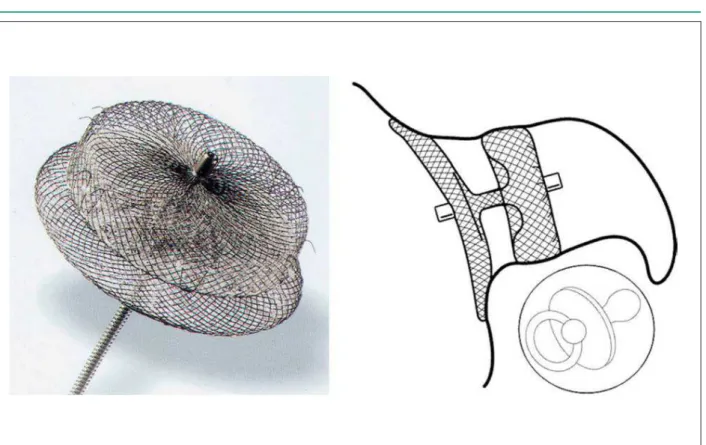
The ACP is a self-expandable nitinol device with a polyester patch within, formed by three parts: a cylindrical lobe with a fixed length of 6.5 mm, to which diameter (16 – 30mm, stepwise by 2mm) the prosthesis size refers; an occlusive disc, 4mm larger than the 16 – 22mm prosthesis, and 6mm larger than the 24 – 30mm devices; and a flexible central connector pine. There are six pairs of barbs attached to the lobe and directed to the disc, all identified by radiopaque marks, to enhance retention of the lobe in the LAA (Figure 1).
Device implantation and follow-up protocol
Before intervention, a pre-evaluation TEE was performed to exclude thrombi in the LAA, oral anticoagulation was suspended, and an antibiotic prophylaxis with cefuroxime was given. All procedures were performed via femoral access, under local anesthesia, and were guided exclusively by angiography (biplane in most cases). Heparinization with 5000 units of heparin was given at the beginning of the procedure. Access to the left atrium was gained through transseptal puncture or passage through a preexisting patent foramen ovale (PFO) or atrial septal defect (ASD -Table 1). After angiography and measurement or estimation of the LAA diameter at the intended implantation site in at least 2 different projections (Figure 2), a device with a diameter at least 4 mm larger than the landing zone diameter was chosen for implantation. In patients with paroxysmal AF in sinus rhythm at the time of implantation, these measurements were taken during atrial diastole. Once the ACP was implanted and some compression of the lobe by the LAA wall was observed (Figure 3), device stability was tested by gently pulling and releasing the delivery cable (Minnesota wiggle maneuver). The lobe has to move in conjunction with the LAA while the disk moves freely with the wire. Control angiographies were performed in various projections (depicting the lobe separated from the disk) prior to device release. In case of unsatisfactory positioning or anchoring, the prosthesis was recaptured, preferentially except for the distal part of the lobe and redeployed in a different angle, or changed for a more suitably sized device. Once adequately positioned and fixed, the ACP was released and a final angiography was performed. Patients with a concomitant PFO or ASD had their defects closed by reloading the same delivery cable and sheath with an adequate septal occluder. Unless an arterial puncture had been performed simultaneously, patients themselves performed compression
Table 1 - Baseline clinical and echocardiographic features
Characteristic Studied population (n = 86)
Age (years) 72.2 ± 10.1 Male gender (%) 65.1 Permanent / paroxysmal AF (%) 57.0 / 43.0
CHADS2-Score 2.6 ± 1.2 (1 – 6)
C (%) 17.4
H (%) 82.6
A (%) 52.3
D (%) 26.7
S (%) 37.2
CHA2DS2-VASc Score 3.6 ± 1.6 (1-7) Contraindication for oral anticoagulation (%)
Relevant bleeding or high
risk of bleeding 69.8 Frequent falls 8.1
Labile INR 4.7
Aversion to oral anticoagulation 15.1
Other 2.3
LVEF (%) 55.5 ± 9.9 (30 – 70)
Figure 1 - The Amplatzer Cardiac Plug (1a) and the “paciier principle” (1b).
Figure 3 - Implantation (3a) and release (3b) of the Amplatzer Cardiac Plug in the left atrial appendage.
differently sized ACPs, a pericardial tamponade requiring emergency pericardial drainage ensued, and the LAA closure was waived. The PFO was closed and the patient was discharged the following day.
Eighty-seven devices were implanted in the 85 patients in whom success was achieved, as two patients received two devices each. In 81 of them adequate positioning and anchoring was obtained with the first device chosen. In the remaining four patients the initial prosthesis was changed for a more suitably sized one. In two cases this was a larger one and in two cases a smaller one. In two patients, both with a bilobulated LAA, an incomplete closure of the LAA was observed after device implantation. In one of them, an additional ACP was implanted, and in the other one the remaining part of the LAA was closed with an Amplatzer vascular plug, with a good final result in both cases. One ACP embolized into the aorta about 15 minutes after being released. This was observed during an incidental percutaneous coronary intervention (PCI) still ongoing, and the device was percutaneously retrieved and replaced by a smaller ACP. The patient was discharged the day after the procedure and the stable position of the second device was ascertained by echocardiography before dismissal. In addition to the pericardial tamponade described above, in one patient a small pericardial effusion with no hemodynamic compromise was observed, with total resolution during follow-up. One patient who underwent simultaneous PCI and TAVI developed acute renal failure
with spontaneous recovery. There were two periprocedural cerebral events, one due to air embolism and the other most probably thromboembolic, both without clinical sequelae at the time of hospital discharge the following day.
Forty-eight patients (55.8%) underwent a simultaneous intervention (ASD or PFO closure, PCI, or TAVI, some in various combinations) at the time of LAA closure. The mean total administered volume of contrast medium was 253.5±114.3 ml, and the mean total fluoroscopy time 19±12 min.
One patient with a bleeding gastrointestinal tumor prohibiting anticoagulation died due to uncontrollable gastrointestinal bleeding six days after ACP implantation. Among the remaining 84 eligible patients, clinical and echocardiographic follow-up was obtained in 69 (82.1%). After 25.9 patient-years there were no strokes and no peripheral thromboembolizations. There were two late deaths, one due to respiratory failure secondary to bronchopneumonia. The other death was cardiac in a patient with known severe three-vessel coronary artery disease. In all but two patients, follow-up TEE demonstrated total occlusion of the LAA. In six patients, a non-mobile thrombus was detected on the device. All of them disappeared on repeat TEE done after reinstitution of oral anticoagulation for three months. In four patients, the presence of a fixed thrombus on the device could not be ruled out. Three of them remained on acetylsalicylic acid. In the fourth one warfarin was resumed and maintained for four months, with no change being observed in the control TEE after
Table 2 - Procedural results
Studied population Multicentric European Experience8
Number of patients 86 143
Acess to left atrium
Transseptal (n, %) 56 (65.1) 121 (84.6)
PFO (n, %) 27 (31.4) 17 (11.9)
ASD (n, %) 3 (3.5) 3 (2.1)
LAA oriice (angiography – mm) 19.5 ± 4.3 19.7 ± 4.3
Success (%) 99 96
ACP size (mm) 23.1 ± 3.9 22.2 ± 3.6
Associated procedures
PFO occlusion (n, %) 27 (31.4) 10 (7.0)
ASD occlusion (n, %) 3 (3.5) 1 (0.7)
PCI (n, %) 22 (25.6) n.a.
TAVI (n, %) 5 (5.8) n.a.
Complications
Periprocedural cerebral events (n, %) 2 (2.3) 3 (2.1)
Cardiac tamponade (n, %) 1 (1.1) 5 (3.5)
Pericardial effusion (n, %) 1 (1.1) 4 (2.8) Device embolization (n, %) 1 (1.1) 2 (1.4)
__________________________________________________________________
* Schmid M, Gloekler S, Saguner A et al. Manuscript submitted to publication.
this time. In one patient a persistent small, hemodynamically non-significant left-to-right shunt at the transseptal puncture site was observed during late follow-up.
Discussion
Adequate levels of oral anticoagulation with warfarin proved effective to reduce stroke by 64% in NVAF patients9.
This means that in a third of patients, the therapy is ineffective. Moreover, multiple studies, including the SPORTIF series, have shown that up to 29% of the international normalized ratios (INRs) are subtherapeutic, 15 – 20% are supratherapeutic10, and that even in patients with optimal
drug compliance, the INR is in its therapeutic range only about 60% of the time11. It must also be considered that the benefits
of anticoagulation are not achievable without incurring the risk of bleeding. There is an annual risk of 3% for major bleeding and 9.6% for hemorrhagic complications in general with the use of warfarin12. Accordingly, its administration
is contra-indicated in up to 44% of the patients with AF, especially those with recurrent major bleeding or previous cerebral bleeding2. Other issues to keep in mind are the
difficult administration profile of the drug and the high rates of patient noncompliance due to the necessary life-style modifications. In clinical practice, therefore, the level of prescription of warfarin varies from 23% to 66% in high-risk patients and 8% to 49% in moderate risk patients5.
New anticoagulant drugs proved to be as or more effective than warfarin, with a safety profile which is at least comparable. Dabigatran, an oral direct thrombin inhibitor, administered at a dose of 150mg twice daily, significantly reduced the rate of peripheral embolization, with similar rates of major bleeding. When given at a dose of 110mg twice daily, it showed similar rates of systemic embolism and significantly lower rates of major hemorrhage13. Rivaroxaban,
an oral direct factor Xa inhibitor, given in a 20mg dose once daily, was non-inferior to warfarin in terms of peripheral embolism and bleeding complications as a whole, and was superior with regard to the occurrence of fatal and cerebral bleedings14. Apixaban, another oral direct factor Xa inhibitor,
when 5mg twice daily was administered, was superior to warfarin with regard to both the prevention of cerebral and peripheral embolism and the occurrence of major bleeding complications15. These drugs, however, also have significant
drug interactions (amiodarone, verapamil, quinidine) and some side effects, especially dyspepsia associated with dabigatran. Some are contraindicated in patients with liver or renal failure; they should be administered with caution in frail patients and in patients older than 75 years, due to enhanced bleeding risks; and there is currently no tested antidote that can be given in cases of major bleeding or emergency surgery16-18. Apart from these unfavorable characteristics and
from their markedly higher cost when compared to warfarin, neither of these drugs is free of bleeding risk, especially in elderly patients and patients with previous major bleedings, and noncompliance, well illustrated by the high rates of drug discontinuity in the RE-LY (21% on dabigatran and 17% on warfarin group)13,19 and ARISTOTELE (25% on apixaban and
27% on warfarin group) trials15. To overcome these limitations,
non-pharmacologic therapeutic strategies for prevention of stroke in NVAF continue to be warranted.
The LAA, a remnant of the embryonic left atrium, is a (multi)lobulated structure of variable anatomy, made of trabecules of pectinate muscles that form crypts in between them. The asymmetric junction that connects it to the left atrium is usually narrower than its body, and is located typically anterior and inferior to the left superior pulmonary vein2,20. In AF, the LAA structure and the marked reduction
of its flow velocities and ejection fraction provide a rich milieu for blood stasis and thrombus formation, making the LAA the most important source of cerebral and peripheral emboli. Accordingly, in a review of 23 studies in which the LAA was examined by autopsy, TEE, or direct intra-operative inspection, intracardiac thrombus was encountered in 17% of NVAF patients, 91% of which in the LAA6. That is why
the LAA has been deemed “our most lethal attachment” 21,
and its occlusion was proposed as a valuable alternative to anticoagulation for embolism prevention in patients with NVAF. LAA occlusion can be performed in three distinctive ways: surgical ligation or exclusion, concomitant to valvular surgery, coronary revascularization, or MAZE procedures; percutaneous epicardial exclusion, either thoracoscopic or via the pericardial sac, a new method that mimics surgical ligation; and percutaneous endovascular occlusion.
Despite the proven effectiveness of surgical occlusion of the LAA, and its inclusion in the guidelinesfor mitral valve surgery 22, its main limitation is high incomplete
occlusion rates, varying from 10%-80%, depending on the employed technique and on the surgeon’s experience. The highest success rate of complete occlusion is achieved with LAA excision, and the lowest, with exclusion by suture or staple ligation7,23.
The familiarity, ease of implantation, and low thrombogenicity of the Amplatzer devices led to the first LAA closure series with an Amplatzer Septal Occluder in Bern, Switzerland. A study describing the results of such off-label procedures in 16 patients showed one device embolization and complete LAA occlusion in all remaining patients after a 5 patient-years follow-up24. However, a longer-time
registry demonstrated that the use of septal occluders for LAA occlusion was associated with lower success and higher embolization rates when compared to the implantation of dedicated devices.*
The first dedicated device for LAA occlusion was the PLAATO System (ev3, Plymouth, MN, no longer available), first implanted in 200125. It consisted of a self-expandable
nitinol cage covered with a non-thrombogenic PTFE membrane. Short-term as well as 5-year results after PLAATO system implantation were good, with a 42% reduction of the stroke rate predicted by the CHADS2 score (3.8% / year
vs. 6.6% / year)26,27.
PROTECT-AF trial proved the non-inferiority of Watchman device implantation in comparison to chronic warfarin therapy in patients with NVAF, with a major event rate (stroke, systemic embolization, or cardiovascular or unexplained death) of 3.0 / 100 patient-years versus 4.9 / 100 patient-years, but at a cost of more complications in the group randomized to device implantation (7.7% vs. 3.7%)29. However, the incidence of
procedure-related complications, mainly pericardial effusions and strokes secondary to air embolization, significantly decreased along the learning curve of the operators30.
There are many structural differences between the Amplatzer Cardiac Plug and the Watchman device. The most important refers to the occlusive disc. The Watchman device is basically a plug that should be precisely implanted to avoid both its protrusion into left atrium as well as the creation of a cul de sacwhere thrombus may form. The ACP consists of two parts joined by a central pin. Being short, the ACP can be implanted in a shallow position in the LAA, as only the proximal 2 cm are needed for its occlusion. The occlusive disk permits the complete closure of the LAA orifice (“pacifier principle”24 Figure
1), surpassing the problems that the myriad of LAA anatomical variations, mostly distally located, may impose. The flexibility of the central pin allows a misalignment between the disc and the lobe of the ACP after implantation, adapting the prosthesis to the LAA axis rather than distorting it31. Also, the more anatomic
surface that derives from the occlusive disc implantation results in a rheology that is closer to normal and also in a more predictable endothelization32. Another significant difference between the
devices is the fabric covering of the Watchman device, which is permeable to blood, hence the recommendation to continue warfarin therapy for six weeks after implantation. The ACP, on the other side, seems to permit cessation of anticoagulation immediately after its implantation8. Moreover, the kit used for
implanting the ACP features a double-curved sheath, facilitating coaxial intubation of the LAA, and it is compatible with other Amplatzer devices, making simultaneous closure of atrial shunts straightforward by simply reloading the sheath with an additional device33. These features associated with the familiarity with the
Amplatzer technique make ACP implantation user-friendlier in comparison to the Watchman device.
Regardless of the implanted device, however, percutaneous LAA occlusion is not a risk-free intervention, given the intrinsic structural vulnerability of the LAA and the possibility, albeit low, of device embolization, or embolization of preexisting thrombi not adequately identified by TEE or preliminary contrast medium injection into the LAA. Therefore, the procedure must only be indicated after assessing the risk of stroke (estimated by the CHADS2 and the CHA2DS2-VASc scores34,35), risk of
bleeding (estimated by the HAS-BLED score36), risk of the
intervention and quality of life.
The mean CHADS2 score of 2.6 of the studied cohort projects a yearly occurrence of 5.2% of embolic events4,34.
No events were observed during the follow-up period. In addition, the total complication rate in this high-risk patient series was lower than that reported in the multicentric European experience with the ACP8 (table 2), as well as in
the PROTECT-AF trial29 but higher than CAP registry with the
Watchman device30. Similarly to all interventional procedures,
the learning curve plays an essential role in LAA closure. The empirical post-implantation medication protocol, namely dual antiplatelet therapy with no further oral anticoagulation, was adopted based on the legendary low thrombogenicity of the Amplatzer septal ocluder devices37.
It can be argued, however, that the thrombogenic potential of a device occluding a septal defect in sinus rhythm is lower than that related to one in fibrillating LA8. Hence, the late
echocardiographic finding of device-associated thrombi in some patients came as no surprise, taking into account the high CHADS2 score of this population, and being aware of previous reports showing similar findings38. Retrospectively
reviewing the images of these procedures it could be noted that, in 70% of them, the disk of the occluder was implanted somewhat inside the LAA rather than at its ostium, as would be ideal. However, these thrombi were firmly attached to the device. They generally disappeared after temporary reinstitution of oral anticoagulation, with no embolic events. The frequency of this finding in the growing experience with ACP implantation may suggest, however, a need to adapt post-implant medication protocols.
In summary, these data allow for concluding that ACP implantation for LAA occlusion is associated with high success and acceptable complication rates, and promising follow-up results. As with every interventional procedure, however, the clinical benefits that derive from the intervention depend on careful patient selection, on having passed the learning curve, and on well-defined, adequate pre- and post-implantation protocols.
Potential Conflict of Interest
The author Ahmed A. Khattab states he serves an attorney-in-fact for ST. JUDE -AGA. Author Bernhard Meier states he receives consulting and lecturing fees from ST. JUDE - AGA.
Sources of Funding
There were no external funding sources for this study.
Study Association
References
1. Roger VL, Go AS, Lloyd-Jones DM, Adams RJ, Berry JD, Brown TM, et al. Heart disease and stroke statistics--2011 update: a report from the American Heart Association. Circulation. 2011;123(4):e18-e209.
2. Singh IM, Holmes Jr DR. Left atrial appendage closure. Curr Cardiol Rep. 2010;12(5):413-21.
3. Fuller CJ, Reisman M. Stroke prevention in atrial fibrillation: atrial appendage closure. Curr Cardiol Rep. 2011;13(2):159-66.
4. Camm AJ, Kirchhof P, Lip GYH, Schoeten U, Savelieva I, Ernst S, et al. Guidelines for the management of atrial fibrillation. The Task Force for the Management of Atrial Fibrillation of the European Society of Cardiology (ESC). Eur Heart J. 2010;31(19):2369-429.
5. Ederhy S, Dufaitre G, Boyer-Chatenet L, Boyer-Chatenet, Meuleman C, Di Angelantonio E, et al. Should all patients with non-valvular atrial fibrillation be anticoagulated? Int J Cardiol. 2010;143(1):8-15.
6. Blackshear JL, Odell JA. Appendage obliteration to reduce stroke in cardiac surgical patients with atrial fibrillation. Ann Thorac Surg. 1996;61(2):755-9. 7. Healey JS, Crystal E, Lamy A, Teoh K, Semelhago L, Hohnloser SH, et al. Left Atrial Appendage Occlusion Study (LAAOS): results of a randomized controlled pilot study of left atrial appendage occlusion during coronary bypass surgery in patients at risk for stroke. Am Heart J. 2005;150(2):288-93.
8. Park JW, Bethencourt A, Sievert H, Santoro G, Meier B, Walsh K, et al. Left atrial appendage closure with Amplatzer cardiac plug in atrial fibrillation: initial European experience. Catheter Cardiovasc Interv. 2011;77(5):700-6. 9. Hart RG, Pearce LA, Aguilar MI. Meta-analysis: antithrombotic therapy to prevent stroke in patients who have nonvalvular atrial fibrillation. Ann Intern Med. 2007;146(12):857-67.
10. White HD, Gruber M, Feyzi J, Kaatz S, Tse HF, Husted S, et al. Comparison of outcomes among patients randomized to warfarin therapy according to anticoagulant control: results from SPORTIF III and V. Arch Intern Med. 2007;167(3):239-45.
11. Mega JL. A new era for anticoagulation in atrial fibrillation. N Engl J Med. 2011;365(11):1052-4.
12. Wysowski DK, Nourjah P, Swartz L. Bleeding complications with warfarin use: a prevalent adverse effect resulting in regulatory action. Arch Intern Med. 2007;167(13):1414-9.
13. Connolly SJ, Ezekowitz MD, Yusuf S, Eikelboom J, Oldgreen J, Parekh A, et al. Dabigatran versus warfarin in patients with atrial fibrillation. N Engl J Med. 2009;361(12):1139-51
14. Patel MR, Mahaffey KW, Garg J, Pan G, Singer DE, Hacke W, et al. Rivaroxaban versus warfarin in nonvalvular atrial fibrillation. N Engl J Med. 2011;365(10):883-91.
15. Granger CB, Alexander JH, McMurray JJ, Lopes RD, Hylek EM, Hanna M, et al. Apixaban versus warfarin in patients with atrial fibrilation. N Engl J Med. 2011;365(10):981-92.
16. Douketis JD. Dabigatran as anticoagulant therapy for atrial fibrillation. Which patients should receive it, which patients may not need it, and other practical aspects of patient management. Pol Arch Med Wewn. 2011;121(3):73-80. 17. Legrand M, Mateo J, Aribaud A, Ginisty S, Eftekhari P, Huy PT, et al. The use of dabigatran in elderly patients. Arch Intern Med. 2011;171(14):1285-6. 18. Jacobs JM, Stessman J. New anticoagulants among elderly patients is caution
necessary?: Comment on «The use of dabigatran in elderly patients» Arch Intern Med. 2011;171:1287-8.
19. Gage BF. Can we rely on RE-LY? N Engl J Med. 2009;361(12):1200-2. 20. Khattab AA, Meier B. Transcatheter left atrial appendage exclusion, gold or
fool´s gold? Eur Heart J Suppl. 2010;12 (Suppl. E):E35-E40.
21. Johnson WD, Ganjoo AK, Stone CD Srivyas RC, Howard M. The left atrial appendage: our most lethal human attachment! Surgical implications. Eur J Cardiothorac Surg. 2000;17(6):718-22.
22. Bonow RO, Carabello BA, Chatterjee K, de Leon AC Jr, Faxon DP, Freed MD, et al. 2008 focused update incorporated into the ACC / AHA 2006 guidelines
for the management of patients with valvular heart disease: a report of the American College of Cardiology / American Heart Association Task Force on Practice Guidelines (Writing Comitee to Revise the 1998 Guidelines for the Management of Patients With Valvular Heart Disease). Circulation. 2008;118(15):e523-661.
23. Dawson AG, Asopa S, Dunning J. Should patients undergoing cardiac surgery with atrial fibrillation have left atrial appendage exclusion? Interact Cardiovasc Thorac Surg. 2010;10(2):306-11.
24. Meier B, Palacios I, Windecker S, Rotter M, Cao QL, Keane D, et al. Transcatheter left atrial appendage occlusion with Amplatzer devices to obviate anticoagulation in patients with atrial fibrillation. Catheter Cardiovasc Interv. 2003;60(3):417-22.
25. Sievert H, Lesh MD, Trepels T, Omran H, Bartorelli A, Della Bella P, et al. Percutaneous left atrial appendage transcatheter occlusion to prevent stroke in high-risk patients with atrial fibrillation: early clinical experience. Circulation. 2002;105(16):1887-9.
26. Ostermayer SH, Reisman M, Kramer PH, Mattheu’s RV, Gray WA, Block PC, et al. Percutaneous left atrial appendage transcatheter occlusion (PLAATO system) to prevent stroke in high-risk patients with non-rheumatic atrial fibrillation: results from the international multi-center feasibility trials. J Am Coll Cardiol. 2005;46(1):9-14.
27. Block PC, Burstein S, Casale PN, Kramer PH, Teirstein P, Williams DO, et al. Percutaneous left atrial appendage occlusion for patients in atrial fibrillation suboptimal for warfarin therapy: 5-year results of the PLAATO (Percutaneous Left Atrial Appendage Transcatheter Occlusion) Study. J Am Coll Cardiol. 2009;2(7):594-600.
28. Gorodnitskiy A, Lucariello RJ, Aizer A, Coppola JT. A novel approach to left atrial appendage occlusion: the Watchman device. Cardiol Rev. 2010;18(5):230-3.
29. Holmes DR, Reddy VY, Turi ZG, Doshi SK, Sievert H, Buchbinder M, et al. Percutaneous closure of the left atrial appendage versus warfarin therapy for prevention of stroke in patients with atrial fibrillation: a randomised non-inferiority trial. Lancet. 2009;374(9689):534-42.
30. Reddy VY, Holmes DR, Doshi SK, Neuzil P, Kar S. Safety of percutaneous left atrial appendage closure: results from the Watchman Left Atrial Appendage System for Embolic Protection in Patients With AF (PROTECT-AF) Clinical Trial and the Continued Access Registry. Circulation. 2011;123(4):417-24.
31. Armaganijan LV, Staico R, Pedra SF, Moreira DA, Braga SL, Feres F, et al. Experiência inicial com o novo Amplatzer Cardiac Plug para oclusão percutânea do apêndice atrial esquerdo. Rev Bras Cardiol Invas. 2011;19(1):14-23.
32. Bass JL. Transcatheter occlusion of the left atrial appendage – experimental testing of a new Amplatzer device. Catheter Cardiovasc Interv. 2010;76(2):181-5.
33. Meier B. Left atrial appendage closure: do we have one ear too many? Cardiac & Vascular Update 2010;3:12-4.
34. Gage BF, Waterman AD, Shannon W, Boechler M, Rich MW, Radford MJ. Validation of clinical classification schemes for predicting stroke: results from the National Registry of Atrial Fibrillation. JAMA. 2001;285(22):2864-70. 35. Olesen JB, Lip GY, Hansen ML, Tolostrup IS, Lindhardsen J, Selmer C,
et al. Validation of risk stratification schemes for predicting stroke and thromboembolism in patients with atrial fibrillation: nationwide cohort study. BMJ. 2011;342:d124.doi: 10.1136/bmj.d124
36. Pisters R, Lane DA, Nieuwlaat R, de Vos CB, Crijns HJ, Lip GY. A novel user-friendly score (HAS-BLED) to assess one-year risk of major bleeding in atrial fibrillation patients: the Euro Heart Survey. Chest. 2010;138(5):1093-100.
37. Krumsdorf U, Ostermayer S, Billinger K, Trepels T, Zadan E, Horvath K, et al. Incidence and clinical course of thrombus formation on atrial septal defect and aptent foramen ovale closure devices in 1,000 consecutive patients. J Am Coll Cardiol. 2004;43(2):302-9.