may-aug. 2015, Vol. 25, No. 61, 251-259. doi:10.1590/1982-43272561201513
Subjective time studies have shown that time perception can be modulated by the visual perception of implied movement of human bodies in static images: the greater the implied movement, the longer the duration for the individual (Nather & Bueno, 2011). This time distortion has been related to embodiment mechanisms and empathy (Nather, Bueno, Bigand, & Droit-Volet, 2011) as a consequence of incorporating visualized implied movement through an increase in the accumulation of pulses of the internal clock
Article
Implied Movement in Static Images Reveals Biological Timing Processing
1Abstract: Visual perception is adapted toward a better understanding of our own movements than those of non-conspeciics. The present study determined whether time perception is affected by pictures of different species by considering the evolutionary scale. Static (“S”) and implied movement (“M”) images of a dog, cheetah, chimpanzee, and man were presented to undergraduate students. S and M images of the same species were presented in random order or one after the other (S-M or M-S) for two groups of participants. Movement, Velocity, and Arousal semantic scales were used to characterize some properties of the images. Implied movement affected time perception, in which M images were overestimated. The results are discussed in terms of visual motion perception related to biological timing processing that could be established early in terms of the adaptation of humankind to the environment.
Keywords: motion perception, time, evolution (biology)
Movimento Implícito em Imagens Estáticas Revelou um Processamento Biológico do Tempo
Resumo: A percepção visual é adaptada para compreender melhor os movimentos da própria espécie do que aqueles de outras espécies.
O objetivo deste estudo foi veriicar se a percepção temporal seria afetada por fotograias de diferentes espécies de animais levando em
consideração a escala evolutiva. Imagens sem (“S”) e com movimento implícito (“M”) de um cachorro, guepardo, chimpanzé e homem foram expostas a estudantes universitários. As imagens S e M de cada espécie foram apresentadas em ordem aleatória ou uma após a outra (S-M ou M-S) para dois grupos de participantes. Escalas Semânticas para Movimento, Velocidade e Arousal foram utilizadas para a caracterização de algumas propriedades das imagens. O movimento implícito afetou a percepção do tempo: as imagens M foram superestimadas. Os resultados foram discutidos em termos da percepção visual de movimento relacionada a um processamento de tempo biológico que pode ter sido estabelecido cedo em termos de adaptação do homem ao meio ambiente.
Palavras-chave: percepção de movimento, tempo, evolução (biologia)
Movimiento Implícito en Imágenes Estáticas Revelo un Procesamiento Biológico de
lo Tiempo
Resumen: La percepción visual es adaptada para comprender mejor los movimientos de la misma especie que aquellos de otras especies. Este estudio examinó si la percepción del tiempo se vería afectada por fotografías de diferentes especies de animales, teniendo en cuenta la escala evolutiva. Imágenes sin (“S”) y con movimiento implícito (“M”) de un perro, guepardo, chimpancé y hombre fueron expuestas a estudiantes universitarios. Las imágenes S y M de cada especie se presentaron en orden aleatorio, o una después de la otra (MS o MS), para 2 grupos de participantes. Escalas semánticas para Movimiento, Velocidad y Arousal fueron utilizadas para la caracterización de las imágenes. Movimiento implícito afectó la percepción del tiempo: M imágenes fueron sobreestimadas. Los resultados se discutieron en términos de la percepción visual del movimiento relacionada con lo
procesamiento biológico del tiempo que pueda haberse ijado al principio de la adaptación del hombre con el medio ambiente.
Palabras clave: percepción de movimiento, tiempo, evolución (biologia)
1 Support: Francisco C. Nather received a Young Researcher Award from the São
Paulo Research Foundation (FAPESP – Protocol no. 2011/17981-9). José Lino O. Bueno received a Research Grant from the National Council for Scientiic and Technological Development (CNPq-PQ – Protocol no. 307485/2011-0). Vinicius Anelli received a FAPESP Undergraduate Research Scholarship (Protocol no. 2012/13067-3) and CAPES/DAAD Exchange Scholarship. Guilherme P. Ennes received a RUSP Undergraduate Research Scholarship (Protocol no. 13.1.1651.59.0).
2 Correspondence address:
Francisco Carlos Nather. Universidade Federal do ABC. Centro de Matemática, Computação e Cognição. Rua Arcturus, 03. CEP 09606-070. São Bernardo do Campo-SP, Brazil. E-mail: [email protected]
Francisco Carlos Nather2 Universidade Federal do ABC, São Bernardo do Campo-SP, Brazil
Vinicius Anelli Universidade de São Paulo,
Ribeirão Preto-SP, Brazil
Guilherme Ennes Universidade de São Paulo,
Ribeirão Preto-SP, Brazil
José Lino Oliveira Bueno Universidade de São Paulo,
(Gibbon, 1977). Through embodiment mechanisms, speciic brain areas are activated, reconstructing the actions by simply observing the static pattern of a past action (Gallese, 2005; Rizzolatti & Craighero, 2004; Rizzolatti & Lupino, 2001). Thus, time distortions in the perceiver may be an index of empathic processes that enable the individual to understand the others’ actions as a function of the timing related to the observed movements (Nather, Bueno, & Bigand, 2013).
Movement perception in static images has been studied with regard to different and speciic aspects of the visual perception of motion using photographic images of humans and animals by considering social and evolutionary contexts. For example, dog, cheetah, monkey, and human photographs were used to discuss the impact of implied motion in static images in terms of motion adaptation through an aftereffect (Pavan, Cuturi, Maniglia, Casco, & Campana, 2011; Winawer, Huk, & Boroditsky, 2008). These studies showed that pictures that imply motion activate “direction-selective” and “velocity-tuned” mechanisms that are involved in real physical motion perception. An increase in the speed (velocity) of the images highly activates brain areas that respond to real motion (Williams & Wright, 2010).
Different studies have used photographs of different biological stimuli or inanimate objects to understand different aspects of motion perception in static images (Downing, Jiang, Shuman, & Kanwisher, 2001). The seminal study by Freyd (1983) showed that individuals mentally represent movement when they are observing actions of other humans in photographs. By analyzing the time required by observers to distinguish two images of the same action, presented one after the other, Freyd showed that when they are presented in chronological order, the participants needed more time to distinguish the images relative to the future position of a human movement. This delay was attributed to the mental movement representation (Chatterjee, Freyd, & Shiffrar, 1996; Shiffrar & Freyd, 1993), which can be interpreted as a delay in time processing. Furthermore, movement perception is implicitly related to the perception of time because real motion occurs at the intersection of time and space.
Different circuits, such as the right visual area MT/V5, underlie both motor and perceptual representations of temporal events in the brain (Bueti, Bahrami, & Walsh, 2008; Bueti, Walsh, Frith, & Rees, 2008). Speciically, the middle temporal (MT) and superior temporal sulcus (STS) of the human brain cortex were activated while observing static images that implied different movements (Proverbio, Riva, & Zani, 2009; Senior et al., 2000; Williams & Wright 2010). Kourtzi and Kanwisher (2000) showed that photographs of humans and different animals that implied more movement distinctly activated the MT/MST areas. An image of an athlete who moved generated more activation of these brain areas than a photograph of the same athlete who was still or another man who was resting. Similarly, the image of a
jumping dolphin in the sea generated less activation of these brain areas than a polar bear lying still.
People can discern the movements of animals of different species because of their distinct biological features that are related to adaptation to different environments. Moreover, the human visual system seems adapted to perceiving the actions of other human beings as a result of the social relevance of movements performed during daily life (Kaiser, Shiffrar, & Pelphrey, 2012; Thompson & Parasuraman 2012). This developed capacity is attributable to learned experiences because people tend to mimic or embody human actions (Blake & Shiffrar, 2007; Pinto & Shiffrar, 2009). Buccino et al. (2004) used static images and short ilms of a dog, monkey (chimpanzee), and man performing different activities. They showed that actions performed by these three species were processed differently. The major brain activation of the STS in the participants was related to the visualized actions of their own motor repertoire (Kaiser et al., 2012). This enhanced sensitivity is essential for the social maintenance of the group (Pinto & Shiffrar, 2009), in which the human brain is more sensitive to human motion than to movements performed by nonhumans. However, motion perception in static and moving images, in terms of biological motion, has not been suficiently analyzed while considering subjective time perception.
Yamamoto and Miura (2012) showed that drawings of a dog and man that implied dynamic movements were perceived in the same way. The dog and man images that implied more movement were estimated longer than those that did not imply movement. However, this study used only two animal species and did not consider the biological relationships between them. The present study tested whether humans are able to recognize the actions of different species in static images by taking into account their species-speciic characteristics. It evaluated whether visual movement perception affects subjective time according to an internal mechanism of time from a biological perspective (biological timing processing).
be closed for images of the same species (monkey and man) than for images of the other two species (cheetah and dog), independent of the implied motion that is present in the photographic images. In the G2 group, the same species images as the G1 group were used, but they were presented in an experimental design that avoided the participants’ observing the S and M images of the same species immediately one after the other. The S and M images of different species were presented randomly to the participants. This procedure was used to further verify the effects of implied body movement in the S and M images on time perception, independent of the species used. This group was speciically related to the visual motion perception of each image rather than to the possible clues provided by S and M image presentation of the same species one after the other. Therefore, if time estimation is affected by implied motion in static images, then the M images of the four species will cause greater time estimation than the S images.
Method
Participants
Forty-seven university students (24 men; 19.72 ± 2.27 years old) from the University of São Paulo with normal or corrected-to-normal vision were randomly invited to participate in the study.
Instruments
The experiment was performed in an isolated, soundproof room at the central library of the University of São Paulo campus in Ribeirão Preto, Brazil, during the day. The room was illuminated with artiicial light (compact luorescent 40 lm W-1 light bulbs). Eight digital photographs of four different mammal species (dog [Canis lupus familiaris], cheetah [Acinonyx jubatus], chimpanzee [Pan troglodytes], and man [Homo sapiens sapiens]) that did or did not imply movement were used as stimuli (M and S images, respectively). The M images showed the animals running, and the S images showed the same animals stopped (standing still). These stimuli were constructed using photographic images from the Internet, which were modiied using Photoshop CS6 software to generate correspondence between their speciic body sizes, brightness, and saturation. All of the images (20 × 30 cm) were presented in the center of a 19” computer screen. The ground was illed with light brown, and the background was white. In addition to the species images, three different images of arrows that occupied the same central position as the different species on the computer screen were used. Eight arrow stimuli (mean size of 10 × 20 cm) were the following: arrows from the middle of the visual ield to the left or right side of the image, from the middle up and down, from the diagonal right to up and down or from the
diagonal to left to up and down. These arrows were used to avoid the rapid comparison of different species images in the different exposure sequences. E-Prime 2.0software installed on an HP notebook computer was used to present the stimuli and record time estimations. Differential Semantic Scales for the locutions “Movement” implied in the image (unipolar Likert-type 1-7 point scale), “Velocity” of the animal species (unipolar Likert-type 1-7 point scale), and “Arousal” evoked (Ranking Manikin 1-5 point scale) (Lang, 1980) were used to better characterize the movement properties of the photographic images. The semantic scale data were obtained using an experimental copybook that was also used to record personal information about the participants.
Procedure
Data collection. The tasks were verbally explained to
the participants. They were placed facing the central region of the computer monitor at a distance of approximately 60 cm. Each stimulus presentation began by pressing the “presentation” key, with a total exposure time of 6 s. At that moment, the monitor was illed with white, indicating that the participant could initiate time estimation. Immediately after the observation time, the participant reproduced the exposure duration by pressing the “initiate” key. The experienced duration of each stimulus was inalized by pressing the “inished” key (reproduction method in the prospective paradigm). The stimuli were presented randomly to the two groups of participants according to their presentation order: G1 (N = 23, 11 men; age: M = 19.72 years, SD = 2.27 years) and G2 (N = 24, 13 men; age: M = 19.88 years, SD = 1.78 years). For each species, there were two images: one that did not imply movement (S image) and one that implied movement (M image). In both groups, the S and M images were presented interspersed by three different arrow (“a”) images, which were also presented for 6 s each. Each experimental session consisted of the presentation of three arrow images presented before each of the species images (S and M), for a total of 32 time estimations: eight for S and M images of the species and 24 for the arrows. In the G1 group, the S and M images of the same species were immediately presented one after the other, and the images were presented randomly to each participant (M after S or S after M). In the G2 group, all of the images were presented randomly, but the two images (S and M) of the same species were not immediately presented one after the other. For example, two complete experimental sessions of arrow images (“a”) and S and M images of the species were the following: aaa
DogS aaa DogM aaa ManM aaa ManS aaa CheetahS aaa
CheetahM aaa MonkeyM aaa MonkeyS (G1 group); aaa
MonkeyM aaa CheetahS aaa ManM aaa DogM aaa ManS
aaa MonkeyS aaa DogS aaa CheetahM (G2 group). The
task. The time estimations for the arrows were not used in the data analysis and were discarded. The participants did not receive feedback about their time estimations, and they were instructed not to count time. After the time estimations, the participants were asked to observe and judge the species images by completing the Differential Semantic Scales for the locutions Movement, Velocity, and Arousal. The scales, similar to the images, were presented randomly to each participant. The participants completed the task of completing the semantic scales in the experimental copybook while observing the images as long as they wanted.
Data analysis.The analysis of the time estimation data
for the G1 and G2 groups was conducted separately. Two-way repeated-measures analysis of variance (ANOVA; 2 × 4) was used to test the statistical interaction between the factors implied movement (S and M images) and species (dog, cheetah, monkey, and man). Sidak’s t-test was used for post hoc comparisons. One-sample t-test was used to compare the participant’s time estimations with the actual exposure time (6 s) of the images for both groups. Factorial analyses of the time estimation data were conducted using KMO > .80 and Bartlett’s test (α < .0001) (Kaiser, 1974). The analysis of the semantic scale data was also conducted separately for the G1 and G2 groups. Two-way repeated-measures ANOVA (2 × 4) was used to test the statistical interaction between the factors implied movement (S and M images) and species (dog, cheetah, monkey, and man) separately for each locution (Movement, Velocity, and Arousal). Sidak post hoc comparisons were used in these analyses. Paired t-test was used to compare the S and M images of each species. Multidimensional analyses (ALSCAL) were conducted for the Movement, Velocity, and Arousal data using a correlation matrix with Euclidian distances and also considering Young’s S-stress (p < .001). Student’s t-test was used to compare the Movement and Velocity semantic scale data between the G1 and G2 groups.
Ethical Considerations
The experiment was approved by the Ethics Committee of the Universidade de São Paulo, Faculdade de Filosoia, Ciências e Letras de Ribeirão Preto, Brazil (CAAE no. 02745412.2.0000.5407).
Results
G1 Group
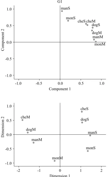
The ANOVA of the time estimation data did not reveal an effect of image (S and M) or species (dog, cheetah, monkey, and man) or an interaction between implied movement and species. The one-samplet-test did not reveal under- or overestimations when the time estimation data were compared with the actual 6 s exposure time (Table 1). The factorial analysis revealed that the time estimations for the S and M images were different (KMO > .80; B(28) = 93.77, p < .0001). The graphical representation of this analysis was
distributed only in the irst quadrant. Images of the dog and cheetah (center) were separate from the monkey and man (borders). Interestingly, in the case of the monkey and man images, the S and M images were mixed according to the implied movement intensities, in which the S images of the monkey and man were positioned close together in the upper left side, and the M images were positioned close together in the bottom right side.
The ANOVA of the Movement scale (2 × 4) revealed signiicant effects of implied movement [F(1; 21) = 219.90, p < .001] and species [F(3; 66) = 13.22; p < .001] and an implied movement × species interaction [F(3; 66) = 13.11; p < .001]. The post hoc comparisons showed that the movement scores for both monkey and man species (primates) were signiicantly less than the others (all p < .01). The paired t-test conirmed that the S and M images of the different species were scored differently (all p < .001). The ANOVA
(2 × 4) of Velocity also veriied signiicant effects of implied movement [F(1; 21) = 77.69; p < .001] and species [F(3;
Table 1
Means and Standard Errors of the G1 and G2 Participants’ Scores on the Semantic Differential Scales (Movement, Velocity, and Arousal Locutions)
Species G1 G2
66) = 24.65; p < .001] and an implied movement × species interaction [F(3; 66) = 6.89; p < .001]. The post hoc comparisons showed that the attributed velocity score for the monkey was less than the other three species (all p < .001), and the man image was less than the dog and cheetah images (both p < .01). The paired t-test showed that the S and M images of the different species were scored differently (all p < .001). Finally, the ANOVA (2 × 4) of Arousal
also revealed signiicant effects of implied movement [F(1; 21) = 22.78; p < .001] and species [F(3; 66) = 25.03; p < .001] and an implied movement × species interaction [F(3; 66) = 3.71; p < .05]. The post hoc comparisons showed that the arousal scores for the cheetah were greater than the other three species (all p < .01). Interestingly, the paired
t-test showed that the S and M images of the dog, cheetah, and man (all p < .01) were scored differently, but the monkey image was not (Table 1).
The multidimensional analysis of Movement, Velocity, and Arousal showed a data distribution that separated the species according to their movements. The S images were separate from the M images in the four quadrants (p < .001). The dog and cheetah images (upper squares) were separate from the monkey and man images (bottom squares). Furthermore, the participants mixed the species according to their movements, with the dog and cheetah S and M images in the left quadrants and monkey and man S and M images in the right quadrants (Figure 1). Altogether, the factorial and multidimensional analyses indicated a tendency by the
Component 2
G1
1.0
0.5
0.0
-0.5
-1.0
-1.0 -0.5 0.0 0.5 1.0
monM manM dogM dogS cheM cheS monS manS
Dimension 2
Component 1
Dimension 1 1.0
0.5
0.0
-0.5
-1.0
-2 -1 0 1 2
monM
monS manS dogS cheS
cheM
dogM
manM
Figure 1. Graphical representation of the mean values of time estimations in the factorial analysis (upper) and Movement, Velocity, and Arousal scales in the multidimensional analysis (lower) in the G1 group. For both analyses, the S and M images of the monkey and man were distributed closer together. In the experimental design for the G1 group, the two images of each species were presented one after the other.
Component 2
G2
1.0
0.5
0.0
-0.5
-1.0
-1.0 -0.5 0.0 0.5 1.0
manS dogM manM cheS dogS cheM mon M monS
Dimension 2
Component 1
Dimension 1 0.4
0.0
-0.4
-0.8
-2 -1 0 1 2
monM cheM
dogM
manM
monS dogS
manS cheS
participants to approximate the monkey and man (primates) images during the time estimations and when completing the differential semantic scales.
G2 Group
The ANOVA (2 × 4) of the time estimation data did not reveal an effect of species (dog, cheetah monkey, and man) or an interaction between implied movement (S and M) and species. However, it revealed a marginal effect of implied movement in the images [F(1; 23) = 3.57; p < .07]. This effect was further revealed by the one-sample t-test, showing that the M images of the dog [t(23) = 1.55; p < .01], cheetah [t(23) = 2.08; p < .05], and man [t(23) = 2.32; p < .05] were overestimated compared with the actual 6 s exposure time (Table 1). The factorial analysis revealed that the time estimations for the S and M images was different (KMO > .84; B(28) = 95.30; p < .0001). However, the graphical representation of this analysis did not show a clear separation of the species as was observed in the G1 group (Figures 1 and 2).
The ANOVA (2 × 4) of the Movement scale data revealed signiicant effects of implied movement [F(1; 23) = 126.43; p < .001] and species [F(3; 69) = 7.95; p < .001] and an implied movement × species interaction [F(3; 69) = 8.19; p < .001]. The post hoc comparisons showed that the movement scores for the monkey images were signiicantly less than both the dog and cheetah images (all p < .001). The paired t-test showed that the S and M images of all of the species were scored differently (all p <
.001). The ANOVA (2 × 4) of Velocity revealed signiicant effects of implied movement [F(1; 21) = 77.69; p < .001] and species [F(3; 66) = 24.65; p < .001] and an implied movement × species interaction [F(3; 66) = 6.89; p < .001]. The post hoc comparisons showed that the velocity scores for the monkey were less than the other three species (all p < .001), and the cheetah was scored with a greater velocity than the other three species (p < .01). The paired t-test conirmed that the S and M images of the different species were scored differently (all p < .001). As in the G1 group, the Movement and Velocity scale data showed that the monkey M image was scored with less implied movement and velocity. For the Arousal scale, the ANOVA (2 x 4) showed signiicant effects of implied movement [F(1; 23) = 77.88; p < .001] and species [F(3; 65) = 10.75; p < .001] and an implied movement × species interaction [F(3; 69) = 10.65; p < .001]. The post hoc comparisons showed that the arousal scores for the monkey were less than the other three species (p < .05). The paired t-test showed that the S and M images of all of the species were scored differently (all p < .001) (Table 1).
The multidimensional analysis showed a data distribution that separated the species according to their movements. The S images were separate from the M images in the four quadrants, as was observed in the G1 group (p < .001). However, the S images in the G2 group were all grouped in the upper right quadrant (Figures 1 and 2).
The analysis of the semantic scale data between the G1 and G2 groups showed that the S images of the dog [t(45) = -1.79; p = .05], cheetah [t(45) = 17.80; p < .01], and man [t(45) = -2.44; p < .01] were scored with more implied movement in the G1 group. For attributed velocity, only the S image of the dog [t(45) = 2.02; p < .05)] was different between the G1 and G2 groups. The same was not observed for the M image data. The semantic scales were completed after the time estimations, and this unexpected result revealed that the participants also scored the implied movement differently from the attributed velocity when they could compare the two movements of the same species prior to observing the next ones.
Discussion
The present study showed that static images of different animal species that implied movement caused different effects on time perception. In the G1 group, the participants could immediately compare the S and M images of each species before observing the next images. The factorial analysis of the G1 group data showed that the time estimations were made according to the species’ characteristics. The images of the dog and cheetah were closely localized but in different areas of the graphical representation, and these two species were separate from the monkey and man. Interestingly, the placement of the S and M images of the primates revealed that the participants did not eficiently distinguish the duration of exposure to images of these species (Figure 1).
In the G2 group, the S and M images of the same species were not immediately presented but after the observation of three images (S or M) of the other species (see Method). The factorial analysis showed that the S and M images of the dog, cheetah, and man were distributed without any deined pattern (graphical area). Therefore, in the G2 group, time was processed according to the implied movement of each image alone, independent of the species. Furthermore, the dog, cheetah, and man M images were overestimated in this group, and the monkey M image was not (Figure 2). The M image of the monkey was scored with less Movement (G1) and less Movement and Velocity (G2) than the M images of the other three species, demonstrating that implied motion was perceived differently by the participants. For example, the Movement scores for the M image of the monkey were less in both the G1 and G2 groups (5.0 points) relative to the other species (6.0-6.5 points). This result may be attributable to the monkey species used (i.e., chimpanzee), which is adapted to moving faster in trees rather than on the ground compared with a dog, cheetah, and man.
(Blake, 1993; Brown, Kaplan, Rogers, & Vallortigara, 2010; Oram & Perrett, 1994; Vallortigara, Regolin, & Marconato, 2005). Therefore, the present results may be discussed based on the framework of different theories that involve aspects of movement perception that are related to a socially tuned brain, mirror neurons, and embodiment. The human brain was evolutionarily developed to understand and respond to movements of conspeciics (Kaiser et al., 2012; Pinto & Shiffrar, 2009; Puce & Perrett, 2003), and internal mood has social relevance to an individual’s behavior (Jokisch, Daum, Suchan, & Troje, 2005; Saygin, 2007). Embodiment mechanisms are directly involved in speciic neural circuits in the brain, activating mirror neurons (Buccino et al., 2001, 2004) that are able to respond to the movements of different species in a likely evolutionary repertory context. However, such indings do not explain the way in which time distortions only in the G2 group are related to biological motion perception.
The visual properties of the S and M images were considered in the present experimental design. Data comparisons between the G1 and G2 groups showed that the S images (less movement) of the dog, cheetah, and man were scored differently with regard to implied movement, and the S image of only the dog was scored differently with regard to attributed velocity (Table 1). Interestingly, the scoring task was performed after the time estimations, and the participants were given as much time as they wanted to complete the scales. Therefore, the properties of movement perception within a speciic sequence of observing the images of the different species could affect their subsequent judgment scores because of making comparisons between prior images. However, this psychological effect was not observed for M images (more movement), indicating that the visual motion perception of body positions in static images occurred independently of the experimental design. The participants were more attentive to movement perception through different means when observing the two images of the same species one after the other (G1 group) or not (G2 group), and these experimental contexts differentially affected their time perception. Notably, the multidimensional analysis showed that the S and M images of the monkey and man (primates) and the dog and cheetah in the G1 group were separate. The same was not observed for the S images of any of the species in the G2 group (Figures 1 and 2).
M image overestimations in the G2 group occurred according to the accumulation of internal clock pulses. More attention was given to the M images of these species because of inherent implicit movements. The internal clock model supposes that timing is derived from the number of pulses generated by one pacemaker that is linked to one accumulator (Gibbon, Church, & Meck, 1984). Attention modulates the accumulation of pulses that are stored in the accumulator, acting as a switch (Zakay & Block, 1996). Because of tuning of the human brain to perceive more
relevant biological movement characteristics, switches in attention increased the number of accumulated pulses while observing the M images, and exposure time is judged to be longer. The greater implied movement in a body image then causes the major acceleration of pulses of the internal marker, thus generating the observed overestimations (Nather et al., 2013). The clock model can explain the time overestimations for the M images in the G2 group, but it does not explain why overestimations were not observed in the G1 group. We propose that the participants in the G1 group compared images of each species separately, and the characteristics of each one received more attention than their implied body movements. In the G2 group, the participants were unable to consider each species alone because they were presented randomly; because of this, their pictorial characteristics were more evident, such as implied movement.
Visual information of the movement of another species differentially impacts time perception in terms of biological danger (e.g., a cheetah represents a predator). An internal time mechanism may be necessary to guarantee survival. Vivid representation of the dynamic movements of prey and man in rock paintings illustrates the impact of motion perception in mankind’s social behavior (d’Huy, 2013). Therefore, the study of biological relationships in terms of psychological processes of motion perception is very important to understand how mankind has adapted to a constantly changing environment and formed distinct habitats. This innovative possibility should be considered in future work, related not only to the psychological processes of timing but also to studies of biology, archeology, and anthropology. Such studies could expose observers to experimental situations in which the movement of different species – more speciically, biological timing processing – is analyzed in terms of survival and evolutionary history.
The present study represents advances in time perception research and biological time processing. We revealed some aspects of the effects of static images of different mammalian species on time perception. Further studies should include other species and a greater number of images of each. We propose that phylogenetically closer species are processed similarly with regard to biology, revealing an internal mechanism of biological time processing.
References
Blake, R. (1993). Cats perceive biological motion. Psychological Science, 4(1), 54-57. doi:10.1111/j.1467-9280.1993.tb00557.x
Blake, R., & Shiffrar, M. (2007). Perception of human motion. Annual Review of Psychology, 58, 47-73. doi:10.1146/annurev.psych.57.102904.190152
Buccino, G., Binkofski, F., Fink, G. R., Fadiga, L., Fogassi, L., Gallese, V., … Freund, H.-J. (2001). Action observation activates premotor and parietal areas in a somatotopic manner: An fMRI study. European Journal of Neuroscience, 13(2), 400-404. doi:10.1111/j.1460-9568.2001.01385.x Buccino, G., Lui, F., Canessa, N., Pastteri, I., Lagravinese,
G., Benuzzi, F., ... Rizzolatti, G. (2004). Neural circuits involved in the recognition of actions performed by nonconspeciics: An fMRI study.
Journal of Cognitive Neurosciences, 16(1), 114-126. doi:10.1162/089892904322755601
Bueti, D., Bahrami, B., & Walsh, V. (2008). Sensory and association cortex in time perception. Journal of Cognitive Neuroscience, 20(6), 1054-1062. doi:10.1162/jocn.2008.20060
Bueti, D., Walsh, V., Frith, C., & Rees, G. (2008). Different brain circuits underlie motor and perceptual representations of temporal intervals. Journal of Cognitive Neuroscience, 20(2), 204-214. doi:10.1162/jocn.2008.20017
Chatterjee, S. H., Freyd, J. J., & Shiffrar, M. (1996). Conigural processing in the perception of apparent biological motion. Journal of Experimental Psychology: Human Perception and Performance, 22(4), 916-929. doi:10.1037/0096-1523.22.4.916
d’Huy, J. (2013). Neural correlates of myths in which an image becomes alive. Leonardo, 46(2), 145-150. doi:10.1162/LEON_a_00529
Downing, P. E., Jiang, Y., Shuman, M., & Kanwisher, N. (2001). A cortical area selective for visual processing of the human body. Science, 293(5539), 2470-2473. doi:10.1126/science.1063414
Freyd, J. J. (1983). The mental representation of movement when static stimuli are viewed. Perception & Psychophysics, 33(6), 575-581. doi:10.3758/BF03202940 Gallese, V. (2005). Embodied simulation: From neurons
to phenomenal experience. Phenomenology
and the Cognitive Sciences, 4(1), 23-48.
doi:10.1007/s11097-005-4737-z
Gibbon, J. (1977). Scalar expectancy theory and Weber’s law in animal timing. Psychological Review, 84(3), 279-325. doi:10.1037/0033-295X.84.3.279
Gibbon, J., Church, R. M., & Meck, W. H. (1984). Scalar timing in memory. Annals of the New York Academy of Sciences, 423,52-77.
Grossman, E., Donnelly, M., Price, R., Pickens, D., Morgan, V., Neighbor, G., & Blake, R. (2008). Brain areas involved in perception of biological motion. Journal of Cognitive Neuroscience, 5(5), 711-720. doi:10.1162/089892900562417
Jokisch, D., Daum, I., Suchan, B., & Troje, N. F. (2005). Structural encoding and recognition of biological motion: Evidence from event-related potentials and source analysis. Behavioral Brain Research, 157(2), 195-204. doi:10.1016/j.bbr.2004.06.025 Kaiser, H. F. (1974). An index of factorial simplicity.
Psychometrika, 39(1), 31-36. doi:10.1007/BF02291575
Kaiser, M. D., Shiffrar, M., & Pelphrey, K. A. (2012). Socially tuned: Brain responses differentiating human and animal motion. Social Neurosciences, 7(3), 301-310. doi:10.1080/17470919.2011.614003
Kourtzi, Z., & Kanwisher, N. (2000). Activation in human MT/MST by static images with implied motion. Journal of Cognitive Neurosciences, 12(1), 48-55. doi:10.1162/08989290051137594
Lang, P. J. (1980). Behavioral treatment and biobehavioral assessment: Computer applications. In J. B. Sidowski, J. H. Johnson, & T. Williams (Eds.), Technology in mental health care delivery systems (pp. 119-137). Norwood, NJ: Ablex.
Mather, G., & West, S. (1993). Recognition of animal locomotion from dynamic point-light displays. Perception, 22(7), 759-766. doi:10.1068/p220759
Nather, F. C., & Bueno, J. L. O. (2011). Static images with different induced intensities of human body movements affect subjective time. Perceptual and Motor Skills, 113(1), 157-170. doi:10.2466/24.25.27.PMS.113.4.157-170 Nather, F. C., Bueno, J. L. O., & Bigand, E. (2013).
Body movement implied by static images modulates eye movements and subjective time estimation. Psychology & Neuroscience, 6(3), 261-270. doi:10.3922/j.psns.2013.3.04
Nather, F. C., Bueno, J. L. O., Bigand, E., & Droit-Volet, S. (2011). Time changes with the embodiment of another’s body posture. PLoS One, 6(5), e19818. doi:10.1371/journal.pone.0019818
Oram, M. W., & Perrett, D. I. (1994). Responses of anterior superior temporal polysensory (STPa) neurons to “biological motion” stimuli. Journal of Cognitive Neuroscience, 6(2), 99-116. doi:10.1162/jocn.1994.6.2.99 Pavan, A., Cuturi, L. F., Maniglia, M., Casco, C., &
Campana, G. (2011). Implied motion from static photographs inluences the perceived position of stationary objects. Vision Research, 51(1), 187-194. doi:10.1016/j.visres.2010.11.004
Pinto, J., & Shiffrar, M. (2009). The visual perception of human and animal motion in point-light displays. Social Neuroscience, 4(4), 332-346. doi:10.1080/17470910902826820
Proverbio, A. M., Riva, F., & Zani, A. (2009). Observation of static pictures of dynamic actions enhances the activity of movement-related brain areas. PLoS One, 4(5), e5389. doi:10.1371/journal.pone.0005389
Puce, A., & Perrett, D. (2003). Electrophysiology and brain imaging of biological motion. Philosophical Transations of the Royal Society of London. Series B, Biological Sciences, 358(1431), 435-445. doi:10.1098/rstb.2002.1221
Rizzolatti, G., & Lupino, G. (2001). The cortical motor system. Neuron, 31(6), 889-901. doi:10.1016/S0896-6273(01)00423-8
Saygin, A. P. (2007). Superior temporal and premotor brain areas necessary for biological motion perception. Brain, 130(Pt 9), 2452-2461. doi:10.1093/brain/awm162
Senior, C., Barnes, J., Giampietro, V., Simmons, A., Bullmore, E. T., Brammer, M., & David, A. S. (2000). The functional neuroanatomy of implicit-motion perception or representational momentum. Current Biology, 10(1), 16-22. doi:10.1016/S0960-9822(99)00259-6
Shiffrar, M., & Freyd, J. J. (1993). Timing and apparent motion path choice with human body photographs. Psychological Science, 4(6), 379-384. doi:10.1111/j.1467-9280.1993.tb00585.x
Thompson, J., & Parasuraman, R. (2012). Attention, biological motion, and action recognition. NeuroImage, 59(1), 4-13. doi:10.1016/j.neuroimage.2011.05.044 Vallortigara, G., Regolin, L., & Marconato, F. (2005).
Visually inexperienced chicks exhibit spontaneous preference for biological motion patterns. PLoS Biology, 3(7), e208. doi:10.1371/journal.pbio.0030208
Williams, A. L., & Wright, M. J. (2010). Static representations of speed and their neural correlates in human area MT/V5. NeuroReport, 20(16), 1466-1470. doi:10.1097/WNR.0b013e32833203c1
Winawer, J., Huk, A. C., & Boroditsky, L. (2008). A motion aftereffect from still photographs depicting motion. Psychological Science, 19(3), 276-283. doi:10.1111/j.1467-9280.2008.02080.x
Yamamoto, K., & Miura, K. (2012). Time dilation caused by static images with implied motion. Experimental Brain Research, 223(2), 311-319. doi:10.1007/s00221-012-3259-5
Zakay, D., & Block, R. A. (1996). The role of attention in time estimation processes. In M. A. Pastor & J. Artieda (Eds.), Time, internal clocks and movement (pp. 143-164). Amsterdam, Netherlands: Elsevier.
Francisco Carlos Nather is a Professor of the Centro de Matemática, Computação e Cognição at the Universidade Federal do ABC.
Vinicius Anelli is a undergraduated student of Biologycal Sciences at the Faculdade de Filosoia, Ciências e Letras de Ribeirão Preto, Universidade de São Paulo.
Guilherme Ennes isa undergraduated student of Music at the Faculdade de Filosoia, Ciências e Letras de Ribeirão Preto, Universidade de São Paulo.
José Lino Oliveira Bueno is a Full Professor of the Faculdade de Filosoia, Ciências e Letras de Ribeirão Preto, Universidade de São Paulo.
Received: July 11, 2014 1st Revision: Dec. 15, 2014 2nd Revision: Feb. 24, 2015 Approved: Feb. 25, 2015
How to cite this article: