M
EDICAL
J
OURNAL
A
RTICLE
Luiz Cesar PeresSudden unexpected infant death syndrome
in Ribeirão Preto, Brazil
Patology Department, Faculdade de Medicina de Ribeirão Preto, São Paulo, Brasil
Address for correspondence:
Luiz Cesar Peres
Departamento de Patologia - FMRP-USP Av. Bandeirantes, 3900
Ribeirão Preto/SP, Brasil - CEP 14049-900 E-mail: [email protected]
Context: Sudden Infant Death Syndrome (SIDS) is the leading cause of death in the first year of life in developed countries. Brazilian and Latin American literature lack studies on the subject, which is largely unknown among health workers. Objective: To identify cases that could be classified as SIDS among children of less than one year of age submitted to autopsy at the Serviço de Verificação de Óbitos do Interior (SVOI), in Ribeirão Preto, SP, Brazil. A retrospective analysis of all autopsies from SVOI in this age group from January 1987 to December 1990 was done. Results: There were 369 autopsies of which 344 (93.2%) deaths were expected and 25 (6.8%) unexpected. From the 25 unexpected cases 16 (64%) deaths could not be explained after autopsy and from these cases only 10 were eligible for the study because they had full organ sampling. There were 7 males and 3 females and the age at death ranged from 1 to 3 months (average: 1.7 months). Two were found dead, 3 died at home, 4 died on the way to hospital and 1 died while being fed. Autopsy diagnoses were aspiration (8 cases), SIDS (1 case) and undetermined (1 case). Aspiration was not confirmed by histology and the only findings were mild pulmonary edema, subcapsular petechiae and intraparenchymatous hemorrhage in thymus. Conclusion: That there were 10 cases of unexpected and unexplained deaths of children less than 1 year-old during the evaluated period with characteristics similar to SIDS which should therefore be classified as such.
Uniterms: SIDS. Sudden death. Autopsy. Child death.
INTRODUCTION
n developed countries the sudden unexpected and unexplained death of children under 1 year of age is considered to be Sudden Infant Death Syndrome (SIDS), and is recognised as the leading cause of post neonatal mortality. In Brazil there are few studies on SIDS and the majority of deaths attributed to it do not follow the criteria originally established in the 1970’s definition,1
as seen in the well-conducted investigation done by Victora
et al (1987)2 in the Porto Alegre region. This has led to
the widespread erroneous view among doctors and the general population that there is no such problem in Brazil. Some regions of the country, especially in the south and southeast, have undergone a striking improvement in general living conditions due to greater economic development in recent years, which is reflected in a rapid decrease in the infant mortality rate. In Ribeirão Preto, the rate has dropped from over 50 deaths per thousand live births in the seventies to less than 20 per thousand in the nineties.3 This reduction has been achieved through
increasing living standards and the reduction in preventable deaths. In this context it may be expected that SIDS will be responsible for at least some of these deaths.
Meanwhile, a branch of the University of São Paulo autopsy service was established in the city of Ribeirão Preto, linked to the Department of Pathology, Ribeirão Preto Medical School, called Serviço de Verificação de
Óbitos do Interior (SVOI), with the aim of investigating natural deaths, whether expected or not, that did not occur under medical observation.
These two facts, a substantial reduction in the infant mortality rate and the systematic autopsy of natural deaths not occurring under medical observation, are the basis for the present investigation.
METHODS
The clinical data and autopsy reports on children between the ages of 1 and 12 months submitted to post-mortem examination at SVOI from January 1987 to December 1990 were reviewed. The age at death, sex, and where and how the deaths occurred were tabulated along with the autopsy findings and the given causes of death. The author revised all slides of organs taken during autopsy, giving special attention to lung morphology.
RESULTS
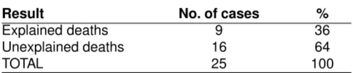
During the evaluated period there were 369 autopsies on children between 1 and 12 months of age, of which 344 (93.2%) deaths were expected and 25 (6.8%) were unexpected (Table 1). After autopsy, 9 (36%) of these cases could be explained by the findings whereas 16 (64%) remained unexplained (Table 2), but in only 10 was there full organ sampling permitting adequate analysis (Table 3). Table 4 shows the clinical and autopsy data of the 10 cases of unexpected and unexplained deaths. There were 7 male and 3 female: the age ranged from 1 to 3 months (average: 1.7 months). Two were found dead at home, 4 died before they could be taken to hospital and 1 died while being fed by the mother. In 8 cases the pathologists who performed the post-mortem examination identified clotted milk in the trachea and main bronchi and the deaths were attributed to aspiration of gastric contents. In one case no cause for the death was seen and in another one the pathologist’s diagnosis was SIDS.
Revision of the slides from the organs taken out during autopsy revealed only mild pulmonary edema in 6 cases, thymic hemorrhage in 3 cases and petechiae over the lungs, heart and thymus in 2 cases. No histological abnormality was seen in 2 cases. Aspiration of clotted milk was not observed in the lungs of any of the cases (Table 4).
The diagnoses attributed by the pathologists who performed the autopsies are shown in Table 4. Aspiration of gastric contents was the most frequent diagnosis with 8 (80%) cases. In one case (10%) the diagnosis was SIDS and in another one (10%) the cause of death remained undetermined.
DISCUSSION
SIDS, the leading cause of post-neonatal death in developed countries,4 has been reported in developing
countries in recent years.5,6 The lack of epidemiological
studies not only in Brazil but all over Latin America has led to serious difficulties in its recognition and study in this part of the world, where it is rarely accepted as an important cause of death of children. Based on what has happened in the industrialized countries, the reduction in infant mortality rate in Brazil, especially due to the reduction in preventable diseases, will probably bring SIDS into focus.
According to the original definition of SIDS by Beckwith in 1970,1 SIDS is characterized by the
unexpected death of a child of less than one year of age Table 3
Histological study of cases of unexplained death
Histological documentation No. of cases %
Complete 10 62.5 Incomplete 6 37.5 TOTAL 16 100
Table 2
Result of post-mortem examination in the cases of unexpected deaths
Result No. of cases %
Explained deaths 9 36 Unexplained deaths 16 64 TOTAL 25 100
Table 1
Autopsies of children between 1 and 12 months of age performed at SVOI from January 1987 to
December 1990.
Characteristics of deaths No. of cases %
which remains unexplained after a post-mortem examination is done. Later on, it was proposed that professional investigations at the scene of death should be required in the definition.7 Although these are important,
they certainly impose tremendous difficulties on SIDS detection and study,8 especially in retrospective
investigations or in countries like Brazil, and thus they have not been required in this study. In the study of Victora et al (1987),2 diagnosis of SIDS was based on clinical
evidence, as the autopsies, performed in over 90% of the cases, did not follow strict criteria.
The majority of the children submitted to post-mortem examination at the SVOI in the studied age group died of expected causes of death, as could easily be predicted. Ten cases, representing 2.7% of the total, died unexpectedly and their deaths remained unexplained after post-mortem investigation. The available data on these cases show that they bear many similarities to SIDS.
In this study males were more common than females. Although not completely understood, this male preponderance is generally recognised in the pertinent literature.9
In this study the mean age at death was 1.7 months and they all occurred in the first three months of life. Although the peak incidence of SIDS is in the third and fourth months, it may occur from the neonatal period10
until the end of the first year.11
SIDS has long been called “cot death” or “crib death” because the victims are usually found dead where they are put to sleep but it is now understood that it may happen anywhere and at any time.12 Some children are found
agonizing, and die before any help can be obtained. Nonetheless, others may die while they are being taken to hospital or even under supervision by parents or doctors. The way children died in this study is thus in accordance with what is classically reported.
The gross and microscopic findings in SIDS are scant, by definition.13,14 Petechiae over thoracic organs are
the most frequent findings. They are interpreted as secondary to increased negative thoracic pressure following vigorous inspiratory movements against a closed epiglottis, indicating that asphyxia was the cause of death.14-16 Petechiae are not exclusive to SIDS and can be
Table 4
Clinical and epidemiological data, diagnoses of the cause of death at the post-mortem examination and microscopic findings for the unexpected and unexplained autopsy cases.
Case Sex Age (months) How, where and when Post-mortem diagnosis Histological death occurred findings
1 m 3 found dead SIDS pulmonary edema 2 m 1 died on way to hospital aspiration thymic
hemorrage, petechiae 3 m 1 found dead aspiration pulmonary
edema 4 m 1 died on way to hospital aspiration no abnormalities 5 m 1 died at home aspiration pulmonary
edema, thymic hemorrhage 6 m 2 died at home aspiration petechiae 7 f 3 died at home aspiration pulmonary edema 8 f 3 aspiration of milk aspiration pulmonary
edema 9 f 1 died on way to hospital undetermined pulmonary
edema, petechiae, thymic
seen at any age, including intrauterine deaths.11
Intraparenchymatous thymic hemorrhage usually accompanies subcapsular petechiae and has the same connotation.14 Our results show that some of the children
had subcapsular thymic petechiae and intrapa-renchymatous hemorrhage, whether associated or not, confirmed by histology. However, the most common finding in our cases was pulmonary edema. This is interpreted as being an agonizing event and not directly implicated in the death.14
Aspiration of gastric contents, usually clotted milk, identified at autopsy and interpreted as the cause of death in 8 of the 10 cases, was not confirmed by histology. It is assumed to be an agonizing or post-mortem event due to the generalized muscular relaxation and for this reason it is only seen in the lumen of the trachea or main bronchi. Aspiration is thought to be responsible for death when the material is found in the distal airway, i.e., bronchioles and alveoli.13,17 To obtain a definite diagnosis of this
condition, it should therefore be done using frozen sections of the lungs during the autopsy, which was not done in our cases. Paraffin-embedded lung sections did not show signs of aspiration in any of our cases.
Aspiration of gastric contents has long concerned health workers and parents. To avoid it, the prone sleeping position has been widely advocated for babies in the past, but many epidemiological surveys in different parts of the world have shown that it is up until now the single most
important risk factor for SIDS.18,19 A striking decrease in
the frequency of SIDS has also been demonstrated when the sleeping position is changed from prone to supine.21-24
Although many hypotheses have been formulated to explain this, the mechanisms involved in the deaths are still unknown. On the other hand, aspiration is not a major problem except in in well-defined risk situations such as pre-term babies, children with gastro-esophageal reflux and those with neurological disorders.13 The risk of death due
to aspiration in children sleeping in the supine position25 is
less than that due to SIDS in children sleeping in the prone position, which is the reason for the change in recommendation given to parents in developed countries.24
CONCLUSION
In spite of the inherent limitations of a retrospective investigation, the present study shows the existence of 10 cases of unexpected death in infancy which remained unexplained after post-mortem examination at SVOI and can therefore be classified as SIDS.
Prospective studies are necessary to determine the epidemiological characteristics of SIDS in Brazil, which, by definition, must require complete post-mortem examinations, including autopsy.
REFERENCES
1. Beckwith JB. Introduction: discussion of terminology. In: Bergman AB, Beckwith JB, Ray CG, editors. Sudden infant death syndrome. Washington DC: University of Washington Press 1970;p.18.
2. Victora CG, Letícia CN, Lombardi C, et al. Quadro epidemiológico das mortes súbitas na infância em cidades gaúchas (Brasil). São Paulo: Ver Saúde Públ 1987;21:490-6.
3. SICAEV. Sistema de coleta e análise de estatísticas vitais. Ribeirão Preto, SP: Secretaria Municipal da Saúde, Boletim 1992: I (1).
4. MMWR. Sudden infant death syndrome - United States, 1983-1994. Morb Mortal Wkly Rep 1996;45:859-63. 5. Wolf BH, Ikeogu MO. Is sudden infant death syndrome a
problem in Zimbabwe? Ann Trop Paediatr 1996;16:149-53. 6. Tan SY, Lau G. Sudden infant death in a tropical environment: Singapore’s experience. Forensic Sci Int 1996;79:11-22.
7. Willinger M, James LS, Catz C. Defining the sudden infant death syndrome (SIDS): deliberations of an expert panel
convened by the National Institute of Child Health and Human Development. Pediatr Pathol 1991;11:677-84. 8. Guntheroth WG, Spiers P, Naeye RL. Redefinition of the
Sudden Infant Death Syndrome: the disadvantages. Pediatr Pathol 1994;14:127-32.
9. McLaughlin AS, Valdes MG, Jacobsen RM, et al. Incidence of sudden infant death syndrome in Olssted County, Minnesota: 1945 through 1992. Mayo Clinic Proc 1995;70:837-43.
10. Rodrigues-Alarcon J, Melchor JC, Linares A, et al. Early neonatal death or near-death syndrome. An epidemiological study of 29 cases. Acta Paediatr 1994;83:704-8.
11. Valdes-Dapena M, Gilbert-Barness, Naeye RL. Sudden infant death syndrome in Potter’s pathology of the fetus and infant. In: Barness-Gilbert E, editor. Mosby-Year Book Inc 1997;12:433-52.
12. Valdes-Dapena M. Sudden and unexpected death in infancy: a review of the world literature. Pediatrics 1967;39(1):123-37. 13. Valdes-Dapena M. Sudden infant death syndrome: overview
of recent research developments from a pediatric pathologist’s perspective. Pediatrician 1988;15:222-30. 14. Berry PJ. Pathological findings in SIDS. J Clin Pathol
15. Krous HF. The microscopic distribution of intrathoracic petechiae in sudden infant death syndrome. Arch Pathol Lab Med 1984;108:77-9.
16. Beckwith JB. Intrathoracic hemorrhages: a clue to the mechanism of death in sudden infant death syndrome? Ann New York Acad Sci 1988;533:37-47.
17. Valdes-Dapena M, Mc-Feeley PA, Hoffman HJ, et al. Histopathology atlas for the sudden infant death syndrome. Armed Forces Institute of Pathology in cooperation with the American Registry of Pathology and the National Institute of child health and human development. Washington DC: 1993.
18. Fleming PJ. Understanding and preventing sudden infant death syndrome. Current Opin Pediatr 1994;6:158-62. 19. Fleming PJ, Blair PS, Bacon C, et al. Environment of infants
during sleep and risk of the sudden infant death syndrome: results of 1993-5 case-control study for confidential inquiry into stillbirths and deaths in infancy. Confidential enquiry
into stillbirths and deaths: regional coordinators and researches. Brit Med J 1996;313:191-5.
20. Taylor JA, Krieger JW, Reay DT, et al. Prone sleeping position and the sudden infant death syndrome in King County, Washington: a case-control study. J Pediatr 1996;128:626-30. 21. Spiers PS, Guntheroth WG. Recommendations to avoid the
prone sleeping position and recent statistics for sudden infant death syndrome in the United States. Arch Pediatr Adolesc Med 1994;148:141-6.
22. Willinger M. SIDS prevention. Pediatr Ann 1995;24:358-64. 23. Irgens LM, Markestad T, Baste V, et al. Sleeping position and sudden infant death syndrome in Norway 1967-1991. Arch Dis Child 1995;72:478-82.
24. Wigfield RE, Fleming PJ, Berry PJ, et al. Can the fall in Avon’s sudden infant death rate be explained by changes in sleeping position? Brit Med J 1992;304:282-3.
25. Faure C. Position and sudden infant death of the infant (editorial). Presse Med 1995;24:1879-81.