Simplifying ectasia screening with corneal
and anterior segment tomography
A tomografia de córnea e segmento anterior na propedêutica do
exame complementar na avaliação de ectasia
Bruno de Freitas Valbon
1, Rodrigo Teixeira Santos
1,2, Isaac Ramos
1, Ana Laura Canedo
1, Leonardo Nogueira
1,
Renato Ambrósio Jr.
11Corneal Tomography and Biomechanics Study Group — Rio de Janeiro (RJ), Brazil.
2Cornea and Refractive Surgery Department, São Paulo School of Medicine, Federal University of São Paulo (UNIFESP) – São Paulo (SP), Brazil.
The authors declare no conflicts of interest
Received for publication: 19/3/2012 - Accepted for publication: 29/5/2012
A
BSTRACTThe methodology currently used for interpretation of the cornea and anterior segment tomography for the diagnosis of corneal ectasia. Description of the clinical interpretation of anterior segment tomography (Pentacam - Oculus, Wetzlar, Germany); case report of the ectopia lentis demonstrating the importance o evaluation of the cornea and anterior segment of the keratoconus. We excluded the disease, analyzing the tomographic analysis of rates of ectasia and observe an asymmetry between nasal and temporal sides in assessing the depth map of the anterior chamber. On the biomicroscopic examination in mydriasis, was found a ectopia lentis.We must be mindful not only to corneal tomography indices to evaluate a diagnostic test, but look at other important information provided by anterior segment tomography.
Keywords: Tomography; Corneal topography; Keratoconus; Ectopia lentis; Anterior eye segment; Dilatation, pathologic; Case reports
R
ESUMODescrevemos a importância da tomografia de córnea e segmento anterior na propedêutica do exame complementar na avaliação de ectasia. Esta descrição da interpretação clínica dos índices da tomografia de córnea e segmento anterior (Pentacam – Oculus, Wetzlar, Germany) neste relato de caso, demonstra a relevância de uma nova tecnologia na avaliação da córnea e segmento anterior na suspeita de ceratocone. O diagnóstico de ceratocone foi excluído pela análise dos índices tomográficos de ectasia. Detectou-se assimetria entre os lados nasal e temporal por meio da avaliação do mapa de profundidade de câmara anterior. No exame biomicroscópico sob midríase foi constatado subluxação do cristalino, sendo assim devemos estar atentos não somente aos índices tomográficos corneanos na avaliação do exame complementar e sim analisarmos outros dados importantes oferecidos pela tomografia de córnea e segmento anterior.
I
NTRODUCTIONT
he evolution of refractive surgery has led to a need for advanced screening and diagnostic methods to investigate ectasia. Since the advent of corneal topography(1), whichexamines in detail the anterior surface of the cornea, new technologies have emerged that improve the characterisation of the cornea on a tomographic level(2).
Corneal tomography(2), through its anterior and posterior
elevation maps, the profile of pachymetric progression and indices such as ART (Ambrosio Relational Thinnest) and the parameter D (deviation), is useful in the screening and differentiation between normal and diseased corneas or those susceptible to disease.
Furthermore, the tomography of the cornea and anterior segment(3) provides data such as Scheimplufg images, lens
densitometry, the positioning of intraocular lenses, and the angle, volume and depth of the anterior chamber.
This article demonstrates the importance of corneal tomography to exclude the suspicion of keratoconus using tomographic indices and to diagnose a subluxated lens using other anterior chamber data, such as nasal-temporal asymmetry.
Case report
Clinical data and diagnostic tests
A 16-year-old male patient with suspected keratoconus was referred to our service to perform a tomography of the cornea and anterior segment. The patient had no personal or family history of ophthalmic or systemic disease. He had undergone corneal topography with simulated keratometry of 45.80@178°/ 48.70 in the right eye (RE) and 45.20@174°/48.50 in the left eye (LE), with inconclusive results. Central pachymetry was 580 and 585 ìm in the RE and LE, respectively.
3D corneal tomography(1,3), through its anterior and
poste-rior elevation maps, the profile of pachymetric and indices such as ART (Ambrosio Relational Thinnest) and the parameter D
(deviation), can help to differentiate between normal and diseased corneas or those susceptible to disease, as the test provides higher sensitivity and specificity.(4)
The test can be interpreted using the Belin/Ambrosio Enhanced Display (BAD) software, which is used to identify or exclude disease in suspected cases and to evaluate corneas susceptible to post-Lasik ectasia.
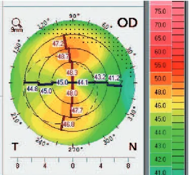
Figure 1 A shows the anterior sagittal topographic map of the RE with simulated keratometry of 44.1 @ 179.8/48.1. Figure 1 B, shows the same map for the LE with simulated keratometry of 45.1 @ 177.8 / 48.2. Both eyes had an increased curvature with a slight superior asymmetry.
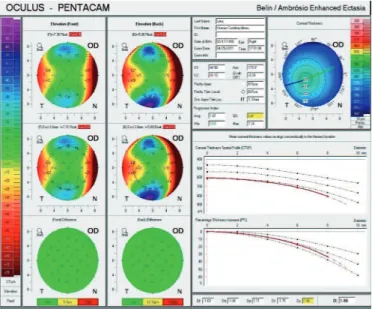
Figures 2 and 3 show anterior and posterior elevation maps, subtraction maps, pachymetric progression curves, and pachymetric progression indices for the RE and LE, respectively. Figure 2 shows normal anterior and posterior elevation maps and subtraction maps moving towards the green area (<12 ìM), which represents comfort and safety. Pachymetric progression charts, especially the Percentage Thickness Increase (PTI) described by Ambrosio et al.(5), show an escape at 6.0 mm, but
still within the 95% confidence interval, thus confirming a nor-mal pachymetric distribution.
While studying deviation indices (parameter D) in BAD, Ambrosio et al. (unpublished data, 2010) showed that the best cut-off value to differentiate eyes with keratoconus from normal eyes is 1.6. Our patient had 1.06, which is below the cut-off value. A study by Ambrosio et al.(6) shows that the ROC curves
for average and maximum ART (Ambrosio Relational Thinnest, which studies the thinnest point on the index of pachymetric progression) are 0.987 and 0.983, respectively. The best cut off values for average and maximum ART to differentiate normal eyes from those with keratoconus are 424 and 339, respectively. Dividing the thinnest point by the average and maximum pachymetric progression indices for the RE we found average and maximum ART values of 578 and 452, respectively, well above the cut off value.
Figure 3 shows the same corneal tomography indices for
the LE. Anterior and posterior elevation maps and subtraction maps show that the LE is within the green area (<12 ìm). PTI(5)
also shows an escape at 6.0 mm, similar to the RE, but also within the 95% confidence interval. The parameter D is 0.96, and dividing the thinnest point by the average and maximum pachymetric progression indices we found average and maximum ART values of 618 and 485, respectively.
According to parameters presented by Ambrosio et al. (4-6), both eyes had no tomographic signs characteristic of
keratoconus; therefore, the condition can be excluded in this case. Supplementing the diagnostic investigation with a tomography of the cornea and anterior segment, an asymmetry between the nasal and temporal sides of the map of anterior
Figure 2. B/A Enhanced ectasia of the right eye Figure 3. B/A Enhanced ectasia of the left eye
Figure 4. Map of anterior chamber depth. Note the asymmetry between the nasal and temporal sides of the right eye
Figure 5. Scheimplufg image showing temporal inferior decentralisation of the lens without mydriasis
Figure 6. Biomicroscopy (without mydriasis)
chamber depth was seen (Figure 4), as well as an inferior decentralisation of the lens in the Scheimpflug image of the RE (Figure 5).
The tomography of the cornea and anterior segment(3)
volu-me, and depth. Measurement of anterior chamber depth(3) is done
considering the distance from the endothelium at the corneal apex to the anterior curvature of lens, in millimeters. In addition, it can be measured manually at any position of the anterior segment, which is extremely important especially in the pre- and postoperative evaluation of candidates to intraocular implants in phakic eyes.
In view of these observations from the map of anterior chamber depth and the Scheimplufg image of the RE, a biomicroscopy was performed (Figure 6), showing no alterations. When the test was repeated with mydriasis, the Scheimpflug image showed a clearly visible lens zonule (Figures 7 A and B) and the biomicroscopy showed an inferior temporal subluxation of the lens (Figures 8 A and B). The patient was referred back to his primary physician with all the necessary recommendations.
C
ONCLUSIONConsidering the constant technological innovations in refractive surgery, the tomography of the cornea and anterior
segment(2) stands out as an important test to detect, monitor and
exclude disease with greater precision.
The term “tomography of the anterior segment” has been used to describe the diagnostic methods which provide a com-plete analysis of the region, either through the Scheimpflug principle, optical coherence or high frequency ultrasound.
In our case, analysis of the anterior segment using Scheimpflug images(3) allowed us to exclude the suspicion of
ectasia based on corneal tomography indices and to diagnose a subluxation of the lens, seen as an asymmetry in the map of an-terior chamber depth.
This article shows that tomography can be very helpful in the diagnostic investigation of diseases of the cornea and anteri-or segment.
R
EFERENCES1. Klyce SD. Computer-assisted corneal topography. High-resolution graphic presentation and analysis of keratoscopy. Invest Ophthalmol Vis Sci. 1984;25(12):1426-35.
Figures 7 A and B. Scheimplufg image with mydriasis. Note the lens zonule
Corresponding author:
Bruno de Freitas Valbon
Av. Conde de Bonfim, nº 211 - Sala 712 – Tijuca
CEP 20520050 – Rio de Janeiro (RJ), Brasil
Tel: 55 (21) 8103-7117
E-mail: [email protected]
2. Ambrosio R Jr, Belin MW. Imaging of the cornea: topography vsto-mography. J Refract Surg. 2010;26(11):874-9.
3. Ambrosio Júnior R, Netto MV, Schor P, Chalita MR, Chamon W, editores. Wavefront, topografia e tomografia da córnea e segmento anterior: atualização propedêutica em cirurgia refrativa. Rio de Janeiro: Cultura Médica; 2006.
4. Belin MW, Khachikian SS, Ambrosio R Jr, Salomão M. Keratoconus/ ectasia detection with the Oculus Pentacam: Belin/Ambrosio enhanced ectasia display. Highligths. 2007;35(6).
5. Ambrosio R Jr, Alonso RS, Luz A, Coca Velarde LG. Corneal thickness spatial prole and corneal-volume distribution: tomographic indices to detect keratoconus. J Cataract Refract Surg. 2006;32(11):1851-9.