1
Primer
Linking Inflammation to Natural Killer T Cell Activation
Mariolina Salio, Vincenzo Cerundolo*
Nuffield Department of Clinical Medicine, Weatherall Institute of Molecular Medicine, University of Oxford, Oxford, United Kingdom
To help us fight infections, the immune system relies on the coordinated activity of innate and adaptive immunity. Innate immunity represents an evolutionarily conserved first line of defense, kicking into action within hours of encountering pathogens, while adaptive immunity involves a higher degree of specificity for pathogen-encoded molecular determinants and long-term memory responses. Components of the innate immune system include epithelial barriers, antimicrobial compounds, and a range of different cell types, including dendritic cells, macrophag-es, neutrophils, and natural killer cells. Conversely, the main components of adaptive immunity are B and T lymphocytes, which survey billions of antigens via cell surface receptors that undergo somatic DNA rearrangements to confer almost unlimited specificities. Activation of innate responses, triggered by the detection of conserved microbial molecules (such as viral nucleic acids or bacterial cell wall components) by pattern-recognition receptors (such as Toll-like receptors), is critical for eliciting antigen-specific immune responses [1]. Over the past ten years it has emerged that a population of unconventional T lymphocytes, called natural killer T cells (NKT cells), operates at the interface between innate and adaptive immune responses; these cells express rearranged antigen-specific surface T cell receptors (although with limited diversity), but are also capable of a very rapid response mode, resulting in the activation of antigen presenting cells and facilitating the development of adaptive immunity.
Whereas most T lymphocytes recognize peptide antigens presented by molecules of the major histocompatibility complex (MHC), NKT cells recognize lipid antigens presented by CD1d molecules, which are functionally related to MHC molecules [2]. Upon activation, NKT cells modulate the activity of CD1d-expressing cells via the costimulatory molecule CD40 ligand and induce interferon (IFN)-c-dependent activation of other cell types, ultimately enhancing antigen specific B and T cell responses (Figure 1) [3]. Because of their effect on multiple cells of the immune system, NKT cells contribute to the regulation of a variety of processes, such as self-tolerance, tumor surveillance, and anti-microbial responses [4].
Auto-Reactivity by Design
During maturation of T lymphocytes the majority of auto-reactive cells are destroyed in the thymus to prevent autoimmu-nity. However, a proportion of NKT cells expressing a semi-invariant T cell receptor, hereafter referred to as semi-invariant NKT cells (iNKT cells), maintain the ability to recognize a range of endogenous lipids in the context of CD1d molecules during both inflammatory and non-inflammatory conditions. It has recently been shown that in the absence of inflammation, iNKT-cell auto-reactivity leads to a special activation state, characterized by impaired calcium signaling, which leads to the secretion of GM-CSF (granulocyte-macrophage colony-stimulating factor), with limited amounts of inflammatory cytokines [5]. In contrast, during microbial infection, inflammatory cytokines, such as interleukin (IL)-12 and IL-18, enhance basal iNKT-cell auto-reactivity and promote secretion of IFN-c[6–9]. In addition, during inflamma-tory responses, iNKT-cell activation can be influenced by
increased expression of surface CD1d molecules by activated antigen presenting cells (APCs) [10,11] and/or by increased expression of enzymes leading to the biosynthesis of endogenous self lipids [7,8]. These multiple mechanisms influencing iNKT-cell activation suggest an important link between inflammation and iNKT cells. Indeed, recent results have highlighted the ability of iNKT cells to abolish the suppressive activity of myeloid-derived suppressor cells (MDSCs), which are expanded during tumor growth and microbial infections and restore antigen-specific immune responses during influenza virus infection (Figure 1) [12]. Although the identity of several bacteria-derived lipids capable of activating iNKT cells has been determined [6,13,14], the identity of the lipids modulating iNKT-cell auto-reactivity has been elusive and is currently being investigated by several groups [15]. Identification of such lipids is important to a fuller understanding of the rules regulating CD1d assembly and loading, and also for the design of novel compounds that can selectively modulate iNKT-cell activation [16,17]. Results published in this issue ofPLoS Biology[18], and in earlier issues ofPLoS ONE [19] andThe Journal of Immunology[20], shed new light on the identity of iNKT cell natural ligands and provide further support for the link between inflammation and iNKT-cell activation.
A Closer Look at the Lipid Repertoire Bound to CD1d Molecules
Analysis of the ability of defined lipids to stimulate iNKT-cell activation has revealed that a restricted number of murine iNKT-cell clones can recognize phosphatidylinositol (PI) [21], phospha-tidylethanolamine (PE), and phosphatidylglycerol (PG) and the ganglioside GM3 [22]. However, reactivity to these lipids is weak and limited to a few iNKT-cell clones. To identify other endogenous natural iNKT-cell agonists, one approach has relied on the use of CD1d+cells with defined genetic defects in the lipid
biosynthetic pathway. The results of these experiments have shown that a class of lipids known as glycosphingolipids (GSL),
Citation:Salio M, Cerundolo V (2009) Linking Inflammation to Natural Killer T Cell Activation. PLoS Biol 7(10): e1000226. doi:10.1371/journal.pbio.1000226
PublishedOctober 27, 2009
Copyright:ß2009 Salio, Cerundolo. This is an open-access article distributed under the terms of the Creative Commons Attribution License, which permits unrestricted use, distribution, and reproduction in any medium, provided the original author and source are credited.
Funding:This work is funded by Cancer Research UK Program grant C399/A6199 to VC. The funders had no role in study design, data collection and analysis, decision to publish, or preparation of the manuscript.
Competing Interests:The authors have declared that no competing interests exist.
Abbreviations:APCs, antigen presenting cells; ER, endoplasmic reticulum; GM-CSF, granulocyte-macrophage colony-stimulating factor; GSL, glycosphingolipid; IFN, interferon; IL, interleukin; iNKT cells, invariant NKT cells; LPC, lysophospha-tidylcholine; MDSCs, myeloid-derived suppressor cells; MHC, Major Histocompat-ibility Complex; NKT cells, natural killer T cells; PC, phosphatidylcholine; PE, phosphatidylethanolamine; PG, phosphatidylglycerol; PI, phosphatidylinositol; PLA2, phospholipase A2.
such as isoglobotrihexosylceramide (iGb3), contributes to the pool of endogenous lipids that leads to human and murine iNKT cell activation [6,23,24]. However, the presence of iGb3 in dendritic cells and developing T lymphocytes remains controver-sial [25–29]. Furthermore, it has become clear that experiments carried out in mice or in cells with defective GSL catabolism need to be carefully controlled, because impaired iNKT-cell positive selection and activation could be due not to the absence of activating lipids, but to the failure of lipid presentation resulting from sequestering of lipids in the lysosomes of these APCs [30,31]. Consistent with this possibility, mice deficient in the lysosomal enzymeb-hexosaminidase, which generates iGb3 from iGb4, have impaired iNKT-cell development [24], whereas iGb3 synthase-deficient mice have normal numbers of iNKT cells [25]. Thus, the role of iGb3 as a self antigen for iNKT cells remains unclear.
In order to better characterize the classes of self-lipids available for recognition by iNKT cells, two groups have independently employed a novel approach to analyze the repertoire of lipids bound to CD1d molecules [18–20]. The Cresswell group engineered human CD1d molecules that could be proteolytically cleaved after recycling physiologically through the endocytic pathway and then compared lipids that were associated with CD1d molecules in different cellular compartments, that is, either retained in the endoplasmic reticulum (ER), secreted in soluble form, or cleaved [20]. By using multiple mass spectrometry methods to identify the major lipid species, the researchers
reported that the ER-retained form of CD1d was predominantly loaded with phosphatidylcholine (PC), the most abundant phospholipid in eukaryotic cells. The only detectable lipid associated with the secreted CD1d molecules was sphingomyelin, which is synthesized in the Golgi and is not present in the ER, whereas the protease-cleaved CD1d molecules were loaded with PC, sphingomyelin, and lysosphospholipids. In contrast, Gumperz and colleagues focused their analysis on lipids associated with secreted human CD1d molecules, as they had previously observed that, in contrast to mouse iNKT cells, autoreactivity of human iNKT-cell clones is largely independent of CD1d endosomal trafficking [32]. In thePLoS ONEpaper, Gumperz and colleagues found a large spectrum of lipids associated with soluble human CD1d molecules [19]. By a high-resolution analysis, a total of 177 lipid species were identified, comprising glycerophospholipids (including common diacylglycerol species, plasmalogens, lysopho-spholipids, and cardiolipins) and sphingolipids (including sphin-gomyelins and GSL, such as the ganglioside GM3). Altogether, these results highlight the possibility that, depending on the cellular localization, CD1d molecules may be loaded with a different spectrum of endogenous ligands.
Given that some of these lipids are known to be bioactive and have been reported to play significant roles in cancer, autoimmune disease, cellular signaling, and cell death [33,34], the Gumperz group has investigated the ability of iNKT cells to recognize and react to synthetic preparations of all the CD1d bound lipids - these
Figure 1. iNKT cells at the interface between innate and adaptive immunity.iNKT cells recognize lipids presented by CD1d molecules. Upon activation, iNKT cells modulate the function of CD1d-expressing cells, such as APCs and B cells. This leads to the priming of antigen-specific T cells, induction of antibody responses, and activation of natural killer cells, a subset of cells acting in the innate immune response. iNKT cells can also inhibit the suppressive function of MDSCs.
findings are reported in this issue ofPLoS Biology[18]. Remarkably, the mono acyl lysophosphatidylcholine (LPC) (Figure 2) was the only antigenic species capable of activating both a panel of iNKT-cell clones and lines and, albeit weakly, freshly isolated peripheral blood lymphocytes.
LPC as an Inflammatory Lipid and Stimulating Signal for iNKT Cells
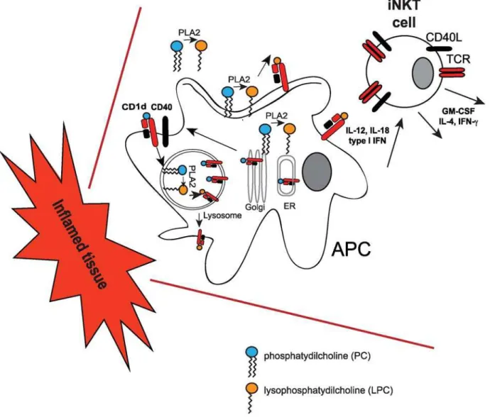
LPC is produced by the phospholipase A2 enzymes (PLA2), which can be localized to a number of intracellular and extracellular sites. Activation of PLA2 by a variety of growth factors, hormones, and cytokines can lead to the release of LPC into the cytoplasm, the lysosome, or at the cell surface [35]. At all these locations, LPC could be available for loading onto CD1d molecules (Figure 2). Of relevance is the observation from the Gumperz group that blocking secreted PLA2 activity in monocytes (and thereby reducing the levels of lipid ligand) with a polyclonal antibody, led to reduced basal iNKT-cell activation without affecting CD1d expression [18]. Since LPC accumulates to high
concentrations in blood and other fluids during chronic inflam-mation, and since Gumperz and colleagues showed that lysopho-spholipids can bind to CD1d molecules previously loaded with other cellular ligands or GSL [18], monitoring the levels of CD1d-bound LPC could represent one of the mechanisms leading to iNKT-cell activation and expansion. Interestingly, Dhodapkar and colleagues previously reported an increase of LPC species in one pathological setting - in the plasma of myeloma patients. This was accompanied by an expansion of a subset of NKT cells [36], which, unlike iNKT cells, express a broader range of T cell receptors. These results suggest that recruitment and expansion of invariant and non-invariant NKT cells could occur more widely in different inflammatory settings and eventually contribute to immune pathology. Indeed, iNKT cells have a chemokine receptor profile that allows them to preferentially ‘‘home’’ to inflamed tissues [37]. It is tempting to speculate that, during inflammation, secreted LPC could be presented by APCs recruited at inflammatory sites, resulting in iNKT-cell activation (Figure 2). However, the efficiency of LPC presentation in vivo remains to be defined. Gumperz and colleagues provide some support for this
Figure 2. iNKT-cell activation by lysosphospholipids.During inflammation cytoplasmic, membrane and secreted phospholipases (such as PLA2) produce lysophospholipids (such as LPC) from cellular phospholipids. Lysosphospholipids can be loaded onto CD1d molecules at the cell surface, in the lysosomes, or during intracellular trafficking through the ER and the Golgi. CD1d-LPC complexes elicit iNKT-cell activation in concert with IL-12, IL-18, and type I IFN secreted by APCs during inflammatory reactions.
notion by showing very weak iNKT-cell stimulation with APCs expressing wild-type CD1d molecules. In contrast, the authors show that recognition of LPC by iNKT cells was enhanced using CD1d molecules unable to recycle from the cell surface to the lysosomes (where the acidic environment could lead to dissociation of the CD1d-lipid complexes) [18]. In addition, higher concen-trations of LPC failed to activate iNKT-cell clones, showing an unexplained inhibitory effect, which could be due to the formation of micelles or less–CD1d-accessible structures [18].
It is known that the length of the lipid hydrocarbon chains determines the stability of lipid binding to CD1d molecules, which in turn influences iNKT-cell activation [38]. Although further studies are warranted to determine the binding affinity of lysopho-spholipids for CD1d molecules and the half-life of these complexes, it is likely that the mono alkyl chain LPC will have a higher rate of dissociation from CD1d molecules than the dual alkyl chain PC. Thus, the combination of limited presentation by recycling CD1d molecules, with the inhibitory effect of high LPC concentrations and possibly the short half-life of CD1d-LPC complexes, could be important features that help to fine-tune iNKT-cell responses in the context of prolonged inflammatory processes.
The crystal structures of human and murine CD1d molecules have revealed the presence of two hydrophobic channels, A9and C9, which are occupied by the lipid tails of iNKT-cell agonists [39]. Interestingly, the results by the Gumperz laboratory in this
issue ofPLoS Biologyhave highlighted a dichotomy in the ability of LPC and PC to stimulate iNKT cells [18]. It will be, therefore, very informative to carry out structural studies comparing CD1d molecules loaded with either lysophospholipids or phospholipids (e.g., mono versus di acyl lipids) to assess whether the differential ability of LPC and PC to activate iNKT cells may be accounted for by variations in the orientation of the polar head as a consequence of different binding of their lipids chains to the A9
and C9hydrophobic channels. This possibility would be consistent with previously published CD1d structures, revealing the ability of different phospholipids to bind CD1d molecules in different orientations [40,41].
In conclusion, the identification of LPC as an endogenous ligand for iNKT cells is an important finding for the understanding of the role that iNKT cells and other immune cells play during inflammation. It will also be interesting to correlate iNKT-cell numbers and activation with changes in activity of the PLA2 isoforms during different inflammatory conditions (for example, upon microbial infections and Toll-like receptor-mediated activation of APCs or during chronic inflammatory processes, such as cancer). Future studies will reveal whether analogues of lysophospholipids could be exploited as novel adjuvants to further harness iNKT cells’ ability to bridge innate and adaptive immune responses or to fine-tune iNKT-cell autoreactivity during autoimmune diseases.
References
1. Palm NW, Medzhitov R (2009) Pattern recognition receptors and control of adaptive immunity. Immunol Rev 227: 221–233.
2. Cohen NR, Garg S, Brenner MB (2009) Antigen presentation by CD1 lipids, T cells, and NKT cells in microbial immunity. Adv Immunol 102: 1–94. 3. Cerundolo V, Silk JD, Masri SH, Salio M (2009) Harnessing invariant NKT
cells in vaccination strategies. Nat Rev Immunol 9: 28–38.
4. Bendelac A, Savage PB, Teyton L (2007) The biology of NKT cells. Annu Rev Immunol 25: 297–336.
5. Wang X, Chen X, Rodenkirch L, Simonson W, Wernimont S, et al. (2008) Natural killer T-cell autoreactivity leads to a specialized activation state. Blood 112: 4128–4138.
6. Mattner J, Debord KL, Ismail N, Goff RD, Cantu C, 3rd, et al. (2005) Exogenous and endogenous glycolipid antigens activate NKT cells during microbial infections. Nature 434: 525–529.
7. Paget C, Mallevaey T, Speak AO, Torres D, Fontaine J, et al. (2007) Activation of invariant NKT cells by toll-like receptor 9-stimulated dendritic cells requires type I interferon and charged glycosphingolipids. Immunity 27: 597–609. 8. Salio M, Speak AO, Shepherd D, Polzella P, Illarionov PA, et al. (2007)
Modulation of human natural killer T cell ligands on TLR-mediated antigen-presenting cell activation. Proc Natl Acad Sci U S A 104: 20490–20495. 9. Brigl M, Bry L, Kent SC, Gumperz JE, Brenner MB (2003) Mechanism of
CD1d-restricted natural killer T cell activation during microbial infection. Nat Immunol 4: 1230–1237.
10. Raghuraman G, Geng Y, Wang CR (2006) IFN-beta-mediated up-regulation of CD1d in bacteria-infected APCs. J Immunol 177: 7841–7848.
11. Skold M, Xiong X, Illarionov PA, Besra GS, Behar SM (2005) Interplay of cytokines and microbial signals in regulation of CD1d expression and NKT cell activation. J Immunol 175: 3584–3593.
12. De Santo C, Salio M, Masri SH, Lee LY, Dong T, et al. (2008) Invariant NKT cells reduce the immunosuppressive activity of influenza A virus-induced myeloid-derived suppressor cells in mice and humans. J Clin Invest 118: 4036–4048.
13. Kinjo Y, Tupin E, Wu D, Fujio M, Garcia-Navarro R, et al. (2006) Natural killer T cells recognize diacylglycerol antigens from pathogenic bacteria. Nat Immunol 7: 978–986.
14. Kinjo Y, Wu D, Kim G, Xing GW, Poles MA, et al. (2005) Recognition of bacterial glycosphingolipids by natural killer T cells. Nature 434: 520–525. 15. Burrows PD, Kronenberg M, Taniguchi M (2009) NKT cells turn ten. Nat
Immunol 10: 669–671.
16. Im JS, Arora P, Bricard G, Molano A, Venkataswamy MM, et al. (2009) Kinetics and cellular site of glycolipid loading control the outcome of natural killer T cell activation. Immunity 30: 888–898.
17. Silk JD, Salio M, Reddy BG, Shepherd D, Gileadi U, et al. (2008) Cutting edge: nonglycosidic CD1d lipid ligands activate human and murine invariant NKT cells. J Immunol 180: 6452–6456.
18. Fox LM, Cox DG, Lockridge JL, Wang X, Chen X, et al. (2009) Recognition of Lyso-phospholipids by human natural killer T lymphocytes. PLoS Biol 7: e1000228. doi:10.1371/journal.pbio.1000228.
19. Cox D, Fox L, Tian R, Bardet W, Skaley M, et al. (2009) Determination of cellular lipids bound to human CD1d molecules. PLoS One 4: e5325. doi:10.1371/journal.pone.0005325.
20. Yuan W, Kang SJ, Evans JE, Cresswell P (2009) Natural lipid ligands associated with human CD1d targeted to different subcellular compartments. J Immunol 182: 4784–4791.
21. Gumperz JE, Roy C, Makowska A, Lum D, Sugita M, et al. (2000) Murine CD1d-restricted T cell recognition of cellular lipids. Immunity 12: 211–221. 22. Wu DY, Segal NH, Sidobre S, Kronenberg M, Chapman PB (2003)
Cross-presentation of disialoganglioside GD3 to natural killer T cells. J Exp Med 198: 173–181.
23. Stanic AK, De Silva AD, Park JJ, Sriram V, Ichikawa S, et al. (2003) Defective presentation of the CD1d1-restricted natural Va14Ja18 NKT lymphocyte antigen caused by beta-D-glucosylceramide synthase deficiency. Proc Natl Acad Sci U S A 100: 1849–1854.
24. Zhou D, Mattner J, Cantu C, 3rd, Schrantz N, Yin N, et al. (2004) Lysosomal glycosphingolipid recognition by NKT cells. Science 306: 1786–1789. 25. Porubsky S, Speak AO, Luckow B, Cerundolo V, Platt FM, et al. (2007) Normal
development and function of invariant natural killer T cells in mice with isoglobotrihexosylceramide (iGb3) deficiency. Proc Natl Acad Sci U S A 104: 5977–5982.
26. Speak AO, Salio M, Neville DC, Fontaine J, Priestman DA, et al. (2007) Implications for invariant natural killer T cell ligands due to the restricted presence of isoglobotrihexosylceramide in mammals. Proc Natl Acad Sci U S A 104: 5971–5976.
27. Christiansen D, Milland J, Mouhtouris E, Vaughan H, Pellicci DG, et al. (2008) Humans lack iGb3 due to the absence of functional iGb3-synthase: implications for NKT cell development and transplantation. PLoS Biol 6: e172. doi:10.1371/ journal.pbio.0060172.
28. Li Y, Teneberg S, Thapa P, Bendelac A, Levery SB, et al. (2008) Sensitive detection of isoglobo and globo series tetraglycosylceramides in human thymus by ion trap mass spectrometry. Glycobiology 18: 158–165.
29. Li Y, Thapa P, Hawke D, Kondo Y, Furukawa K, et al. (2009) Immunologic glycosphingolipidomics and NKT cell development in mouse thymus. J Proteome Res 8: 2740–2751.
30. Gadola SD, Silk JD, Jeans A, Illarionov PA, Salio M, et al. (2006) Impaired selection of invariant natural killer T cells in diverse mouse models of glycosphingolipid lysosomal storage diseases. J Exp Med 203: 2293–2303. 31. Schumann J, Facciotti F, Panza L, Michieletti M, Compostella F, et al. (2007)
Differential alteration of lipid antigen presentation to NKT cells due to imbalances in lipid metabolism. Eur J Immunol 37: 1431–1441.
32. Chen X, Wang X, Keaton JM, Reddington F, Illarionov PA, et al. (2007) Distinct endosomal trafficking requirements for presentation of autoantigens and exogenous lipids by human CD1d molecules. J Immunol 178: 6181–6190. 33. Brites P, Waterham HR, Wanders RJ (2004) Functions and biosynthesis of
plasmalogens in health and disease. Biochim Biophys Acta 1636: 219–231. 34. Exton JH (1994) Phosphatidylcholine breakdown and signal transduction.
35. Burke JE, Dennis EA (2009) Phospholipase A2 structure/function, mechanism, and signaling. J Lipid Res 50 Suppl: S237–242.
36. Chang DH, Deng H, Matthews P, Krasovsky J, Ragupathi G, et al. (2008) Inflammation-associated lysophospholipids as ligands for CD1d-restricted T cells in human cancer. Blood 112: 1308–1316.
37. Kim CH, Johnston B, Butcher EC (2002) Trafficking machinery of NKT cells: shared and differential chemokine receptor expression among V alpha 24(+)V beta 11(+) NKT cell subsets with distinct cytokine-producing capacity. Blood 100: 11–16.
38. McCarthy C, Shepherd D, Fleire S, Stronge VS, Koch M, et al. (2007) The length of lipids bound to human CD1d molecules modulates the affinity of NKT cell TCR and the threshold of NKT cell activation. J Exp Med 204: 1131–1144.
39. Silk JD, Salio M, Brown J, Jones EY, Cerundolo V (2008) Structural and functional aspects of lipid binding by CD1 molecules. Annu Rev Cell Dev Biol 24: 369–395.
40. Giabbai B, Sidobre S, Crispin MD, Sanchez-Ruiz Y, Bachi A, et al. (2005) Crystal structure of mouse CD1d bound to the self ligand phosphatidylcholine: a molecular basis for NKT cell activation. J Immunol 175: 977–984.