Drug resistance profiles and clonality of sporadic
Shigella sonnei
isolates
in Ankara, Turkey
Birgul Kacmaz
1, Ozlem Unaldi
2, Nedim Sultan
3, Riza Durmaz
2,4 1Central Microbiology Laboratory, Medical Faculty, Gazi University, Ankara, Turkey. 2Molecular Microbiology Research and Application Laboratory, National Public Health Agency, Ankara, Turkey.
3
Department of Clinical Microbiology, Medical Faculty, Gazi University, Ankara, Turkey. 4
Department of Clinical Microbiology, Medical Faculty, Kirikkale University, Kirikkale, Turkey.
Submitted: March 21, 2013; Approved: March 14, 2014.
Abstract
The aims of this study were to investigate drug resistance rates, types of extended spectrum beta lactamases (ESBLs), and molecular epidemiological characteristics of 43Shigella sonneiisolates. Ampicillin-sulbactam, amoxicillin-clavulanate, chloramphenicol, and ciprofloxacin were the most active antibiotics. Five isolates harboredblaSHV-12,blaTEM-1andblaCTX-M-15. More than 90% of the iso-lates had an indistinguishable pulsotype.
Key words:Shigella sonnei, extended spectrum beta-lactamase, plasmid profile, pulsed-field gel electrophoresis.
Shigellosis is a global human health problem caused by a group of bacteria calledShigella. There are four spe-cies ofShigellanamedS. sonnei, S. flexneri, S. dysenteriae,
andS. boydiiall of them capable of causing disease. In Tur-key,S. sonneiis the most frequently reportedShigella spe-cies (56.5%), followed by S. dysenteriae (22.9%), S.
flexneri (14.5%) and S. boydii (6.1%)
(http://www.saglik.gov.tr/istatistikler/temel2005/tablo-65. htm.). Similarly,S. sonneiis the predominant serogroup in Ankara, the capital city of Turkey (Saranet al., 2013).
The increasing prevalence of antibiotic-resistant strains ofShigellahas become a major concern worldwide. World Health Organization (WHO) recommends that all patients with bloody diarrhea should be treated with either ciprofloxacin or one of the following three drugs:
pivme-cillinam, azithromycin, and ceftriaxone
(http://whqlibdoc.who.int/publica-tions/2005/9241592330.pdf). However, there are many re-ports indicating fluoroquinolone resistant and extended spectrum beta-lactamase (ESBL)-producingShigella iso-lates from certain countries in 2009-2011 years. (Nandyet al., 2010; Sabra et al., 2009; Varghese and Aggarwal, 2011).
Molecular typing and antimicrobial susceptibility of
S. sonneiisolates can provide very useful data for the man-agement of both isolated cases and outbreaks (DeLappeet al., 2003; Lianget al., 2007; Na-Ubolet al., 2006). In this study, we investigated the rate of antimicrobial resistance ofS. sonneistrains collected in Ankara, the capital city of Turkey, from 2004 to 2006 and analyzed ESBL-producing isolates by PCR and DNA sequencing. Clonal relationship among the S. sonnei isolates was assessed using pulsed field gel electrophoresis (PFGE) and plasmid profile analy-sis.
A total of 43S. sonnei isolates were cultured from stool samples of patients with acute diarrhea. Isolates were collected at the Clinical Microbiology Laboratory of Gazi University Medical School Hospital in Ankara, Turkey during the three-year period between 2004 and 2006. Only one isolate was obtained from each patient. Twenty-three of the 43 patients (53.5%) were male. The ages of the pa-tients ranged from 5 to 75 years, with a mean of 14.1. The isolates were identified to species levels using conventional biochemical tests and group-specific polyvalent antisera, followed by phase specific antisera (Denka Seiken UK Ltd., Derbyshire, UK). Susceptibilities of the isolates to
Send correspondence to B. Kacmaz. Central Microbiology Laboratory, Medical Faculty, Gazi University, Ankara, Turkey. E-mail: [email protected].
ampicillin, chloramphenicol, ciprofloxacin, ampicillin-sul-bactam, amoxicillin-clavulanate, ceftriaxone, cefixime, and trimethoprim-sulfamethoxazole were determined by disk diffusion method according to the criteria recom-mended by the Clinical Laboratory Standards Institute (CLSI) (CLSI, 2010). ESBL production was determined by the double disk synergy method (Jarlieret al., 1988).
ESBL-producingS. sonneiisolates were tested for the presence of b-lactamase-encoding genes blaTEM, blaSHV andblaCTX-M by PCR. Approximately ten fresh colonies were suspended in sterile water, boiled for 10 min, and cen-trifuged for 2 min at 13000 x g. The supernatant was used as DNA source. Universal primers described previously for the b-lactamase-encoding genes were used (Ma et al., 2005; Sidjabatet al., 2009). The reaction mixture contained either 2.5 mM MgCl2forblaSHVPCR or 1.5 mM MgCl2for
blaTEM and blaCTX-M PCR, each of the deoxynucleoside triphosphates at a concentration of 200mM, 5mL of 10 PCR buffer [750 mM Tris-HCl (pH 8.8), 200 mM (NH4)2SO4, 0.1% (v/v) Tween 20], 10 pmol of each primer, and 2.5 U of Taq DNA polymerase (Thermo Scientific-Fermentas Cor-poration, Vilnius, Lithuania) in a final reaction volume of 50 mL. Amplification of blaTEM and blaCTX-M was per-formed using the following conditions: initial denaturation at 94 °C for 4 min, followed by 40 cycles of 94 °C for 15 s, 55 °C for 45 s, and 72 °C for 1 min, with a final extension step at 72 °C for 7 min. Amplification conditions forblaSHV were as follows:initial denaturation at 94 °C for 5 min, fol-lowed by 25 cycles of 94 °C for 30 s, 55 °C for 30 s, and 72 °C for 30 s, with a final extension step at 72 °C for 7 min. The amplified DNA products were analyzed by 1.5% agarose gel electrophoresis in 1x TBE buffer and stained with ethidium bromide.
PCR products ofblaTEM,blaSHV, andblaCTX-Mgenes were purified using Agencourt Ampure (Beckman Coulter Company, Massachusetts, USA). Sequencing reactions were performed with the primers used for PCR reactions. Each sequence reaction mixture consisted of 3.5-5mL puri-fied amplicon, 5 pmol primer, and 4mL of Dye terminator cycle sequencing quick start kit (Beckman Coulter Com-pany, Massachusetts, USA). The sequence reaction was done as follows: initial denaturation at 94 °C for 3 min fol-lowed by 30 cycles consisting of denaturation at 96 °C for 20 s, annealing at 55 °C for 20 s, and elongation at 60 °C for 4 min. The PCR products were purified with Dye-Ter-minator removal kit (Beckman Coulter Company, Massa-chusetts, USA) and 20 mL of purified product was sequenced in Beckman Coulter 8000 instrument. The re-sulting DNA sequences were compared with the GenBank
sequence databases using BLAST
(http://blast.ncbi.nlm.nih.gov/Blast.cgi).
Plasmid profiles analysis of theS. sonneiisolates was carried on plasmids obtained by using the alkaline-lysis method described by Kado and Liu (1981) with some
modi-fications. Our modifications included culturing the isolates in ampicillin containing medium and exclusion of the phe-nol/chloroform prufication step. Molecular sizes of plasmids were determined by using super coiled DNA lad-der (Life Technologies, Carlsbad, CA, USA).
PFGE typing of the 43 S. sonnei isolates was per-formed by following the protocol of Durmazet al.(2009). Briefly, bacterial cells were embedded into low melting agarose including sodium dodecyl sulfate. Cells in plugs were digested with lysozyme and proteinase K. Genomic DNA was restricted by 20 U of XbaI (Thermo Scien-tific-Fermentas Corporation, Vilnius, Lithuania). DNA fragments were separated by using a CHEF-DR II system (Bio-Rad Laboratories, Nazareth, Belgium). The DNA band profiles were analyzed with GelCompar software (version 3.0; Applied Maths, Sint-Martens-Latem, Bel-gium). A 1% band tolerance was used for comparison of DNA profiles. Clonal relationship among isolates was eval-uated by using the criteria of Tenoveret al.(1997).
All 43 isolates were confirmed as S. sonnei using group-specific polyvalent antisera and phase specific anti-sera. According to the serotyping results, 28 (65%) of the 43 isolates showed phase 1, eight (19%) showed phase 2, and seven (16%) showed both phase 1 and 2 profiles (+ 1). According to the antibiotic resistance profiles, four anti-biotypes were designated as shown in Table 1. Since more than 83% of the isolates were classified in only one anti-biotype (type 2), it was very difficult to discuss usefulness of the antibiotyping on our isolates collected during the three-year period. In this study,S. sonneiisolates showed the low rate of susceptibility against trimethoprim-sulfa-methoxazole (5%). In agreement with our results, previous studies from Turkey and other countries also indicated small susceptibility rates (3.5-30%) (Akcaliet al., 2008; Altun and Gur, 2011; Jafariet al., 2009; Saranet al., 2013; Zafar et al., 2009). The studies from Iran and Israel re-vealed 57% and 87% resistance to ampicillin, respectively (Ashkenaziet al., 2003; Jafariet al., 2009). In contrast to the results reported from those countries, low resistance rate was found in the current (12%) and previous studies (17% and 20%) from our country (Akcali et al., 2008; Altun and Gur, 2011). In accordance with some studies (Akcaliet al., 2008; Altun and Gur, 2011; DeLappeet al., 2003), we found that allS. sonneiisolates were susceptible to ampicillin-sulbactam, chloramphenicol, and ciproflo-xacin. However, other works have reported different resis-tance rates (9% to 100%) for chloramphenicol (Ashkenazi
et al., 2003; Jafariet al., 2009).
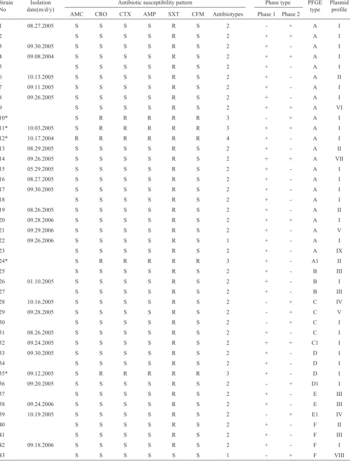
Table 1- Distribution ofS. sonneistrains according to epidemiologic, phenotypic and molecular typing characteristics.
Strain No
Isolation date(m/d/y)
Antibiotic susceptibility pattern Phase type PFGE type
Plasmid profile AMC CRO CTX AMP SXT CFM Antibiotypes Phase 1 Phase 2
1 08.27.2005 S S S S R S 2 - + A I
2 S S S S R S 2 + + A I
3 09.30.2005 S S S S R S 2 + - A I
4 09.08.2004 S S S S R S 2 + + A I
5 S S S S R S 2 + - A I
6 10.13.2005 S S S S R S 2 + - A II
7 09.11.2005 S S S S R S 2 + - A I
8 09.26.2005 S S S S R S 2 + - A I
9 S S S S R S 2 + + A VI
10* S R R R R R 3 - + A I
11* 10.03.2005 S R R R R R 3 + + A I
12* 10.17.2004 R R R R R R 4 + - A I
13 08.29.2005 S S S S R S 2 + - A II
14 09.26.2005 S S S S R S 2 + + A VII
15 05.29.2005 S S S S R S 2 + - A I
16 08.27.2005 S S S S R S 2 + - A I
17 09.30.2005 S S S S R S 2 + - A I
18 S S S S R S 2 + - A I
19 08.26.2005 S S S S R S 2 + - A II
20 09.28.2006 S S S S R S 2 + + A I
21 09.29.2006 S S S S R S 2 + - A V
22 09.26.2006 S S S S R S 1 + - A I
23 S S S S R S 2 + - A IX
24* S R R R R R 3 + - A1 II
25 S S S S R S 2 + - B III
26 01.10.2005 S S S S R S 2 + - B I
27 S S S S R S 2 + - B III
28 10.16.2005 S S S S R S 2 - + C IV
29 09.28.2005 S S S S R S 2 - + C V
30 S S S S R S 2 - + C I
31 08.26.2005 S S S S R S 2 + - C I
32 09.24.2005 S S S S R S 2 + + C1 I
33 09.30.2005 S S S S R S 2 + - D I
34 S S S S R S 2 + - D I
35* 09.12.2005 S R R R R R 3 + - D I
36 09.20.2005 S S S S R S 2 - + D1 I
37 S S S S R S 2 + - E III
38 09.24.2006 S S S S R S 2 + - E III
39 10.19.2005 S S S S R S 2 - + E1 IV
40 S S S S R S 2 + - F II
41 S S S S R S 2 + - F III
42 09.18.2006 S S S S R S 2 + - F I
43 S S S S S S 1 - + F VIII
is the first report of the presenceblaSHV-12b-lactamase gene in aS. sonneistrain isolated in Turkey.
Nine different plasmid profiles (I-IX) were detected. The size of the plasmids ranged from 2 to 10 kb. Two plasmids with approximately 3- and 5- kb in size were pres-ent in more than 90% of the isolates. Twpres-enty-five of the 43
S. sonneiisolates were classified in plasmid profile I (Ta-ble 1). Plasmid profiles of fourblaCTX-M-15-producing iso-lates were the same (profile I). Although it was indicated that plasmid profile analysis is one of the appropriate meth-ods for molecular characterization ofShigellaisolates re-covered over short time periods (12), we could not find any epidemiological link among the isolates classified in the same plasmid profile. For instance, the predominant plasmid profile I included more than half of the isolates col-lected from 2004 to 2006.
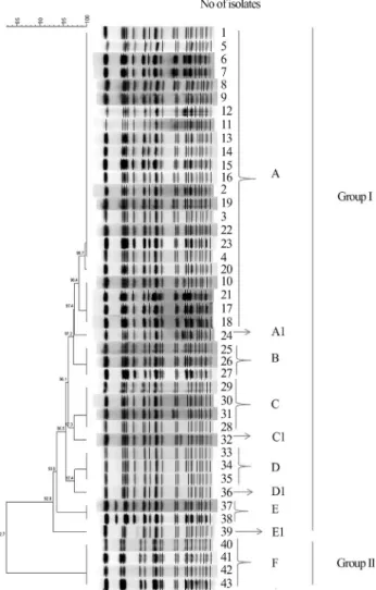
PFGE typing of the 43S. sonneiisolates yielded 10 different profiles, six (A, B, C, D, E, and F) were indistin-guishable profiles including 23, 3, 4, 3, 2, and 4 isolates, re-spectively and the remaining four (A1, C1, D1, and E1) were closely related profiles having 1 to 2 band-differences from their related clones. Genetic heterogeneity among different PFGE profiles was very low. According to similarity per-centage of > 82%, two major PFGE profiles were deter-mined, Group 1 included 39 isolates, and Group 2 included four isolates. There were 1-2 band-differences among PFGE profiles in major group I, while there were four dif-ferences between major group 1 and group 2 (Figure 1).
We observed limited correlation between the typing results and epidemiological characteristics. Plasmid profile I was found in five distinct PFGE types (PFGE types A, B, C, D, and F) and a common PFGE type (PFGE type A) were found in six different plasmid profiles (profile I, II, V, VI, VII, and IX). In parallel to our data, Pichelet al.(2007) found similar plasmid profiles in distinct PFGE clones and five different plasmid profiles in one outbreak clone. Four CTX-M-15-producing isolates showed three different PFGE types, two plasmid profiles, and two antibiotypes. PFGE type A was predominant both in phase 1 and phase 1+2 serotypes. Twenty-three (82%) of the 28 isolates sified in the phase 1 serotype, 5 (62%) of the 8 isolates clas-sified in the phase 2, and 6 (86%) of the 7 isolates clasclas-sified in the phase 1+2 showed the type 2 antibiotic resistance phenotype. There were no correlation between PFGE types and antibiogram types, however at least half of the isolates of each antibiotype showed PFGE profile A. Although the previous studies revealed that PFGE was the most discrimi-natory method among subtyping methods (DeLappeet al., 2003; Na-Ubol et al., 2006; Pichel et al., 2007), the S. sonneiisolates analyzed in the current study showed very low genetic diversity and there was no direct epidemiologi-cal relationship among the isolates having indistinguish-able PFGE profiles. For instance, 23 isolates classified in PFGE profile “A” were isolated from the first week of Sep-tember 2004 to the last week of SepSep-tember 2006. On the
other hand, the strains isolated from patient 2 and patient 7 with an interval of three days showed different PFGE pro-files “E1” and “C”, respectively. In parallel to our results, a study carried on theShigellaisolates from Ankara has also indicated high relationship among the tested isolates (Saran
et al., 2013). These findings support the comment that PFGE is a powerful tool for shigellosis outbreak tion. However it has some disadvantages for the investiga-tion of clonal relainvestiga-tionships among S. sonnei strains circulating over a period of months or years (Lianget al., 2007).
In conclusion, this is the first report indicating the presence ofblaSHV-12inS. sonneiin Turkey. High genetic homology among the sporadicS. sonneiisolates may indi-cate ongoing cross-transmission for a long period rather than a recent transmission.
References
Akcali A, Levent B, Akbas E, Esen B (2008) Typing ofShigella sonneistrains isolated in some provinces of Turkey using antimicrobial resistance and pulsed field gel electrophoresis methods. Mikrobiyol Bul 42:563-572.
Altun B, Gur D (2011) Antimicrobial resistance profiles of Shigellaspp. isolated from feces samples in Hacettepe Uni-versity Ihsan Dogramaci Children’s hospital between 1999-2010. Mikrobiyol Bul 45:609-616.
Ashkenazi S, Levy I, Kazaronovski V, Samra Z (2003) Growing antimicrobial resistance ofShigellaisolates. J Antimicrobial Chemotherapy 51:427-429.
Clinical and Laboratory Standards Institue (2010) Performance standards for antimicrobial susceptibility testing; Twentieth informational supplement M100-S20 Vol.30 No.1. DeLappe N, O’Halloran F, Fanning S, Corbett-Feeney G, Cheasty
T, Cormican M (2003) Antimicrobial resistance and genetic diversity ofShigella sonneiisolates from western Ireland, an area of low incidence of infection. J Clin Microbiol 41:1919-1924.
Durmaz R, Otlu B, Koksal F, Hosoglu S, Ozturk R, Ersoy Y, Aktas E, Gursoy NC, Caliskan A (2009) The optimization of a rapid pulsed-field gel electrophoresis protocol for the typ-ing of Acinetobacter baumannii, Escherichia coli and Klebsiellaspp. Jpn J Infect Dis 62:372-377.
Izumiya H, Tada Y, Ito K, Morita-Ishihara T, Ohnishi M, Tera-jima J, Watanabe H (2009) Characterization of Shigella sonneiisolates from travel-associated cases in Japan. J Med Microbiol 58:1486-1491.
Jafari F, Hamidian M, Rezadehbashi M, Doyle M, Salman-zadeh-ahrabi S, Derakhshan F, Zali MR (2009) Prevalence and antimicrobial resistance of diarrheagenic Escherichia coliandShigellaspecies associated with acute diarrhea in Tehran, Iran. Can J Infect Dis Med Microbiol 20:e56-e61. Jamala W, Rotimia VO, Palb T, Sonnevendb A, Dimitrovc TS
(2010) Comparative in vitro activity of tigecycline and other antimicrobial agents againstShigellaspecies from Kuwait and the United Arab of Emirates. J Infect Public Health 3:35-42.
Jarlier V, Nicolas M H, Fournier G, Philippon A (1988) Extended broad-spectrum beta-lactamases conferring transferable re-sistance to newer beta-lactam agents inEnterobacteriaceae: Hospital prevalence and susceptibility patterns. Rev Infect Dis 10:867-878.
Kado CL, Liu ST (1981) Rapid procedure for detection of large and small plasmids. J Bacteriol 145:1365-1373.
Liang SY, Watanabe H, Terajima J, Li CC, Liao JC, Tung SK, Chiou CS (2007) Multilocus variable-number tandem-repeat analysis for molecular typing ofShigella sonnei. J Clin Microbiol 45:3574-3580.
Ma L, Alba J, Chang F, Ishiguro M, Yamaguchi K, Siu LK, Ishii Y (2005) A novel SHV-derived extended-spectrum b
-lacta-mase (SHV-57) that confers resistance to ceftazidime but not cefazolin. Antimicrob Agents Chemother 49:600-605. Nandy S, Mitra U, Rajendran K, Dutta P, Dutta S (2010) Subtype
prevalence, plasmid profiles and growing fluoroquinolone resistance inShigellafrom Kolkata, India (2001-2007): A hospital-based study. Trop Med Int Health 15:1499-1507. Na-Ubol M, Samosornsuk S, Seidlein Lv, Tapchaisri P, Ali M,
Clemens JD, Chaicumpa W (2006) Molecular characteris-tics ofShigellaspp. isolated from patients with diarrhoea in a new industrialized area of Thailand. Epidemiol Infect 134:997-1003.
Pichel M, Gonzalez Fraga S, Terragno R, Mulki J, Gentile A, Kremer C, Mola AM, Noseda R, Binsztein N (2007) Analy-sis of clonal relationship amongShigella sonneiisolates cir-culating in Argentina. Epidemiol Infect 135:681-687. Sabra AH, Araj GJD, Kattar MM, Abi-Rached RY, Khairallah
MT, Klena JD, Matar GM (2009) Molecular characteriza-tion of ESBL-producingShigella sonneiisolates from pa-tients with bacilliary dysentery in Lebanon. J Infect Dev Ctries 3:300-305.
Saran B, Erdem B, Tekeli FA, Sahin F, Aysev AD (2013) Charac-terization ofShigellastrains isolated in Ankara, Turkey by antimicrobial resistance models, plasmid profile analysis and pulsed-field gel electrophoresis. Mikrobiyol Bul 47:35-48.
Sidjabat HE, Paterson DL, Adams-Haduch JM, Ewan L, Pasculle AW, Muto CA, Tian G, Doi Y (2009) Molecular epidemiol-ogy of CTX-M-producingEscherichia coliisolates at a ter-tiary medical center in western Pennsylvania. Antimicrob Agents Chemother 53:4733-4739.
T.C. Saglik Bakanligi Temel Saglik Hizmetleri Genel Müdürlügü. 2006. Temel Saglik Hizmetleri Genel Müdürlügü Çalisma Yilligi 2005. Available at: http://www.saglik.gov.tr/istatistikler/temel2005/tablo-65.ht m. Accessed 4 June 2013.
Tenover FC, Arbeit RD, Goering RV (1997) How to select and in-terpret molecular strain typing methods for epidemiological studies of bacterial infections: A review for healthcare epi-demiologists. Infect Control Hosp Epidemiol 18:426-439. Varghese SR, Aggarwal A (2011) Extended spectrum
beta-lactamase production inShigellaisolates- A matter of concern. Ind J Med Microbiol 29:76-78.
World Health Organization. 2005. Guidelines for the control of shigellosis, including epidemics due toShigella dysenteriae type 1 Available at: http://whqlibdoc.who.int/publica-tions/2005/9241592330.pdf. Accessed 4 June 2013. Zafar A, Hasan R, Nizami SQ, Seidlein LV, Soofi S, Ahsan T,
Chandio S, Habib A, Bhutto N, Siddiqui FJ, Rizvi A, Clem-ens JD, Bhutta ZA (2009) Frequency of isolation of various subtypes and antimicrobial resistance ofShigellafrom urban slums of Karachi, Pakistan. Int J Infect Dis 13:668-672.