BIOFILM FORMATION BY Listeria monocytogenes ON STAINLESS STEEL SURFACE AND BIOTRANSFER POTENTIAL
Maíra Maciel Mattos de Oliveira1; Danilo Florisvaldo Brugnera1; Eduardo Alves2; Roberta Hilsdorf Piccoli1*
1
Departamento de Ciência dos Alimentos, Universidade Federal de Lavras, Lavras, MG, Brasil; 2Departamento de
Fitopatologia,Universidade Federal de Lavras, Lavras, MG, Brasil.
Submitted: January 24, 2009; Returned to authors for corrections: May 17, 2009; Approved: August 23, 2009.
ABSTRACT
An experimental model was proposed to study biofilm formation by Listeria monocytogenes ATCC 19117
on AISI 304 (#4) stainless steel surface and biotransfer potential during this process. In this model, biofilm
formation was conducted on the surface of stainless steel coupons, set on a stainless steel base with 4
divisions, each one supporting 21 coupons. Trypic Soy Broth was used as bacterial growth substrate, with
incubation at 37 °C and stirring of 50 rpm. The number of adhered cells was determined after 3, 48, 96,
144, 192 and 240 hours of biofilm formation and biotransfer potential from 96 hours. Stainless steel
coupons were submitted to Scanning Electron Microscopy (SEM) after 3, 144 and 240 hours. Based on the
number of adhered cells and SEM, it was observed that L. monocytogenes adhered rapidly to the stainless
steel surface, with mature biofilm being formed after 240 hours. The biotransfer potential of bacterium to
substrate occurred at all the stages analyzed. The rapid capacity of adhesion to surface, combined with
biotransfer potential throughout the biofilm formation stages, make L. monocytogenes a potential risk to
the food industry. Both the experimental model developed and the methodology used were efficient in the
study of biofilm formation by L. monocytogenes on stainless steel surface and biotransfer potential.
Key words: Listeria monocytogenes, biofilm, biotransfer potential.
INTRODUCTION
The term biofilm was created to describe the sessile form
of microbial life, characterized by adhesion of microorganisms
to biotic or abiotic surfaces, with consequent production of
extracellular polymeric substances (35). Microbial adhesion
and biofilms are of great importance for the food industry and
occur on a high variety of food contact surfaces (29). In food
processing industries, surfaces of stainless steel equipment and
utensils are recognized as the major microbial adhesion and
biofilm formation sites (13).
Surface-adhered microbial cells contaminate food products
during the processing. This ability of transferring
microorganisms through contact with food is termed
biotransfer potential. Viable microorganisms adhered to
surfaces will present a biotransfer potential even if the number
of cells present is low or if it varies within a particular area (23,
30).
Several microorganisms are capable of participating in the
adhesion processes and biofilm formation. In the food industry,
these microorganisms can be classified as spoilage and
pathogenic. Among the pathogenic microorganisms, L.
monocytogenes is one of the most outstanding. This bacterium
is an emergent pathogen of ubiquitous distribution in nature,
surviving under adverse environmental conditions. Developing
in different substrates, it is capable of colonizing biotic and
abiotic surfaces (19, 39). Studies have shown the capacity of L.
monocytogenes to persist in the environment for years (28, 43).
Researches on the presence of L. monocytogenes on the surface
of equipment and utensils, report its occurrence in meat and
dairy processing industries (11, 15, 27). According to Chae et
al. (10), the occurrence of foodborne outbreaks as well as
sporadic cases caused by this bacterium, can be attributed to its
increased ability of surviving in food processing environments
through biofilm formation.
Listeriosis is considered an atypical foodborne disease
because of its high severity, non enteric nature and long
incubation period (26). Acquired through the ingestion of
contaminated food, listeriosis can affect mainly
immunocompromised individuals, the elderly, pregnant women
and newborns (25). However, there are records of listeriosis
outbreaks, characterized by gastrointestinal symptoms
accompanied by fever, involving healthy individuals (7, 18,
31). Listeriosis manifests as febrile gastroenteritis (37),
meningitis, encephalitis, mother-to-fetus infections and
septicemia, resulting in death in 25–30% of cases (25). Thus,
the high risk of food contamination by sessile cells of L.
monocytogenes, with consequent infection dissemination
makes it necessary to develop control strategies aimed to delay,
reduce, or even eliminate the accumulation of this bacterium on
industrial surfaces. According to Oliveira et al. (36), it has
been recognized that a greater understanding of the interaction
between microorganisms and food processing surfaces is
required to control these problems.
The association of L. monocytogenes to surfaces has been
mainly analyzed in the laboratory. However, such studies still
need to be standardized, since they are difficult to carry out in
situ, in food processing environments (33). The difficulty
found in investigating microbial biofilms in nature and the
precarious experimental conditions found in most laboratories
led to the development of different experimental models of
biofilm formation in vitro (38). These systems allow the study
of biofilms under defined and controlled conditions and are
necessary for the execution of reproducible experiments (22).
This work proposes the use of an experimental model to
study biofilm formation by L. monocytogenes ATCC 19117 on
AISI 304 (#4) stainless steel surface and biotransfer potential.
MATERIALS AND METHODS
Experiment execution sites
The experiment was carried out at the Federal University
of Lavras (Lavras – MG, Brazil), in the Food Microbiology
Laboratory of the Department of Food Science and Electron
Microscopy and Ultra Structural Analysis Laboratory.
Microorganism used, standardization, inoculum
preparation and storage
The microorganism used was L. monocytogenes ATCC
19117, acquired from the Culture Collection Section of the
Medical Biology Division of the Adolfo Lutz Institute (São
Paulo - SP, Brazil). To standardize the number of cells, the
strain was initially inoculated in an Erlenmeyer flask
containing 150 mL of Trypic Soy Broth (TSB) (Himedia®,
Mumbai, Maharashtra, India), incubated at 37 °C. The growth
curve was determined by performing periodic absorbance
readings (600 nm) and serial dilutions in saline solution [NaCl
0.9% (p/v)]. Then, from the saline solution, and using Trypic
Soy Agar (TSA) (Himedia®, Mumbai, Maharashtra, India) as
culture medium, spread plating methodology was improved to
determine the Log CFU.mL-1. Throughout the experiment, the
strain was stored under refrigeration in freezing culture
medium (15 mL glycerol, 0.5 g bacteriological peptone, 0.3 of
yeast extract and 0.5 g NaCl, per 100 mL of distilled water,
with the final pH adjusted to 7.2 ± 7.4). For strain reactivation
transferred to test tubes containing TSB, with two subcultures
at 37 °C for 24 hours. The culture was striated in TSA added to
Petri dishes and incubated at 37 °C for 24 hours. Of the
colonies formed on the TSA surface, some were removed and
transferred into an Erlenmeyer flask containing 150 mL of
TSB, which was incubated at 37 °C until reaching the number
of cells necessary for the experiment, approximately 9.17 Log
CFU.mL-1 (OD600nm=0.895).
Biofilm formation experimental model
The experimental model of biofilm formation by L.
monocytogenes (Figure 1A) was elaborated based on a system
first used by Bagge et al. (3) and Gram et al. (21), with
modifications. In the present study, the experimental model
consisted of the following items: AISI 304 (#4) stainless steel
base, with 4 divisions, each supporting 21 AISI 304 (#4)
stainless steel coupons (1 x 8 x 18 mm), vertically displaced
(Figure 1B); 1000 mL beaker; magnetic bar and magnetic
agitator to allow the free circulation of the substrate inside the
beaker. The beaker was sealed with a Petri dish and plastic
film. AISI 304 (#4) stainless steel was chosen for being the
most utilized in the food industry.
Figure 1. (A) Experimental model of biofilm formation, using the culture medium Trypic Soy Broth (TSB) as substrate. (B) Base
and AISI 304 (#4) stainless steel coupons used in the biofilm formation experimental model.
Preparation of the coupons and stainless steel base
In order to initiate the bacterial cell adhesion stage, the
coupons and stainless steel base were previously hygienized
and sterilized. First they were cleaned with acetone 100%,
washed by immersion in alkaline detergent [NaOH 1% (w/v),
pH 13.2] for 1 hour, rinsed with sterilized distilled water, dried
and cleaned with alcohol 70% (v/v). After the hygienization,
they were washed with sterilized distilled water, dried for 2
hours at 60 ºC and autoclaved at 121 ºC for 15 minutes (41).
Bacterial cell adhesion to stainless steel coupon surface
Initially, 1000 mL of TSB previously sterilized and 70 mL
of TSB containing the bacterial culture were added to the
approximately 8 Log CFU.mL-1.The stainless steel base
containing the coupons was placed inside the beaker, which
was sealed and incubated at 37 ºC under 50 rpm agitation.
Every 48 hours, the coupons were removed from the base and
immersed three times into a saline solution to remove the
planktonic cells, and again placed in a new sterilized base,
which was immersed in 1000 mL of TSB in a beaker
containing a magnetic bar. Both the TSB and the beaker with
the magnetic bar had been also previously sterilized. The
system was sealed and incubated at 37 ºC under 50 rpm
agitation. This procedure was repeated every 48 hours,
completing 240 hours of incubation, to form a mature biofilm.
The substitution of the stainless steel base, beaker,
magnetic bar and culture medium (TSB) every 48 hours as well
as the removal of the planktonic cells aimed to simulate the
permanence of the stainless steel surface-adhered cells, after
the incorrectly conducted hygienization procedure, in the food
industry.
Enumeration of the adhered bacterial cells
The number of bacterial cells adhered to the stainless steel
coupons was determined after 3, 48, 96, 144, 192 and 240
hours of cultivation. Initially, the coupons were immersed three
times in saline solution to remove the planktonic cells,
followed by the removal of the adhered cells using previously
sterilized standardized swabs (15 mm x 25 mm). The swabs
were transferred to test tubes containing 10 mL of saline
solution and stirred in vortex for one minute. Serial dilutions of
up to 10-6 were made in test tubes containing 9 mL of saline
solution. Aliquots of 100 µL of each dilution were inoculated
in Petri dishes containing TSA, using the spread plate
technique. Afterwards, the Petri dishes were incubated at 37 ºC
for 24 hours. For each measuring period, two randomly
collected coupons were used as replicates. Three repetitions of
the experiments were conducted, and in each, results were
expressed by the coupons average in Log CFU.cm-2.
Scanning Electron Microscopy (SEM)
Stainless steel coupons were submitted to SEM after 3,
initially immersed in a fixing solution (modified Karnovsky's:
glutharaldehyde 2.5%, formaldehyde 2.5% in sodium
cacodylate buffer 0.05M, pH 7.2, CaCl2 0.001M) for a
minimum of 24 hours, washed with sodium cacodylate buffer
three times for 10 minutes, fixed in osmium tetroxide (1% in
distilled water) for 1 hour at ambient temperature in an exhaust
hood, washed three times in distilled water and dehydrated in
acetone gradient (25%, 50%, 75%, 90% and 100%, three
times). The coupons were later transferred to the critical point
apparatus (Bal-tec CPD 030) to complete drying, mounted on stubs and sputter-coated with gold (Bal-tec CPD 050) (5). At the end of this procedure, the coupons were examined in a
scanning electron microscope (EVO 040 Leo) to obtain the
micrographs.
Biotransfer potential evaluation
With the aim of determining the number of planktonic
cells present, aliquots of 1 mL of TSB were removed from the
beaker at 0, 3, 48, 96, 144, 192 and 240 hours of stainless steel
coupons incubation. After 48 hours, these aliquots were
immediately removed before the exchange of the culture
medium (TSB). Serial dilutions up to 10-10 were carried out in
test tubes containing 9 mL of saline solution. Aliquots of 100
µL of each dilution were inoculated in Petri dishes containing
TSA, using the spread plate technique. The Petri dishes were
incubated at 37 ºC for 24 hours. The evaluation of the biofilm
cell detachment to the culture medium was conducted by the
values obtained at 96, 144, 192 and 240 hours, considering that
up to 48 hours the number of planktonic cells referred to the
initial inoculum, since the culture medium was not yet replaced
by another sterile. Thus, the ability to detach and contaminate
the sterile substrate, showed by sessile cells, was considered as
biotransfer potential, which was demonstrated by the presence
of planktonic cells in the substrate after contact with
contaminated surfaces. The experiment was repeated three
times and the result was expressed in Log CFU.mL-1.
Determination of the initial adhesion capacity
Initial adhesion capacity was determined in each repetition
after 3 hours by the number of cells of the initial inoculum in
Log CFU.mL-1. The result was multiplied by 100 (8, 10).
RESULTS AND DISCUSSION
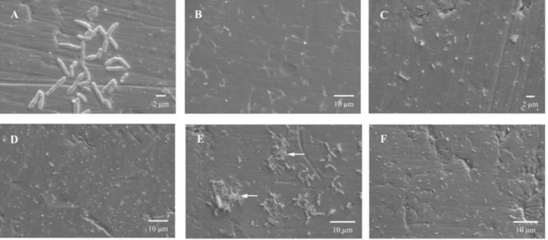
L. monocytogenes adhered to the stainless steel surface,
presented a count of 4.89 Log CFU.cm-2 after 3 hours of
contact (Table 1). As observed by the plate count method, via
SEM, a rapid adherence of L. monocytogenes to the surface
was also verified. After 3 hours of contact, the distribution of
the surface-adhered cells occurred irregularly. At this stage,
two different situations were observed. In some areas, several
cells were adhered to the surface. Most were in the process of
binary fission, indicating possible posterior formation of
microcolonies (Figure 2A). However, in some places, the
bacterial adherence observed was not so evident (Figure 2B).
Table 1. Number of planktonic (Log CFU.mL-1) and sessile (Log CFU.cm-2) cells of Listeria monocytogenes, quantified during
biofilm formation on AISI 304 (#4) stainless steel surface, with incubation at 37 °C and using the culture medium Trypic Soy
Broth (TSB) as substrate.
Time (hours)
TSB (Log CFU.mL-1)
Stainless steel (Log CFU.cm-2)
3 8.97 ± 0.16 4.89 ± 0.03
48 8.85 ± 0.65 4.08 ± 0.67
96 9.95 ± 0.62 4.64 ± 0.57
144 9.55 ± 0.17 4.63 ± 0.60
192 9.74 ± 0.11 4.52 ± 0.47
240 9.36 ± 0.03 5.64 ± 1.07
Results referring to the average of three repetitions ± the standard deviation.
Bacterial adhesion capacity occurs as a function of the
initial inoculum (time 0) and it is a parameter that evaluates the
ability of free cells, originating from a liquid medium, to
adhere to solid surfaces, which corresponds to the first stage of
biofilm development. Initial adhesion capacity, measured
during 3 hours, was 58.75 ± 0.90 % and corresponded to an
inoculum of 8.26 ± 0.18 Log CFU.mL-1 (OD600nm = 0.873 ±
0.04).
The adhesion of bacteria to surfaces occurs in two stages:
reversible followed by irreversible adhesion (32). During
reversible adhesion, bacteria are easily removed by applying
minimum force (13). Irreversible adhesion initiates after 20
minutes to a maximum of 4 hours of contact at 4-20º C (23, 45)
and presents serious risks to the food industry, since the
removal of irreversibly adhered cells is difficult and requires
the application of strong mechanical force or chemical
interruption of the adhesion using surfactants, sanifiers or heat
(44). Thus, there is a high probability that the irreversibly
adhered cells will remain even after hygienization. This is one
of the main reasons for biofilm formation on surfaces in
contact with food. This risk is aggravated with respect to L.
monoctogenes, since this study observed that this bacterium
has the capacity of rapidly adhering to stainless steel, being
able to reach an irreversible stage in a few hours.
Even with the addition of a new culture medium, without
inoculum after 48 hours up to 192 hours, the number of
surface-adhered bacterial cells remained practically constant
(Table 1). During this period, only bacterial adhesion was
observed, i.e., there was no mature biofilm formation. It was
observed through SEM that after 144 hours of contact, the
distribution of the L. monocytogenes cells adhered to the
was found to be still lower (Figure 2C) than in others (Figure
2D). At this stage, no presence or formation of microcolonies
were observed, contrary to the earlier stage (3 hours). This can
be explained by the possible variability between the stainless
steel coupons regarding bacterial adherence. It was also
observed at this stage (144 hours) that, although the number of
surface adhered cells was similar to that found after 3 hours of
biofilm formation (Table 1), the result obtained by SEM
(Figures 2C and 2D) differed completely from that found at 3
hours (Figures 2A and 2B). Thus, it can be concluded that even
with the similarity between the number of cells adhered after 3
and 144 hours (Table 1), cell display on the surface may be
changed with longer contact time between the cells and the
adhesion surface, making it more uniform.
Similar results were observed by Kalmokoff et al. (24),
who studied biofilm formation by different strains of L.
monocytogenes on stainless steel surface. After 72 hours of
contact at 21 °C using the Brain Heart Infusion broth as
substrate, the authors observed under SEM that most of the
strains did not form biofilm under these conditions, but rather
adhered uniformly to the surface. Despite the differences in the
density of the adhered cells among the strains, few cellular
groupings were observed.
It was possible to observe a large difference between the
size of L. monocytogenes sessile cells in Figures 2A and 2C,
since both the scanning electron micrographs showed the same
magnification. This fact can be explained by the difference in
size that L. monocytogenes cells can have, especially with
regard to length. According to Adams and Moss (1), L.
monocytogenes is a Gram-positive rod, with 0.4 to 0.5 µm in
diameter and 0.5 to 2.0 µm in length. The difference in size
between bacteria belonging to this species occurs, mainly due
to their stage of development.
After 240 hours, an increase in the number of adhered
cells was observed, with a count of 5.64 Log CFU.cm-2 (Table
1). The differentiation between adhesion and biofilm formation
has been proposed as a function of the amount of cells present
per cm2. One of the most currently cited values is that proposed
by Andrade et al. (2), who studied adhesion of Enterococcus
faecium to stainless steel surface and emphasized that in order
for biofilm formation to occur counts above 7 Log CFU.cm-2
are necessary. However, to differentiate adhesion from biofilm,
the bacterial species involved must be observed, since it is
known that distinct species will present different adhesion
behaviors and biofilm formation. Thus, this study considered
only the propositions specifically made for L. monocytogenes
regarding the number of adhered cells necessary for mature
biofilm formation, and not only bacterial adhesion.
L. monocytogenes has the capacity to adhere rapidly to
stainless steel surfaces (6, 40). However, it is not capable of
forming thick biofilms made up of several layers (9 to 12 Log
CFU.cm-2), but rather of adhering to surfaces at levels ranging
from 4 to 6 Log CFU.cm-2 (21), which is in agreement with the
values obtained in this study. Ronner and Wong (40), studying
the development of biofilms by L. monocytogenes on stainless
steel surface, obtained counts above 5 Log CFU.cm-2, such as
that found after 240 hours of cultivation in this experiment,
indicating biofilm formation and not only bacterial adhesion.
Chae and Schraft (9) promoted biofilm formation by L.
monocytogenes Murray and 7148 on the surface of glass
coupons for 240 hours of incubation at 37 °C using TSB as
substrate. Counts of approximately 6 Log CFU.cm-2 were
observed for L. monocytogenes Murray and 5 Log CFU.cm-2
for L. monocytogenes 7148. These data are compatible with
those found in this work for stainless steel.
As observed after 3 hours, two different situations were
verified by applying SEM after 240 hours concerning the
distribution of surface-adhered cells. In the first situation, the
formation of small microcolonies was observed, with the
presence of extracellular polymeric substances (Figure 2E).
Such observation, together with the count of bacterial cells
adhered to the surface at 240 hours (5.64 Log CFU.cm-2),
emphasizes the formation of mature biofilm at this stage rather
than just bacterial adhesion. However, it must be pointed out
that the biofilm formed does not totally cover the surface. In
contrast, in some places, the cells were uniformly distributed
on the surface (Figure 2F) as observed after 144 hours (Figures
does not occur identically on the entire surface.
In this study, the production of extracellular polymers by
L. monocytogenes was found to occur after 240 hours of
biofilm formation. The production of extracellular polymers by
this species is little studied (10). Borucki et al. (4), observed
the formation of exopolysaccharides by different strains of L.
monocytogenes. According to the authors, the strains with a
greater biofilm formation capacity were those producing the
most exopolysaccharides, indicating that the production of
extracellular polymers, such as that observed at 240 hours in
this study (arrows in the Figure 2E), is a key factor for L.
monocytogenes biofilm maturation.
Figure 2. Scanning electron micrographs showing the adherence of Listeria monocytogenes on AISI 304 (#4) stainless steel
surface, after 3 (A and B), 144 (C and D) and 240 hours (E and F) of contact at 37 °C, using Trypic Soy Broth (TSB) culture
medium as substrate. (A) Surface-adhered cells, most of which in process of binary fission. (B) Visualization of a larger coupon
area showing little cell adherence. (C) Lower cellular density. (D) Higher cellular density. (E) Mature biofilm with the presence of
extracellular polymeric substances, as indicated by the arrows. (F) Uniformly-adhered cells.
Mature biofilm formation occurs from 72 to 144 hours
after initial adhesion, and may reach 240 hours (22). Maturity
occurs mainly through population density increase as well as
by pronounced production and deposition of extracellular
polymers, increasing biofilm thickness (12). These
extracellular polymers are produced by the cells established
within the biofilm structure (48) and are composed of several
substances, including polysaccharides, proteins and nucleic
acids (34, 47). The matrix of extracellular polymeric
substances is responsible for the morphology, structure,
cohesion and functional integrity of the biofilm. Its
heterogeneous and complex chemical composition determines
most of the physical-chemical and biological properties (16). In
the food industry, it confers resistance to the commonly applied
hygienization procedures (20), making it difficult for the
mature biofilms to be completely removed from these surfaces.
One of the great biofilm formation issues in the food
industry or other areas is cell detachment, which makes it a
constant source of microorganism contamination in food,
biotransfer potential of microorganisms is interesting. This can
be observed from the values found after 96 hours of biofilm
formation. The planktonic cell count found (Table 1) indicates
that the detachment of L. monocytogenes cells from the
stainless steel surface was practically constant during the stages
analyzed (96, 144, 192 and 240 hours). This shows that such
capacity is independent from total biofilm maturation,
contradicting previous reports by several authors (13, 14, 42,
46). The high values found in all the stages analyzed (>9 Log
CFU.mL-1) can be attributed to the existence of adequate
growth conditions, such as temperature and necessary
nutrients. To these factors is added the planktonic condition of
the cell, which is completely immersed in the culture medium,
rapidly metabolizing the nutrients dispersed in the substrate,
making it easier to obtain energy and allowing cell division to
occur quickly.
Few studies about the biotransfer potential have been
made. Flint et al. (17), observed the detachment of Bacillus
stearothermophilus cells, present on stainless steel surface, into
milk passing over the biofilm. Nevertheless, there were no
studies evaluating L. monocytogenes biotransfer potential.
Rapid surface adhesion capacity, combined with
biotransfer potential throughout the stages of biofilm
formation, make L. monocytogenes a potential risk to the food
industry. Once present in the raw material, L. monocytogenes
will adhere rapidly to the surface of stainless steel equipment
and utensils, being able to multiply, forming mature biofilms
quickly. Biotransfer potential combined with survival and
multiplication capacity in different substrates will cause this
bacterium to rapidly reach infecting doses.
Based on the results obtained, we can infer that the
experimental model developed and the methodology applied
were efficient in studying biofilm formation by L.
monocytogenes on stainless steel surface, as well to evaluate
the biotransfer potential. However, we can observe that the
study of bacterial biofilm formation, besides applying plate
count of the number of the surface-adhered cells, must include
microscopy methods that allow observation of not only
bacterial population increases but also of fundamental aspects,
such as the arrangement of cells on the surface and the
presence of extracellular polymeric substances responsible for
the cohesion and protection of the cells present in the biofilm
structure.
Despite previous studies (3, 21) who also demonstrated
successfully bacterial biofilm development using experimental
models similar to that one adopted in this research, this work
emphasizes the use of the experimental model developed as a
new tool to assess the biotransfer potential, which had not yet
been demonstrated.
Thus, it was concluded that this is a useful technique to be
employed in future studies based on the evaluation of
biotransfer potential to different substrates, bacterial adherence
and biofilm formation on stainless steel surface, as well as in
studies aimed at developing methods to remove the adhered
cells.
ACKNOWLEDGEMENTS
The authors thank the National Counsel of Technological
and Scientific Development (CNPq) for the first author’s
scholarship, and the Research Support Foundation of the State
of Minas Gerais (FAPEMIG) for the financial support.
REFERENCES
1. Adams, M.R.; Moss, M.O. (2004). Food microbiology. The Royal Society of Chemistry, Cambridge, England.
2. Andrade, N.J.; Bridgeman, T.A.; Zottola, E.A. (1998). Bacteriocidal activity of sanitizers against Enterococcus faecium attached to stainless steel as determined by plate count and impedance methods. J. Food Prot. 61 (7), 833-838.
3. Bagge, D.; Hjelm, M.; Johansen, C.; Huber, I.; Gram, L. (2001). Shewanella putrefaciens adhesion and biofilm formation on food processing surfaces. Appl. Environ. Microbiol. 67 (5), 2319-2325. 4. Borucki, M.K.; Peppin, J.D.; White, D.; Loge, F.; Call, D.R. (2003).
Variation in biofilm formation among strains of Listeria monocytogenes. Appl. Environ. Microbiol. 69 (12), 7336–7342.
5. Bossola, J.J.; Russell, L.D. (1998). Electron microscopy. Jones and Bartlett, Boston, 670p.
monocytogenes cells and their adhesion to stainless steel. J. Food Prot. 62 (9), 994–998.
7. Carrique-Mas, J.J.; Hokeberg, I.; Andersson Y.; Arneborn, M.; Tham, W.; Danielsson-Tham, M.L.; Osterman, B.; Leffler, M.; Steen, M.; Eriksson, E.; Hedin, G.; Giesecke, J. (2003). Febrile gastroenteritis after eating on-farm manufactured fresh cheese—an outbreak of listeriosis? Epidemiol. Infect. 130 (1), 79–86.
8. Chae, M.S.; Schraft, H. (2000). Comparative evaluation of adhesion and biofilm formation of different Listeria monocytogenes strains. Int. J. Food Microbiol. 62 (1), 103–111.
9. Chae, M.S.; Schraft, H. (2001). Cell viability of Listeria monocytogenes biofilms. Food Microbiol. 18 (1), 103-112.
10. Chae, M.S.; Schraft, H.; Hansen, L.T.; Mackereth, R. (2006). Effects of physicochemical surface characteristics of Listeria monocytogenes strains on attachment to glass. Food Microbiol. 23 (3), 250–259.
11. Chambel, L.; Sol, M.; Fernandes, I.; Barbosa, M.; Zilhão, I.; Barata, B.; Jordan, S.; Perni, S.; Shama, G.; Adrião, A.; Faleiro, L.; Requena, T.; Peláez, C.; Andrew, P.W.; Tenreiro, R. (2007). Occurrence and persistence of Listeria spp. in the environment of ewe and cow's milk cheese dairies in Portugal unveiled by an integrated analysis of identification, typing and spatial–temporal mapping along production cycle. Int. J. Food Microbiol. 116 (1), 52–63.
12. Cheng, C.; Zhang, Z.; Chen, S.; Bryersand, J.D.; Jiang, S. (2007). Inhibition of bacterial adhesion and biofilm formation on zwitterionic surfaces. Biomaterials. 28 (29), 4192-4199.
13. Chmielewski, R.A.N.; Frank, J.F. (2003). Biofilm formation and control in food processing facilities. Int. J. Food Sci. Tech. 2 (1), 22-32. 14. Christensen, B.E.; Characklis, W.G. (1990). Physical and chemical
properties of biofilms. In: Characklis, W. G.; Marshall, K. C. (eds). Biofilms. John Wiley and Sons, Inc, New York, p.93-130.
15. Cruz, C.D.; Silvestre, F.A.; Kinoshita, E.M.; Landgraf, M.; Franco, B.D.G.M.; Destro, M.T. (2008). Epidemiological survey of Listeria monocytogenes in a gravlax salmon processing line. Braz. J. Microbiol. 39 (2), 375-383.
16. Flemming, H.C.; Windenger, J. (1999). Extracellular polymeric substances (EPS): the biofilm construction material. In: Weber, J.; Sand, W. (eds). Biofouling and Materials: EDMZ, COST 520 Workshop, p.2-18.
17. Flint, S.; Palmer, J.; Bloemen, K.; Brooks, J.; Crawford, R. (2001). The growth of Bacillus stearothermophilus on stainless steel. J. Appl. Microbiol. 90 (2), 151-157.
18. Frye, D.M.; Zweig, R.; Sturgeon, J.; Tormey, M.; LeCavalier, M.; Lee, I.; Lawani, L.; Mascola, L. (2003). An outbreak of febrile gastroenteritis associated with delicatessen meat contaminated with Listeria monocytogenes. Clin. Infect. Dis. 35 (8), 943–9.
19. Gandhi, M.; Chikindas, M.L. (2007). Listeria: a foodborne pathogen that knows how to survive. Int. J. Food Microbiol. 113 (1), 1–15.
20. Gilbert, P.; Mcbain, A.J. (2003). Potential Impact of Increased Use of Biocides in Consumer Products on Prevalence of Antibiotic Resistence. Clin. Microbiol. Rev. 16 (2), 189-208.
21. Gram, L.; Bagge-Ravn, D.; Ng, Y.Y.; Gymoese, P.; Vogel, B.F. (2007). Influence of food soiling matrix on cleaning and disinfection efficiency on surface attached Listeria monocytogenes. Food Control. 18 (10), 1165-1171.
22. Heydorn, A.; Ersbøll, B.K.; Hentzer, M.; Parsek, M.R.; Givskov, M.; Molin, S. (2000). Experimental reproducibility in flow-chamber biofilms. Microbiology. 146 (10), 2409-2415.
23. Hood, S.K.; Zottola, E.A. (1995). Biofilms in food processing. Food Control. 6 (1), 9-18.
24. Kalmokoff, M.L.; Austin, J.W.; Wan, X.-D.; Sanders, G.; Banerjee, S.; Farber, J.M. (2001). Adsorption, attachment and biofilm formation among isolates of Listeria monocytogenes using model conditions. J. Appl. Microbiol. 91(4), 725-734.
25. Khelef, N.; Lecuit, M.; Buchrieser, C.; Cabanes, D.; Dussurget, O.; Cossart, P. (2006). Listeria monocytogenes and the genus Listeria. In: Dworkin, M.; Falkow, S.; Rosenberg, E.; Schleifer, K.H.; Stackebrandt, E.(eds). The prokaryotes: an evolving electronic resource for the microbiological community. Springer, New York, USA, p.404-476. 26. Loguercio, A.P.; Silva, W.P.; Aleixo, J.A.G.; Costa, M.M.; Vargas, A.C.
(2001). Listeria monocytogenes: um importante patógeno de origem alimentar. Hig. Aliment. 15 (80/81), 39-48.
27. López, V.; Villatoro, D.; Ortiz, S.; López, P.; Navas, J.; Dávila, J.C.; Martínez-Suárez, J.V. (2008). Molecular tracking of Listeria monocytogenes in an Iberian pig abattoir and processing plant. Meat Sci. 78 (1/2), 130-134.
28. Lunden, J.M.; Autio, T.J.; Korkeala, H.J. (2002). Transfer of persistent Listeria monocytogenes contamination between food-processing plants associated with a dicing machine. J. Food Prot. 65 (7), 1129–1133. 29. Marques, S.C.; Rezende, J.G.O.S.; Alves, L.A.F.; Silva, B.C.; Alves, E.;
Abreu, L.R.; Piccoli, R.H. (2007). Formation of biofilms by Staphylococcus aureus on stainless steel and glass surfaces and its resistance to some selected chemical sanitizers. Braz. J. Microbiol. 38 (3), 538-543.
30. Midelet, G.; Carpentier, B. (2004). Impact of cleaning and disinfection agents on biofilm structure and on microbial transfer to a solid model food. J. Appl. Microbiol. 97 (2), 262–270.
31. Miettinen, M.K.; Siitonen, A.; Heiskanen, P.; Haajanen, H.; Björkroth, K.J.; Korkeala, H.J. (1999). Molecular epidemiology of an outbreak of febrile gastroenteritis caused by Listeria monocytogenes in cold-smoked rainbow trout. J. Clin. Microbiol. 37 (7), 2358-2360.
33. Møretrø, T.; Langsrud, S. (2004). Listeria monocytogenes: biofilm formation and persistence in food-processing environments. Biofilms. 1 (2), 107–121.
34. Nielsen, P.H.; Frølund, B.; Keiding, K. (1996). Changes in the composition of extracellular polymeric substances in activated sludge during anaerobic storage. App. Microbiol. Biotechnol. 44 (6), 823–830. 35. Nikolaev, Y.A.; Plakunov, V.K. (2007). Biofilm -“City of Microbes” or
an Analogue of Multicellular Organisms? Microbiology. 76 (2), 125-138. 36. Oliveira, K.; Oliveira, T.; Teixeira, P.; Azeredo, J.; Oliveira, R. (2007).
Adhesion of Salmonella Enteritidis to stainless steel surfaces. Braz. J. Microbiol. 38 (2), 318-323.
37. Ooi, S.T.; Lorber, B. (2005). Gastroenteritis due to Listeria monocytogenes. Clin. Infect. Dis. 40 (9), 1327-1332.
38. Palmer Jr, R.J. (1999). Microscopy flowcells: perfusion chambers for real-time study of biofilms. Meth. Enzymol. 310, 160-166.
39. Pan, Y.; Breidt Jr., F.; Kathariou, S. (2006). Resistance of Listeria monocytogenes biofilms to sanitizing agents in a simulated food processing environment. Appl. Environ. Microbiol. 72 (12), 7711–7717. 40. Ronner, A.B.; Wong, A.C.L. (1993). Biofilm development and sanitizer
inactivation of Listeria monocytogenes and Salmonella typhimurium on stainless-steel and buna-N rubber. J. Food Prot. 56 (9), 750– 758. 41. Rossoni, E.M.M.; Gaylarde, C.C. (2000). Comparison of sodium
hypoclorite and peracetic acid as sanitizing agents for stainless steel food processing surfaces using epifluorescence microscopy. Int. J. Food
Microbiol. 61 (1), 81-85.
42. Sauer, K.; Camper, A.K.; Ehrlich, G.D.; Costerton, J.W.; Davies, D.G. (2002). Pseudomonas aeruginosa displays multiple phenotypes during development as a biofilm. J. Bact. 184 (4), 1140–1154.
43. Senczek, D.; Stephan, R.; Untermann, F. (2000). Pulsed-field gel electrophoresis (PFGE) typing of Listeria strains isolated from a meat processing plant over a 2-year period. Int. J. Food Microbiol. 62 (1/2), 155–159, 2000.
44. Sinde, E.; Carballo, J. (2000). Attachment of Salmonella spp. and Listeria monocytogenes to stainless steel, rubber and polytetrafluorethylene: the influence of free energy and the effect of commercial sanitizers. Food Microbiol. 17 (4), 439-447.
45. Smoot, L.M.; Pierson, M.D. (1998). Effect of environmental stress on the ability of Listeria monocytogenes Scott A to attach to food contact surfaces. J. Food Prot. 61 (10), 1293-1298.
46. Trachoo, N. (2003). Biofilms and the food industry. Songklanakarin J. Sci. Technol. 25 (6), 807-815.
47. Tsuneda, S.; Aikawa, H.; Hayashi, H.; Yuasa, A.; Hirata, A. (2003). Extracellular polymeric substances responsible for bacterial adhesion onto solid surface. FEMS Microbiol. Lett. 223 (2), 287–292.