Regulation of Na
+and K
+homeostasis in plants: towards improved salt stress
tolerance in crop plants
Diego M. Almeida
1, M. Margarida Oliveira
1and Nelson J. M. Saibo
1 1Genomics of Plant Stress Unit, Instituto de Tecnologia Química e Biológica António Xavier, Universidade
Nova de Lisboa and Instituto de Biologia Experimental e Tecnológica, Oeiras, Portugal.
Abstract
Soil salinity is a major abiotic stress that results in considerable crop yield losses worldwide. However, some plant genotypes show a high tolerance to soil salinity, as they manage to maintain a high K+
/Na+
ratio in the cytosol, in con-trast to salt stress susceptible genotypes. Although, different plant genotypes show different salt tolerance mecha-nisms, they all rely on the regulation and function of K+
and Na+
transporters and H+
pumps, which generate the driving force for K+
and Na+
transport. In this review we will introduce salt stress responses in plants and summarize the current knowledge about the most important ion transporters that facilitate intra- and intercellular K+
and Na+
ho-meostasis in these organisms. We will describe and discuss the regulation and function of the H+
-ATPases, H+
-PPases, SOS1, HKTs, and NHXs, including the specific tissues where they work and their response to salt stress.
Keywords: Salinity, sodium, potassium, proton pumps, ion transporters.
Received: April 13, 2016; Accepted: August 18, 2016.
Salt stress effects on plant growth and yield
Soil salinity is a major environmental constrain to crop production, affecting millions of hectares of land throughout the world and costing billions of dollars every year (Munns, 2005; Munns and Tester, 2008; Shabala and Cuin, 2008). High salinity affects over 6% of the world’s total land area. Most of this affected land has arisen from natural causes, such as rainfall, windblown salt from ocean, tsunamis, and rock weathering. Apart from natural causes, soil salinization is commonly associated to land clearing by removal of deep root vegetation, thus accumulating more water and consequently raising the levels of salty ground-water, or irrigation practices, such as the use of water with high salt concentration. Currently it is estimated that 20% of the total irrigated land is salt-affected. Given that irri-gated land produces at least twice as much as rain-fed land and is responsible for one third of the world’s food produc-tion, it raises awareness for salinity as a serious problem for crop productivity (Munns, 2005; Munns and Tester, 2008).High soil salinity is a condition characterized by a high concentration of soluble salts, in which NaCl is the most soluble and widespread salt. Soils are classified as sa-line when the electrical conductivity (EC) is 4 dS/m (» 40 mM NaCl) or higher. At this soil salt concentration,
growth and yield of most crops are significantly reduced. Rice, as well as most crop plants, is a glycophyte and there-fore it can only tolerate relatively low concentrations of salt. Among cereal crops, rice is the most salt sensitive one, showing salt stress symptoms and reduced yield even when the EC is lower than 4.0 dS/m (Munns and Tester, 2008). The salinity threshold for rice is 3.0 dS/m with a 12% re-duction in yield per dS/m beyond this threshold (Gaoet al., 2007). However, some degree of genotype diversity for salt stress tolerance is available in rice germplasm. Among 180,000 rice genotypes screened by the International Rice Research Institute (IRRI, 2013), 17% showed acceptable tolerance at an EC of 10 dS/m at seedling stage (Gregorioet al., 2002).
High salinity affects plants in two distinct phases. The first phase is the osmotic effect, which is independent of the accumulation of salt in the shoot. Salts dissolved in the soil solution reduce the soil water potential. This makes the wa-ter uptake from roots thermodynamically hampered and in-duces water deficit (Pardo, 2010; Royet al., 2014). A water deficit signal is rapidly transmitted (within minutes) from roots to shoots and will cause intracellular turgor reduction and decreased cell expansion (Munns, 2005; Munns and Tester, 2008). This signal also promotes the biosynthesis of abscisic acid (ABA), which leads to a lower stomatal con-ductance (Munns, 2005; Munns and Tester, 2008; Royet al., 2014). The lower stomatal conductance causes a lower carbon assimilation, biomass production and decreased yield. The second phase of salinity is ionic specific; this is due to the accumulation to toxic concentrations of sodium
DOI: http://dx.doi.org/10.1590/1678-4685-GMB-2016-0106
Send correspondence to Nelson J.M. Saibo. Genomics of Plant Stress Unit, Instituto de Tecnologia Química e Biológica António Xavier, Universidade Nova de Lisboa and Instituto de Biologia Ex-perimental e Tecnológica, Av. da República, 2780-157 Oeiras, Por-tugal. E-mail: saibo@itqb.unl.pt
(Na+) and/or chloride (Cl-) ions, especially in the older leaves, inducing tissue necrosis and early leaf senescence (Royet al., 2014). For most plant species Na+appears to reach a toxic concentration earlier than Cl-(Tester and Dav-enport, 2003). For rice Na+has been shown to be the pri-mary toxic ion (Chi Lin and Huei Kao, 2001; Tsaiet al., 2004). Both osmotic and ionic effects disturb aerobic me-tabolism and induce the accumulation of reactive oxygen species (ROS) beyond the plant’s capacity for cellular oxi-dant detoxification, which in turn negatively affects cellu-lar structures and metabolism (Chaves and Oliveira, 2004; Chaveset al., 2009).
A deleterious effect imposed by salt stress, during the second phase, is ion imbalance (Munns and Tester, 2008). Potassium (K+) is an essential macronutrient that plays im-portant functions related to enzyme activation, osmotic ad-justment and turgor generation, regulation of membrane potential, and cytoplasmatic pH homeostasis (PPI, 1998; Barragan et al., 2012). Due to similarity in physicoche-mical properties between Na+and K+(i.e., ionic radius and ion hydration energy), the former competes with K+for ma-jor binding sites in key metabolic processes in the cyto-plasm, such as enzymatic reactions, protein synthesis and ribosome functions (Marschner, 1995; PPI, 1998). Na+ in-hibits the enzyme activity of many enzymes that require K+ for functioning (Duggleby and Dennis, 1973). With over 50 different cytoplasmic enzymes being activated by K+, dis-ruption of the K+homeostasis leads to severe metabolism impairment, both in root and leaf tissues (Marschner, 1995; PPI, 1998). It has been suggested that plant survival under salt stress requires a high cytosolic K+/Na+ratio in the cyto-plasm. The restriction of Na+accumulation in shoots under salt stress has been correlated with salt stress tolerance in rice (Luttset al., 1996) and maize (Zea maysL.) (Tester and Davenport, 2003).
Sodium uptake from soil, sensing and signaling
mechanisms
The very low membrane potential across the plasma membrane of root cells (more negative inside) promotes the passive transport of Na+ into the cells, and especially so when the sodium concentration increases in the soil solu-tion. In contrast, Na+efflux (i.e., removal from the cell) is not passive and requires energy expenditure (Maathuiset al., 2014). The passive Na+ uptake into root cells at high soil salinity is mainly mediated by a family of Non-Se-lective Cation Channels (NSCCs family), for which the molecular identity remains largely unknown (Blumwaldet al., 2000; Kronzucker and Britto, 2011) (Figure 1). In addi-tion to the Na+flow across cellular membranes to enter into root cells (symplast flow), it has been reported that, at least in some species, interruptions in the endodermis (passage cells) allow the movement of water and solutes (i.e., Na+) through the cell wall and intercellular spaces. This type of transport into the xylem stream, without crossing the plas-ma membrane, is referred as “apoplast flow” (Yeoet al.,
1987; Kronzucker and Britto, 2011) (Figure 1). Casparian strips and suberine layers in the root endoderm and exo-dermal layers provide some barrier to apoplast flow (Yeoet al., 1987). In many plant species, such as rice, the apoplast flow is considered to be the major port of Na+entry (»50% of total Na+uptake) (Yeoet al., 1987), especially at high sa-linity levels, and is responsible for a significant amount of Na+transported to the shoot (Yeoet al., 1987; Kronzucker and Britto, 2011). Na+ions taken up by the roots are then transported to shoots via xylem vessels by bulk flow (Fig-ure 1). This is driven by the tension in the xylem, which causes the continuous movement of water from the root through the plant to the surrounding atmosphere during transpiration (Nobel, 2009).
Sodium has also a strong inhibitory effect on K+ up-take by cells, probably by inhibiting K+transporters, such as AKT1 (hyperpolarization-activated inward-rectifying K+ channel), a major player in K+ acquisition by plants (Hirschet al., 1998; Fuchset al., 2005), and HAK5 (car-rier-type HUP/HAK/KT transport) (Nieves-Cordones et al., 2010), both present in the plasma membrane of root cells. Additionally, membrane depolarization caused by large cytosolic Na+ influx results in increased K+ efflux, possible through depolarization-activated outward-rectifying K+ channels (e.g., GORK) (Adams and Shin, 2014) and also NSCCs (Sunet al., 2009).
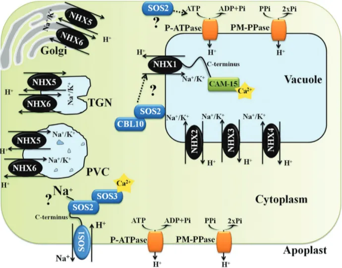
Very little is known about how Na+is sensed in most cellular systems. In theory, Na+can be sensed either outside or inside the cell, or both. Extracellular Na+may be sensed by a membrane receptor, whereas intracellular Na+may be sensed either by membrane proteins or by any of the Na+ sensitive enzymes in the cytoplasm (Condeet al., 2011). The plasma membrane Na+/H+ antiporter SOS1 (SALT OVERLY SENSITIVE 1) has been described as a possible Na+sensor (Shiet al., 2000). Its transport activity is essen-tial for Na+efflux from cells (Quinteroet al., 2002), but its unusually long cytoplasmatic tail is thought to be involved in Na+sensing (Shiet al., 2000) (Figure 2). However, this mechanism it is not fully clear.
In plant cells, Ca2+acts as a second messenger con-necting a wide range of extracellular stimuli with various intracellular responses (Condeet al., 2011). Salt stress orig-inates a fast and transient increase in free cytosolic Ca2+, likely released from the vacuole (Pottosin et al., 2009), which is decoded by Ca2+ sensors, such as calmodulin (CaM), calcineurin B-like proteins (CBLs) and inte-racting protein kinases (CIPKs). When acting as a CBL-CIPK complex, these Ca+sensors are often designed as cal-cium-dependent protein kinases (CDPKs) (Yang and Poo-vaiah, 2003; Condeet al., 2011). Cytosolic Ca2+sensors in turn trigger many signal transduction pathways involved in the regulation of ion channels activity (e.g., NSCCs are strongly blocked by external Ca+2), as well as enzymatic ac-tivity and gene transcription, ending up in ion homeostasis (Pardo and Quintero, 2002; Yamaguchiet al., 2005;
nez-Atienzaet al., 2007; Condeet al., 2011; Adams and Shin, 2014).
Mechanisms of salt tolerance in plants
Salt stress frequently affects plant habitats and many species evolved different mechanisms to cope with it. The mechanisms for salt tolerance can be classified into three main categories. The first one is osmotic stress tolerance, which is regulated by long distance signals that reduce shoot growth (Royet al., 2014) and involves biosynthesis and accumulation of compatible solutes to maintain water uptake (Peleget al., 2011). The second mechanism is ion exclusion, in which Na+transporters reduce the accumula-tion of toxic Na+within roots and leaves. This system oper-ates by controlling the Na+loading into the xylem and Na+ retrieval from the xylem, before reaching the
photosyn-thetic tissues in the shoot (Figure 1). Finally, the third mechanism is tissue tolerance, in which high salt concen-tration is found in leaves, but Na+is compartmentalized at the cellular and intracellular level (especially in the vacu-ole) reducing the deleterious effect of Na+in the cytosol and driving water uptake to cells (Figure 1) (Munns and Tester, 2008). In most cases, the plant salt stress tolerance relies on the three mechanisms together, rather than on only one mechanism in particular (Munns and Tester, 2008; Roy
et al., 2014; Pireset al., 2015).
Sodium transporters and plant salt stress
tolerance
The study of salt stress tolerance in plants usually fo-cuses on the control of Na+movement, namely on: Na+ exclu-sion from roots, Na+ long distance transport, and Na+
Figure 1- Schematic representation showing key plasma and tonoplast membrane transporters, channels and pumps mediating Na+and K+homeostasis
in plants under salt stress (adapted from Royet al., 2014). Na+ions enter the cells via Non Selective Cation Channels (NSCCs) and possibly via other cat-ion transporters not shown (symplast flow - blue arrow) and through the cell wall and intercellular spaces (apoplast flow - red arrow). The Na+/H+
antiporter SOS1 extrudes Na+at the root soil interface, thus reducing the Na+net influx of Na+. At the xylem parenchyma cells, HKT1-like proteins re-trieve Na+from the xylem sap, thereby restricting the amount of Na+reaching the photosynthetic tissues. To translocate Na+back to the root, ions
un-loaded from xylem may be transported into phloem via additional HKT1-like protein. In addition, HKT1-like proteins also load Na+into shoot phloem,
and then Na+is transferred into roots via phloem, preventing Na+accumulation in shoots. SOS1, localized in the xylem parenchyma cells, is also sug-gested to mediate Na+efflux from xylem vessels under high salinity. Incoming Na+, in root and shoots, is stored in the large central vacuole by
compartmentalization at both cellular and tissue level (Munns, 2005; Condeet al., 2011; Royet al., 2014). These processes are mediated by membrane transporters, reason why the ma-nipulation of their activity has an enormous potential to im-prove plant performance under high salinity (Brini and Khaled, 2012). Here, we focus on the specific membrane transporters involved in the above outlined tolerance pro-cesses. In contrast to animal cells, higher plants do not have Na+-ATPases or K+/Na+-ATPases and, rely on H+-ATPases and H+-pyrophosphatases (PPases) to create the pro-ton-motive force necessary to drive Na+ transport across membranes (Condeet al., 2011). The plasma membrane lo-calized SOS1 (Martínez-Atienzaet al., 2007; Jiet al., 2013) and the vacuole membrane (tonoplast) localized NHX1 (Jiang
et al., 2010; Fukudaet al., 2011) are two Na+/H+antiporters involved in Na+ exclusion back to the soil and in Na+ compartmentalization in the vacuole, respectively. In addi-tion, members of the HKT1 family of HKTs (High Affinity Potassium Transporters) are involved in the control of Na+ long distance transport by reabsorption of Na+from the xylem sap into the root cells, preventing the large accumulation of
Na+in the above-ground tissues (Ruset al., 2004) (Figure 1). It is noteworthy that the HKT1 Na+exclusion mechanism from the transpiration stream has been frequently indicated as a strong trait in salt tolerance of different cereals, such as rice (Renet al., 2005) and durum wheat (Triticum turgidum
L. subsp.durum) (Jameset al., 2006).
In the following sections, the role that different Na+ transporters and H+-pumps play in plant salt stress response is discussed.
H
+-Pumps and the plant response to salt stress
Proton gradients are crucial for the transport of ions and solutes across the different plant cell membranes. Three primary proton transport proteins are found in plant cells: (1) plasma membrane (PM) and (2) vacuolar H+-ATPases, which couple ATP hydrolysis with proton transport, and (3) PM and vacuolar H+-PPase, which couple pyrophosphate hydrolysis with proton transport (Gaxiolaet al., 2007; Fuglsanget al., 2010). The H+-Pumps generate an electrochemical potential gradient across membranes,
Almeidaet al. 329
Figure 2- Schematic representation of a hypothetical Arabidopsis cell indicating subcellular localizations, functions, and regulations of NHXs antiporters (NHX1-6), plasma membrane H+-ATPase (P-ATPase), tonoplast H+-ATPase (V-ATPase), tonoplast H+-PPase (V-PPase) and SOS1 (adapted
which is the motive force for a large set of secondary trans-ports.
Plasma membrane H+
-ATPase
The PM H+-ATPase belongs to a class known as P-type ATPases (P-ATPases), and is encoded by a large gene family (Gaxiolaet al., 2007; Fuglsanget al., 2010). The pump is formed by a single subunit protein, which con-tains 10 trans-membrane helices and a large cytoplasmatic domain (Fuglsanget al., 2010). Arabidopsis and rice geno-mes encode 11 and 10 P-ATPases, respectively (Axelsen and Palmgren, 2001; Arangoet al., 2003).
The proton motive force created by P-ATPases is largely responsible for a negative potential across the plas-ma membrane, which is essential for root nutrient uptake, stomatal aperture, phloem loading, and cell growth (Blum-waldet al., 2000; Gaxiolaet al., 2007; Mansour, 2014). Be-sides regulation of many physiological processes, the P-ATPases have a critical role in plant adaptation to salt stress conditions. Higher P-ATPases activity under salt stress conditions repolarizes the NaCl-induced depolariza-tion of PM. This response has been strongly associated with salt stress tolerance (Mansour, 2014). The maintenance of the PM potential under salt stress through P-ATPases activ-ity has a great effect on reduction of Na+influx via depolar-ization-activated NSCCs and K+ efflux via KORs and NSCCs, which help to restore higher K+/Na+levels (Sunet al., 2009). The higher P-ATPases activity under salt stress also energizes the active transport that exclude Na+from root cells, a process dependent of the SOS1 Na+/H+ anti-porter (Gaxiolaet al., 2007). Furthermore, it was reported that higher activation of P-ATPases is often found in halo-phytes and salt tolerant genotypes, which may correlate with salt stress tolerance (Mansour, 2014). For instance, in ricecalluslines, a higher activation of P-ATPases occurred in salt-tolerant lines as compared to less tolerant ones (Pons
et al., 2011).
The salt-dependent activation of PM H+-pump is as-sociated with increased levels of gene expression as well as post-translational modifications of the enzyme present in a preexisting pool (Gaxiola et al., 2007; Mansour, 2014). However, it is likely that regulation of the pump activity oc-curs mostly at post-translational level (Gaxiolaet al., 2007; Fuglsanget al., 2010). The pump activity can be modulated by phosphorylation/dephosphorylation of the penultimate amino acid residue of the cytoplasmatic C-terminus do-main, a threonine residue. The phosphorylated threonine residue promotes binding of the activating 14-3-3 protein (Fuglsanget al., 2010).
Stomatal aperture involves regulation of osmotic pressure within the guard cells, a process powered by P-ATPases activity and responsive to a wide variety of ex-ternal signals (Gaxiolaet al., 2007). Blue light perception in guard cells is mediated by phototropins, which intitiate a signal transduction signal pathway that involves an up-stream protein phosphatase I and a downup-stream protein
kinase that phosphorylates the penultimate C-terminus amino acid residue of the P-ATPase (Takemiyaet al., 2006; Gaxiolaet al., 2007). Under drought and salt stress condi-tions, stomatal closure is induced by ABA through a mech-anism that involves production of hydrogen peroxide (H2O2) and dephosphorylation of the P-ATPases (McAinsh
et al., 1996; Zhanget al., 2001; Gaxiolaet al., 2007).
Vacuolar H+
-ATPase
Among the three proton-pumps found in plant cells, the vacuolar H+-ATPase (V-ATPase) is the most compli-cated one (Gaxiolaet al., 2007). The V-ATPase was first found associated with endomembrane system where it aci-difies and generates a proton force motive within diverse cell compartments (e.g., vacuole, endoplasmic reticulum
andtrans-Golgi network) (Ratajczak, 2000). However,
V-ATPases have also been associated with cell plasma mem-brane (Hanitzschet al., 2007). The ability of the V-ATPase to maintain the cytosolic pH homeostasis and to acidify the endomembrane compartments is crucial during essential processes, such as cell growth and elongation (Hanitzschet al., 2007).
Vacuolar H+-ATPases are multisubunit enzymes composed of two subcomplexes (V1and V0): the peripheral
V1complex consists of eight subunits (A, B, C, D, E, F, G
and H) responsible for ATP hydrolyses, and the V0
mem-brane-integral complex consists of up to six subunits (a, c, c’, c”, d and e) responsible for proton translocation (Gaxio-laet al., 2007) (Figure 3). In plants, the subunit c’ is not found and many of the V-ATPase subunits are encoded by gene families. In Arabidopsis and rice, the 13 subunits which compose the vacuolar H+-ATPases (A, B, C, D, E, F, G, H, a, c, c”, d and e) are encoded by a total of 27 genes and 22 genes respectively (known as VHA genes). If all possi-ble isoform combinations are used, we will have hundreds of different V-ATPase complexes (Sze et al., 2002; Hanitzschet al., 2007).
By convention, the subunits of V1and V0complexes
are distinguished with capital and case letters, respectively. The V1complex consists of: (1) a globular hexameric head
with three alternating copies of subunits A and B forming a ring, (2) a central rotational stalk composed of single copies of subunits D and F, and (3) a outer stalk made of subunits C, E, G and H. Subunits A and B mediate the hydrolysis of ATP at three reaction sites associated with subunit A. Both the central rotational stalk and fixed outer stalk connect the V1 complex to the membrane inserted V0 complex. The
proton transporting V0complex consists of six or more c
subunits, also forming a ring structure. In addition, each V0
The plant vacuole plays a very important role in the maintenance of cellular metabolism due to its role in long term storage of toxic ions, long or short term storage of mineral and/or organic acids and in pH and Ca2+ cyto-plasmatic homeostasis. Furthermore, the V-ATPase is the most abundant H+-pump in the tonoplast and it has been shown that its activity is modulated to cope with environ-mental and metabolic changes (Ratajczak, 2000). For in-stance, under salt stress, a general increase of V-ATPase activity has been reported in many plant species (Matsu-moto and Chung, 1988; Silva and Gerós, 2009). The V-ATPase provides the driving force necessary for Na+ vacu-ole compartmentalization, a process related on the NHX1 antiporter activity (Jianget al., 2010; Bassil and Blumwald, 2014).
The ability to respond to high salinity via changes in the expression of the V-ATPase subunits encoding genes might be a prerequisite and a characteristic of salt stress tol-erance in plants. It has been reported that the transcript lev-els of some subunits are up-regulated in response to high salinity (Narasimhanet al., 1991; Kirschet al., 1996; Silva and Gerós, 2009). However, the expression of V-ATPase genes does not always involve a fixed stoichiometry of mRNAs for the different subunits (Silva and Gerós, 2009). Other factors may also account for the regulation of V-ATPase activity. For instance, the VHA-A subunit from barley (Hordeum vulgare L.) was shown to interact to 14-3-3 proteins, well known activators of PM ATPases, in a phosphorylation-dependent way. That interaction was sug-gested to activate V-ATPase activity (Klychnikov et al., 2007).
Plasma membrane and vacuolar H+
-PPase
H+-pyrophosphatases (H+-PPase) are highly hydro-phobic single subunit proteins that generate proton gradient across the vacuole, Golgi and plasma membrane using the
energy of hydrolysis of pyrophosphate (PPi) molecules (Gaxiola et al., 2007). Plants have two phylogenetically distinct types of H+-PPases: type I and type II. Type I H+-PPases depend on cytosolic K+for their activity and are moderately sensitive to inhibition by Ca2+, and type II H+-PPases are K+insensitive but extremely Ca2+sensitive. The Arabidopsis genome encodes two H+-PPases: a type I H+-PPase (AVP1) and a type II H+-PPase (AVP2) (Drozdowiczet al., 2000). The rice genome also encodes two H+-PPases: OVP1 and OVP2 (Sakakibaraet al., 1996). However, more isoforms have been proposed (Choura and Rebai, 2005). Phylogenetic analysis of V-PPase sequences showed that rice H+-PPases are likely to be type I H+ -PPases (Drozdowiczet al., 2000). Type I H+-PPases are mainly suggested to acidify the vacuole (Gaxiola et al., 2007). However these H+-pumps were also found in the plasma membrane (Ratajczaket al., 1999; Alexandersson
et al., 2004). Arabidopsis type II H+-PPase, AVP2, has
been shown to localize exclusively to Golgi apparatus (Mitsudaet al., 2001).
The expression levels of the H+-PPases are strictly regulated at transcriptional level in response to various en-vironmental conditions or developmental stages. It has been shown that the pollen-specificcis-acting region of the
AVP1gene is involved in the regulation of the gene expres-sion during pollen development. AtCAM15, AtCAMTA 1 (calmoduline-binding transcription factors) (Mitsudaet al., 2003), AtVOZ1, and AtVOZ2 (Arabidopsis thaliana Vas-cular plant One Zinc finger protein) (Mitsudaet al., 2004) were identified as binding to thecis-acting region of the
AVP1gene (Silva and Gerós, 2009; Fuglsanget al., 2010). Salt stress was reported to increase H+-PPase activity (Maeshima, 2000). However, a comprehensive mechanism of H+-PPase gene expression and post-translational regula-tion is still needed. It is likely that the protein C-terminus plays an essential role in supporting the physiological func-tion of H+-PPase activity (Fuglsanget al., 2010).
Given the importance of the pH homeostasis in the cytosol for cell metabolism, it is likely that the activity of all three H+-pumps (P-ATPase, V-ATPase and H+-PPase) is regulated by common regulatory mechanisms. 14-3-3 proteins, which are known to regulate many membrane lo-calized proteins, particularly cell ion pumps (Bunneyet al., 2002), may be involved in such mechanisms.
SOS1 and the plant response to salt stress
Comparisons of unidirectional Na+fluxes and rates of net accumulation of Na+in root indicate that 70-99% of the Na+transported into the root is extruded back to the apo-plast (Munns, 2005; Tester and Davenport, 2003). For rice, that value is indicated as 96% (Munns, 2005), meaning that over time Na+ will accumulate in roots and being trans-ferred via the transpiration stream to the shoot, later accu-mulating there. Since it is important to maintain low cytoplasmatic Na+concentrations for growth and survival under saline conditions, plants have developed a direct
Almeidaet al. 331
Figure 3- Structural model of the plant V-ATPase adapted from Gaxiola
et al.(2007). The peripheral V1complex (blue) and the membrane integral
V0complex (orange) are linked through a peripheral stalk formed by
sub-units a, C, E, G and H. Hydrolysis of ATP is coupled with H+transport to
mechanism to extrude Na+ from cells across the plasma membrane to the soil or apopoplast. Small differences in Na+exclusion capacity create major changes in Na+net ac-cumulation (Tester and Davenport, 2003; Munns, 2005; Brini and Khaled., 2012). However, the role of cellular Na+ efflux is not intuitive in multicellular plants, as Na+ trans-port out of one cell would negatively impact the surround-ing neighbor cells. So, the role of Na+ efflux has to be considered in specific tissues and in the context of the whole plant (Zhu, 2003). Sodium efflux is catalyzed by the plasma membrane Na+/H+ antiporter encoded by SOS1
(Salt Overly Sensitive1=AtNHX7) gene, identified in sev-eral plants including Arabidopsis (Wuet al., 1996), rice (Martínez-Atienzaet al., 2007), wheat (Xu et al., 2008), and tomato (Xuet al., 2008). SOS1 uses the proton gradient established by P-ATPase and/or plasma membrane H+ -PPase to exchange Na+ for H+ across the membrane (Shi and Zhu, 2002; Qiuet al., 2004; Jiet al., 2013). Activity of the ArabidopsisSOS1promoter is detected ubiquitously in virtually all tissues, but it appears to be more active in: (1) root epidermal cells (particularly at the root tip), suggesting that meristem requires special protection, since the root tip cells have very small vacuoles and thus are incapable of vacuolar Na+ compartmentalization, and (2) root paren-chyma cells lining the vasculature (Shi and Zhu, 2002; Kronzucker and Britto, 2011). TheSOS1gene expression pattern, together with the results of ion analysis insos1 mu-tant plants, suggest that SOS1 has several roles: (1) Na+ efflux from roots; (2) slowing down Na+accumulation in the cytoplasm in order to gain time for Na+storage in the vacuole; and (3) control of long-distance Na+transport be-tween roots and leaves by loading and unloading Na+into and from the xylem (Zhu, 2003; Condeet al., 2011). SOS1 may mediate active loading of Na+to the xylem under mild salinity (25 mM NaCl). However, at high salinity (100 mM NaCl), expression ofSOS1is induced and SOS1 may func-tion in Na+retrieval from the xylem (Shiet al., 2002). Such a role for SOS1 in long-distance transport is important for the coordination between transpiration Na+ flow and Na+ vacuolar sequestration in leaves. However, a thermody-namic analysis by Munns and Tester (2008) indicated that the Na+removal from the xylem is unlikely to be mediated by a Na+/H+antiporter such as SOS1, because its operation “in reverse” under high Na+ conditions is thermodynami-cally unfavorable. Instead, class I HKTs have been shown to be involved in xylem unloading of Na+(Renet al., 2005; Jameset al., 2006; Davenportet al., 2007). Thus, the role of SOS1 in long-distance Na+transport remains unclear. Nev-ertheless, many reports suggest that SOS1 plays a critical role in Na+exclusion, thus maintaining cellular ion homeo-stasis and allowing plants to survive and grow under salt stress conditions (Shiet al., 2003; Cuinet al., 2011) (Table 1).
The transcript level ofSOS1is upregulated by high salinity (Shiet al., 2000). Analysis of the 2 Kb upstream of
theSOS1,CIPK24/SOS2andCBL4/SOS3transcription
ini-tiation sites revealed that the promoter of these genes con-tains several binding elements for transcription activation of the bZIP, NAC, WRKY, and TCP classes (Ji et al., 2013). However, transcription factors (TFs) mediating pro-moter activity of SOS genes have not yet been identified. Up-regulation ofSOS1transcript levels under high salinity is suggested to be regulated at the post-transcriptional level, asSOS1promoter activity is not up-regulated by salt stress, but the SOS1 gene expression driven by the constitutive Cauliflower mosaic virus 35S promoter is (Shiet al., 2003). This may indicate that theSOS1transcript is unstable in the absence of salt stress and that the salt stress causes a post-transcriptional stabilization of the transcript (Shi et al., 2003). More recently, it was suggested that the Na+ stress inducedSOS1mRNA stability is mediated by ROS (Chunget al., 2008). In addition, regulation ofSOS1 tran-script levels by high salinity is partly under the control of SOS2 and SOS3 (Shiet al., 2000). CIPK24/SOS2 is a pro-tein kinase and CBL4/SOS3 is a calcium sensor that, together with SOS1, are the three key components compris-ing the Salt Overlay Sensitive (SOS) signalcompris-ing pathway identified in Arabidopsis (Wu et al., 1996) and rice (Martínez-Atienzaet al., 2007). At the cellular level, the SOS signaling pathway has been proposed to mediate cel-lular signaling under salt stress to maintain the ion homeo-stasis (Jiet al., 2013).
Activation of the Na+/H+antiport activity of SOS1 by salt stress is controlled by SOS3 and SOS2 (Zhu, 2003; Jiet al., 2013). In response to an external stimulus, such as high Na+ concentration, transient increases in cytoplasmatic Ca2+ occur and that is decoded by the calcineurin B and neuronal Ca2+ sensor-like protein SOS3. Activation of SOS3 requires N-myristoylation and Ca2+ bound on EF-hand Ca2+binding sites. Activated SOS3 physically inter-acts with the auto-inhibitory domain of SOS2, a member of the SnRK (sucrose non-fermenting-related serine/threo-nine kinase) family, which activates the kinase and facili-tates the localization of the SOS2-SOS3 complex to the plasma membrane. The SOS2-SOS3 complex associates with the Na+/H+ antiporter SOS1, phosphorylating its C-terminal auto-inhibitory domain, which becomes activated and thus pumps Na+out of the cell (Pardo, 2010; Brini and Khaled, 2012; Hasegawa, 2013; Jiet al., 2013) (Figure 2).
inter-Almeida
et
al.
333
Table 1- List of NHX antiporters including information about species, transport selectivity, tissue localization, sub-cellular localization and plant function for each NHX antiporter described in this review. No in-formation available (N/A), Plasma membrane (PM),trans-Golgi network (TGN), and prevacuolar compartment (PVC).
Transporter Species Transport selectivity Tissue localization Subcellular localization Function in planta Refs.
AtNHX1 Arabidopsis Na+/K+ Roots: Vascular tissues Shoots: Floral and vascular tissues, guard cells, trichome.
Tonoplast K+homeostasis and pH
regulation
Rodríguez-Rosaleset al., 2009; Bassilet al., 2011b; Yokoiet al., 2002
AtNHX2 Arabidopsis Na+/K+ Roots Shoots: High in guard cells Tonoplast K+homeostasis and pH
regulation
AtNHX3 Arabidopsis Na+/K+ Mainly in roots Tonoplast N/A
AtNHX4 Arabidopsis Na+/K+ Shoots: Mainly in mature pollen
and seeds
Tonoplast N/A
AtNHX5 Arabidopsis Na+/K+ Roots Shoots: High in guard cells TGN, PVC pH homeostasis in TGN, PVC
AtNHX6 Arabidopsis Na+/K+ Roots Shoots: High in guard cells TGN, PVC pH homeostasis in TGN, PVC
AtNHX7/SOS1 Arabidopsis Na+ Roots: Epidermal cells (particu-larly root tip), parenchyma cells lining the vasculature Shoots
PM Na+efflux Martínez-Atienzaet al., 2007; Shi
and Zhu, 2002; Kronzucker and Britto, 2011
AtNHX8 Arabidopsis N/A N/A PM N/A N/A
OsNHX1 Rice Na+/K+ Roots: Stela, emerging parts of
lat-eral roots. Shoots: Basel part of seedling shoot, vascular bun-dle, flag leaf sheaths, pani-cles, guard cells, trichome.
Tonoplast N/A Fukadaet al., 2004; Fukadaet
al., 2011; Bassilet al., 2012
OsNHX2 Rice Na+/K+ Shoots: Flag leaf sheaths, panicles. Tonoplast N/A
OsNHX3 Rice Na+/K+ Shoots: Flag leaf sheaths, panicles. Tonoplast N/A
OsNHX4 Rice N/A N/A Tonoplast N/A
OsNHX5 Rice Na+/K+ Roots: Stela, emerging parts of
lat-eral roots, root tip. Shoots: Basel part of seedling shoot, vascular bundle, flag leaf sheaths, pani-cles, pollen grain.
TGN, PVC N/A
acts with SOS2 to prevent the SOS3 binding to SOS2 and kinase activation. Such ABI2-SOS2 interaction may repre-sent an integrating node between salt stress and ABA sig-naling (Ohtaet al., 2003; Hasegawa, 2013).
The SOS pathway may also regulate the Na+vacuolar compartmentalization. Interaction of SOS2-CBL10 may result in localization of the kinase complex at the vacuolar membrane where it is possibly involved in the regulation of Na+/H+exchange at the tonoplast, presumably by regula-tion of NHX antiporter(s) activity (Qiuet al., 2004; Kimet al., 2007). However, no NHX antiporter has already been shown to be directly regulated by SOS2 and/or by the SOS2-complex. In addition, SOS2 has been suggested to regulate the V-ATPase activity. SOS2 was found to interact with the B1 and B2 subunits of the V-ATPase in the ab-sence of CBL proteins, and tonoplast vesicles from the Arabidopsis sos2-2 mutant showed reduced ATPase and H+-translocation activities (Batelliet al., 2007).
Potassium homeostasis has also been shown to be modulated by the SOS signaling pathway. The protein CBL10 has been indicated to directly interact with AKT1 channel and negatively regulate its activity in roots (Renet al., 2013). It is well known that plant salt stress tolerance is closely related to maintenance of high K+/Na+cytosolic ra-tio under stress (Tester and Davenport, 2003). The possibil-ity that CBL10 functions as an interconnecting regulator of SOS1 and AKT1 may indicate that CBL10 plays a crucial role in ion homeostasis (K+/Na+) under salt stress by regu-lating both K+and Na+uptake/exclusion (Renet al., 2013).
HKTs and the plant responses to salt stress
Another important determinant of salt stress tolerance in plants is the activity of the HKT (high affinity potassium transporter) proteins (Munns and Tester, 2008; Royet al., 2014). The HKT family is quite diverse, and this diversity reflects their large amplitude of functions (Munns and Tes-ter, 2008; Almeidaet al., 2013; Royet al., 2014). The HKT family is divided in two distinct classes according to their transport characteristics. The main distinguishing feature is the amino acid sequence that constitutes the first pore do-main (PD) (Plattenet al., 2006). Members of class I trans-porters (HKT1) have a serine (S), forming an S-G-G-G motif, where most of the members of class II (HKT2) have a G in the position occupied by the S in class I transporters, forming a G-G-G-G motif (Maseret al., 2002). The pres-ence of either S or G at this position is critical for the cation specificity of transporter. The presence of an S (HKT1) is characterized by a preference for Na+ conductance over other cations, whereas the presence of a G (HKT2) is char-acterized by transport of Na+and/or K+depending on the external concentrations of these two ions (Plattenet al., 2006; Kronzucker and Britto, 2011). However, there are notable exceptions, in particular HKT2;1 from cereals, in which the G has reverted to S (Kronzucker and Britto, 2011), but it has been clearly shown to be involved in medi-ating Na+and K+entry into roots (Munns and Tester, 2008;
Kronzucker and Britto, 2011). The main role of HKT1 is believed to be Na+retrieval from the transpiration stream avoiding the over accumulation of Na+ in the photosynthetic tissues.
HKT1 family
The best characterized member of HKTs class I is AtHKT1;1 from Arabidopsis. Disruption ofAtHKT1;1, the only member of HKT family in Arabidopsis, caused a higher accumulation of Na+in the shoots but reduced con-centration in roots, with little effect on the net Na+uptake
(Rus et al., 2004; Pardo, 2010; Kronzucker and Britto,
2011)AtHKT1;1is preferentially expressed in the plasma membrane of xylem parenchyma cells and phloem cells of both roots and leaves, where it is suggested to regulate the Na+distribution between roots and shoots (Sunarpiet al., 2005; Molleret al., 2009; Pardo, 2010; Kronzucker and Britto, 2011) (Figure 1 and Table 2). Two complementary functions for AtHKT1;1 have been proposed. In the phloem recirculation model, AtHKT1;1 loads Na+ into shoot phloem cells to be transferred to roots via the downward stream, preventing Na+ overaccumulation in the shoot. However, the overall Na+ retranslocation potential via phloem should not exceed 10% of the total Na+loaded in the shoot xylem transpiration stream (Berthomieu et al., 2003). Another function of AtHKT1;1 is to unload Na+ from the xylem transpiration stream, thereby restricting the amount of Na+reaching the photosynthetic tissues and sup-porting salt stress tolerance.
Analysis of several QTLs of salt tolerance in rice (Renet al., 2005; Kronzucker and Britto, 2011) and wheat (James et al., 2006; Byrt et al., 2007; Kronzucker and Britto, 2011) has provided further evidence for the impor-tance ofHKTclass 1 genes in controlling Na+accumulation in leaves upon salt stress. In rice, QTL analyses showed that higher shoot K+content of the salt-toleranceindica geno-type, Nona Bokra, cosegregated with an allelic variant of
SKC1(Shoot K+Content 1) with higher activity as com-pared to that of the salt-sensitive japonica genotype, Koshihikari (Renet al., 2005). SCK1, now referred to as
OsHKT1;5(OsHKT8) is a plasma membrane, K+
independ-ent, and Na+selective transporter that is preferentially ex-pressed in the parenchyma cells surrounding xylem vessels (Renet al., 2005; Pardo, 2010; Almeidaet al., 2013) (Table 2). The Nona BokraOsHKT1;5has four amino acids differ-ent from the Koshihikari protein, and this difference has been associated with greater Na+transporter activity and in-creased ability for maintenance of K+/Na+homeostasis un-der salt stress (Renet al., 2005). Rice contains four more HKT1 members in the genome, OsHKT1;1, OsHKT1;2,
OsHKT1;3, OsHKT1;4 (Ren et al., 2005; Huang et al.,
2006; Wuet al., 2009; Cotsaftiset al., 2012; Almeidaet al., 2013) (See Table 2 for further information). OsHKT1;4
(salt-tolerant) and Nipponbare (salt-susceptible). All
OsHKT1;4splicing forms are translated into protein,
nev-ertheless only the longer splicing form seems to be trans-lated into a functional protein (Cotsaftis et al., 2012). Interestingly, Pokkali is able to maintain a much higher ra-tio of funcra-tional OsHKT1;4 transcripts in younger leaf sheaths as compared to Nipponbare. In addition, transcript levels of the functional transcripts were inversely corre-lated with the individual leaf blade Na+ concentration in both genotypes (Cotsaftis et al., 2012). At this point it seems that the longerOsHKT1;4splicing form is the key transporter controlling the sheath-to-blade transfer of Na+ in rice shoots (Cotsaftiset al., 2012).
In wheat, QTL analyses using durum wheat (Triticum
turgidumL. subsp. durum) breeding Line 149 led to the
identification of two loci,Nax1, andNax2, which decreased Na+accumulation in the leaf blade (Jameset al., 2006; Byrt
et al., 2007; Kronzucker and Britto, 2011). In addition,
bread wheat (Triticum aestivum), which is an allohexaploid (2n = 6s = 42, genome AABBDD), was found to be more salt tolerant than the allotetraploid pasta wheat (AABB genomes). It was shown that the D genome carries a locus (Kna1) responsible for maintenance of high K+/Na+ ratio during salt stress justifying the salt tolerance of bread wheat (Dubcovskyet al., 1996; Byrtet al., 2007; Kronzucker and Britto, 2011). The process controlled by theNax2andKna1
loci reduces net root xylem loading of Na+, while theNax1
locus reduces Na+ accumulation in the leaf blade by re-stricting Na+loading into root xylem and partitioning Na+ into the leaf sheath (Jameset al., 2006; Hasegawa, 2013).
Almeidaet al. 335
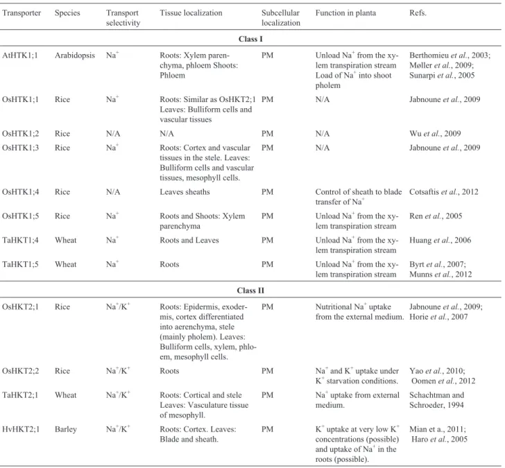
Table 2- List of HKT transporters including information about class, species, transport selectivity, tissue localization, subcellular localization and plant function for each HKT transporter described in this review. No information available (N/A), Plasma membrane (PM).
Transporter Species Transport
selectivity
Tissue localization Subcellular
localization
Function in planta Refs.
Class I
AtHTK1;1 Arabidopsis Na+ Roots: Xylem
paren-chyma, phloem Shoots: Phloem
PM Unload Na+from the
xy-lem transpiration stream Load of Na+into shoot
pholem
Berthomieuet al., 2003; Mølleret al., 2009; Sunarpiet al., 2005
OsHTK1;1 Rice Na+ Roots: Similar as OsHKT2;1
Leaves: Bulliform cells and vascular tissues
PM N/A Jabnouneet al., 2009
OsHTK1;2 Rice N/A N/A PM N/A Wuet al., 2009
OsHTK1;3 Rice Na+ Roots: Cortex and vascular
tissues in the stele. Leaves: Bulliform cells and vascular tissues, mesophyll cells.
PM N/A Jabnouneet al., 2009
OsHTK1;4 Rice N/A Leaves sheaths PM Control of sheath to blade
transfer of Na+
Cotsaftiset al., 2012
OsHTK1;5 Rice Na+ Roots and Shoots: Xylem
parenchyma
PM Unload Na+from the
xy-lem transpiration stream
Renet al., 2005
TaHKT1;4 Wheat Na+ Roots and Leaves PM Unload Na+from the
xy-lem transpiration stream
Huanget al., 2006
TaHKT1;5 Wheat Na+ Roots PM Unload Na+from the
xy-lem transpiration stream
Byrtet al., 2007; Munnset al., 2012
Class II
OsHKT2;1 Rice Na+/K+ Roots: Epidermis,
exoder-mis, cortex differentiated into aerenchyma, stele (mainly pholem). Leaves: Bulliform cells, xylem, phlo-em, mesophyll cells.
PM Nutritional Na+uptake
from the external medium.
Jabnouneet al., 2009; Horieet al., 2007
OsHKT2;2 Rice Na+/K+ Roots PM Na+and K+uptake under
K+starvation conditions.
Yaoet al., 2010; Oomenet al., 2012
TaHKT2;1 Wheat Na+/K+ Roots: Cortical and stele
Leaves: Vasculature tissue of mesophyll.
PM Na+uptake from external
medium.
Schachtman and Schroeder, 1994
HvHKT2;1 Barley Na+/K+ Roots: Cortex. Leaves:
Blade and sheath.
PM K+uptake at very low K+
concentrations (possible) and uptake of Na+in the
roots (possible).
Using high-resolution mapping,Nax1andNax2were iden-tified as members of theHKT1;4gene family andKna1as member of the HKT1;5 gene family (Table 2). Because both Nax genes are originated from a wheat relative,
Triticum monococcum, that was crossed with a durum
wheat, they were namedTmHKT1;4-A2andTmHKT1;5-A, respectively. TheNax2region of the breeding Line 149 was found to correspond to theKna1region of the bread wheat andKna1was namedTaHKT1;5-D(Almeidaet al., 2013).
Nax1 and Nax2 genes do not exist in modern bread or durum wheat genotypes, and introgression ofNax1orNax2
into bread wheat led to reduced leaf blade Na+ accumula-tion and increased leaf blade Na+exclusion relative to the parent respectively. The combination of Nax1and Nax2
further decreased Na+ accumulation in the leaf blade (Jameset al., 2011), showing that these genes clearly have similar functions as AtHKT1;1 in Arabidopsis and
OsHKT1;5andOsHKT1;4in rice (Renet al., 2005; James
et al., 2006; Almeidaet al., 2013) (Table 2). Moreover, field trials with durum wheat, carrying the Nax2 gene, growing under high saline soils showed a 25% increase in grain yield and reduced Na+ accumulation in flag leaf as compared to a near isogenic line without theNax2locus (Munnset al., 2012). Altogether, these results indicate that
HKT1mediated Na+exclusion from shoot is an effective mechanism for enhancing salt stress tolerance in crop plants.
Concerning,HKT1 transcriptional regulation, some transcriptional regulatory elements have been identified in
theAtHKT1promoter. The tandem repeat regions (R1 and
R2) found in the distalAtHKT1 promoter region located about 3.9 kb upstream of the translational start codon were responsible for expression ofAtHKT1in roots (Ruset al., 2004; Baeket al., 2011). The repeat sequence R2 which is closest to ATG acts as an enhancer element ofAtHKT1 ex-pression. Its inactivation caused reducedAtHKT1 expres-sion in root and higher Na+accumulation in shoot (Ruset al., 2004; Baeket al., 2011). TheAtHKT1promoter con-tains a highly methylated GC region (250 bp) at 2.6 kb upstream of the translational start codon. Interestingly, methylation in the leaf is higher than in roots, which sug-gests that higher methylation in this promoter region is re-quired to maintainAtHKT1expression at low levels and perhaps in a correct pattern of expression in the different tissues (Baeket al., 2011). Furthermore, this region con-tains a putative small RNA target site, which was suggested to be involved in methylation guided by small RNAs (Baek
et al., 2011).
HKT2 family
HKT class 2 proteins are generally found in monocot-yledonous species, and no HKT class 2 homologs have been identified in dicotyledonous species (Plattenet al., 2006; Adams and Shin, 2014). Four HKT class 2 members have been characterized in detail: OsHKT2;1 and OsHKT2;2 in rice, TaHKT2;1 in wheat, and HvHKT2,1 in
barley (Hordeum vulgareL.) (Table 2). These transporters have common properties thought to be shared by all HKT2 class 2 members, such as a role in Na+uptake from external medium under K+ limiting conditions (Almeida et al., 2013).
The two characterized rice members of this HKT family, OsHKT2;1 and OsHKT2;2, have been reported to mediate Na+uptake from soil under K+limiting conditions (Table 2).OsHKT2;1gene expression is induced by K+ de-ficiency (Horieet al., 2001; Yaoet al., 2010). OsHKT2;1 is an atypical HKT class 2 member, which has an S residue in the first PD and mediates high-affinity Na+uptake. How-ever, OsHKT2;1 can also mediate K+transport depending on the external concentration of both K+and Na+(Jabnoune
et al., 2009; Yao et al., 2010; Almeida et al., 2013).
OsHKT2;1 is known to be highly involved in “nutritional” absorption of Na+and its relevance in Na+uptake during salt stress may be limited since it has a micromolar affinity for Na+and its activity is rapidly downregulated at high Na+ concentration. Interestingly, RNA levels of at least three otherOsHKTsgenes have been shown to be inhibited by an external Na+concentration as low as 30 mM (Horieet al., 2001). On the other hand, OsHKT2;2 has only been found in the salt-tolerant Nona Bokra and Pokkali genotypes, be-ing absent in the rice salt-sensitive Nipponbare genotype, which suggests that the presence of OsHKT2;2 is an evolu-tionary advantage for salt-tolerant genotypes (Horieet al., 2001; Almeida et al., 2013). OsHKT2;2 is expressed in roots among other tissues and transporting both K+ and Na+, but under salinity only Na+is transported (Kaderet al., 2006, Oomenet al., 2012) (Table 2). Other OsHKT class 2 members have also been identified (OsHKT2;2/1, OsHKT2;3, OsHKT2;4), however these will be not de-scribed. For further information, see Almeidaet al.(2013) and Kronzucker and Britto (2011).
In wheat, TaHKT2;1 seems to have a function in root Na+influx similar to rice OsHKT2;1 (Horieet al., 2009).
TaHKT2;1is expressed in the root cortex and is induced by K+ deficiency (Schachtman and Schroeder, 1994). In planta, TaHKT2;1 has been suggested to have a role in Na+ transport with a possible role in root Na+ uptake, though TaHKT2;1 was also reported to transport K+(Almeidaet al., 2013) (Table 2).
In barley, a relative salt-tolerant species,HvHKT2;1,
is preferentially expressed in root cortex and to a much lower level in leaf blade and sheaths, and it is induced by K+ deficiency in roots and shoots and by high Na+ concentra-tion in shoots.HvHKT2;1mediates both K+and Na+ trans-port (Haroet al., 2005; Mianet al., 2011; Almeidaet al., 2013) (Table 2). Transgenic barley lines over-expressing
content in plants growing in limiting K+ conditions, sug-gesting thatHvHKT2;1may also play a role in K+ absorp-tion or re-absorpabsorp-tion at very low K+concentrations (Mianet al., 2011).
NHX and the plant response to salt stress
At the cellular level, high amounts of Na+can be tol-erated by intracellular partitioning so that the concentration in the cytoplasm is kept as low as 10-30 mM (Munns and Tester, 2008). This strategy can be used by plants for the al-leviation of excessive cytosolic Na+ by sequestrating Na+ into the vacuole, which typically makes up to 80-90% of the cell volume. Other organelles, such as endosomal compart-ments, plastids and mitochondria, may also accumulate Na+ and thus contribute to the overall subcellular Na+ sequestra-tion (Zhu, 2003). The vacuolar sequestrasequestra-tion of Na+ that occurs in all tissues is not only important for Na+ detoxifi-cation in the cytosol, but it is also a critical mechanism of osmotic adjustment to maintain water uptake from saline solutions (Zhu, 2003; Munns and Tester, 2008; Bassilet al., 2012).
An increased vacuolar Na+ concentration requires a coordinated increase in the osmotic pressure of the other subcellular components, including the cytosol, to maintain the osmotic pressure and thereby the volume. This can be achieved by an increase in the K+concentration to a sub-toxic level, as well as by the synthesis and accumulation of compatible solutes (e.g., proline, sucrose, glycine betaine, etc.). Nevertheless, the latter represents a major drawback due to the high energetic cost associated with solute synthe-sis (Munns and Tester, 2008; Maathuiset al., 2014).
The tonoplast controls the movement of inorganic and organic solutes to and from the cytoplasm through a wide range of pumps, carriers and ion channels (Condeet al., 2011). Cation/H+ antiporters mediate the transport of Na+into the vacuole, driven by the electrochemical gradi-ent of protons generated by the V-ATPase and V-PPase en-zymes (Jiang et al., 2010; Bassil and Blumwald, 2014). This Na+/H+exchange is mediated by members of a family of transporters referred to as Na+/H+antiporters (NHXs) in plants or Na+/H+exchange (NHEs) in animals (Jianget al., 2010; Bassil and Blumwald, 2014). In addition, plant NHX antiporters mediate both Na+/H+ and K+/H+ exchange, therefore affecting both salinity tolerance and K+nutrition (Venemaet al., 2002; Leidiet al., 2010).
Diversity of plant NHX antiporters
Plant NHX proteins belong to a large superfamily of monovalent cation/proton antiporters (CPAs) made up of two subgroups, CPA1 and CPA2. The CPA2 family in-cludes members of the less known Cation/H+Exchangers (CHXs) and K+efflux antiporters (KEA). The CPA1 family includes members of the NHX-type, which are ubiquitous in all eukaryotic organisms (Rodríguez-Rosales et al., 2009; Bassilet al., 2012). In Arabidopsis, NHX-type
anti-porter family members comprise eight members that are di-vided into two distinct classes; two divergent members lo-cated at the plasma membrane (SOS1/AtNHX7 and AtNHX8), and six intracellular members located either at the tonoplast (AtNHX1-AtNHX4) or the endosomal mem-brane (Golgi,trans-Golgi network and prevacuolar com-partments) (Rodríguez-Rosaleset al., 2009; Bassilet al., 2012; Regueraet al., 2015). In rice, six NHX-type anti-porter family members were identified as belonging to two distinct classes with different cellular localizations: one in the plasma membrane (SOS1) (Martínez-Atienza et al., 2007), and five intracellular members that are either in the tonoplast, OsNHX1 to OsNHX4, or in the prevacuolar compartment OsNHX5 (Fukuda et al., 2011) (Figures 1 and 2, Table 1). In Arabidopsis, the most abundant mem-bers of NHX-types are AtNHX1 and AtNHX2, accounting for a significant amount of the K+-Na+/H+antiport activity in tonoplast vesicles (Barraganet al., 2012). Detailed infor-mation regarding AtNHXs and OsNHXs tissue localization is described in Table 1.
NHX gene expression under stress conditions
In Arabidopsis seedlings,AtNHX1and2were shown to be induced by salt stress (NaCl), hyperosmotic stress (mannitol) and ABA treatment, whilstAtNHX5was only induced by salt stress (NaCl) (Yokoiet al., 2002). In rice seedlings, salt stress (NaCl), hyperosmotic stress (man-nitol) and ABA treatment increased the transcript levels of
OsNHX1,2,3and5 (Fukudaet al., 2011). These reports show thatNHXgenes are components of the plant salt stress response. Interestingly, treatment with a high KCl concen-tration induced the expression ofOsNHX1and2(Fukudaet al., 2011), andAtNHX1(Yokoiet al., 2002).AtNHX1and2
were induced by ABA but not by NaCl in the ABA-deficientaba2-1 mutant, showing that NaCl induction of these members depends on ABA signaling (Yokoiet al., 2002). AtNHX1 and 2 promoter sequences do not have ABA-responsive elements (ABRE). Nevertheless, the pro-moter of each gene contains MYC/MYBcis-regulatory ele-ments, suggesting thatAtNHX1and 2 are outputs of the ABA-dependent pathway regulated by these transcription factors (Yokoi et al., 2002). On the other hand, the
OsNHX1 promoter (up to 1.8 kb upstream of the
trans-lational start codon) shows several ABA-responsive ele-ments (ABRE), as well as drought responsive eleele-ments MYC/MYBcis-regulatory elements (Almeida et al., un-published results), indicating that OsNHX1, similar to
AtNHX1, is also transcriptionally regulated by an
ABA-dependent pathway. Interestingly, the SOS pathway also appears to regulate the activity of vacuolar Na+/H+ anti-porters (Yokoiet al., 2002; Qiuet al., 2004). The activity of AtNHX1 is possibly regulated through interaction with the protein kinaseSOS2(Qiuet al., 2004) (Figure 2). It was re-ported that the vacuolar Na+/H+ antiporter activity of Arabidopsis membrane vesicles was significantly reduced in vesicles obtained fromsos2null mutants, as compared to
wild type controls, and could be stimulatedin vitroby the addition of activated SOS2 protein (Qiuet al., 2004). The activity was further inhibited by AtNHX1 antibodies. How-ever, phosphorylation of AtNHX1 by SOS2 was not shown. It was further shown that SOS2 also interacts with several V-ATPase subunits (Figure 2), and that vesicles isolated fromsos2null mutants show a considerable lower V-ATPase acidification (Batelliet al., 2007). Thus, com-parisons of antiporter activity in vesicles fromsos2mutant and wild type is complex, as the proton force motive that drives ion transport is not similar in both cases (Batelliet al., 2007).
NHX regulation and structural organization
Transcriptional regulation
Although multiple functional studies on NHX-type proteins, especially NHX1, have been carried out, the de-tails of how NHX1 is transcriptionally regulated remain poorly explored. Adleret al.(2010) reported that NHX1 from the relatively salt-tolerant crop, sugar beet (Beta vulgarisL.) is regulated under salt stress by one or more MYB transcription factors, which could not be identified yet. Despite the importance of rice, only one study reported the identification of a TF interacting with theOsNHX1 pro-moter. Using a chromatin immunoprecipitation assay, an
OsbZIP71TF was identified by Liuet al.(2014) as directly binding to the OsNHX1 promoter. It was shown that
OsbZIP71 gene expression was strongly induced by
drought, polyethylene glycol (PEG), and ABA treatments, but repressed by salt treatment. Transgenic rice lines over-expressingOsbZIP71(p35S::OsbZIP71) showed improved tolerance to drought, salt and PEG-induced drought stresses, suggesting thatOsbZIP71plays an important role in ABA-mediated drought and salt tolerance in rice (Liuet al., 2014). However, the authors did not show whether the identified TF is relevant for OsNHX1 activation under stress.
Post-translational modifications
The cation selectivity of Arabidopsis NHX1 appears to be regulated by its C-terminal tail through the interaction with the vacuolar lumen-localized calmodulin-like protein 15 (AtCaM15), in a Ca2+ and pH-dependent manner (Yamaguchiet al., 2003). Under control physiological con-ditions, when the vacuole pH is acidic (pH 5.5) and the Ca2+ concentration is high, AtCaM15 is bound to the AtNHX1 C-terminal tail, resulting in a higher K+/H+exchange activ-ity over Na+/H+activity (Yamaguchiet al., 2003). On the other hand, salt stress often causes alkalization of the vacu-ole, which reduces AtCaM15 binding to AtNHX1. This leads to an increased Na+/H+exchange activity over K+/H+ activity and subsequent enhanced vacuolar Na+ sequestra-tion (Yamaguchi et al., 2003; Rodríguez-Rosales et al., 2009). In addition, phosphoproteomic studies in Arabidopsis and rice suggested that NHX antiporters are
regulated by phosphorylation (Whitemanet al., 2008a,b). In rice, the vacuolar OsNHX3 was reported to be phospho-rylated at residue S471 located in the C-terminus. The same residue is conserved among the three other rice vacuolar NHXs members (OsNHX1, 2 and 4) (Bassilet al., 2012). Sequence comparison analyses between rice NHXs and Arabidopsis NHX3, 5 and 6 revealed that the Arabidopsis NHXs members contained the same S residue at a similar position (Bassilet al., 2012). So far, the biological role of such post-translational modifications has not yet been func-tionally characterized. Negrãoet al.(2013) identified a sig-nificant nonsynonymous mutation at OsNHX1, serine 477 to asparagine (S477N), present in the rice salt-susceptible genotypes IR 29 and IR 64, but also in the salt-tolerant ge-notype FL 478 (a recombinant inbred line derived from an IR 29 x Pokkali cross). The loss of an S residue can imply the loss of a putative phosphorylation site, and S477 sits in a cluster of S residues with high phosphorylation probabil-ity and is itself a potential phosphorylation target (Negrão
et al., 2013). This residue is located in the C-terminus of the OsNHX1 protein, and it was suggested by the authors that the nonsynonymous mutation may affect the phosphorylation of the OsNHX1 C-terminal possibly by SOS2, which in turn results in lower activation of OsNHX1 exchanger activity. However, further studies are needed to test this hypothesis.
Topology
The crystallographic structure of NHXs antiporters is not yet available. Epitope tagging and protease protection assays applied to full length expressed AtNHX1 in a yeast heterologous system unveiled that AtNHX1 has nine trans-membrane domains, with an additional three “buried” do-mains that do not entirely span the membrane (Yamaguchi
et al., 2003). This study also showed that the hydrophilic C-terminus is oriented to the vacuolar lumen, a feature that is strikingly different from the proposed cytosolic C-termi-nus orientation of animal NHEs (Yamaguchiet al., 2003; Bassilet al., 2012). However, another study performed by Sato and Sakaguchi (2005), using only protein fragments, showed that AtNHX1 contains eleven transmembrane do-mains and a cytosolic C-terminus, resembling an overall membrane topology of the human NHE. The C-terminus of NHX members is highly divergent, even among closely related members within the same species. Because the C-terminus regulates the antiporter activity, it has been sug-gested that divergent C-terminal sequences may constitute a novel way to differentially regulate individual members (Bassilet al., 2012).
Function of the NHX antiporters
Salt tolerance
ho-mologous or heterologous systems) confer salt stress toler-ance in a wide range of plant species. Constitutive over-expression of AtNHX1 appears to increase salt stress tolerance significantly in yeast (Aharon et al., 2003), Arabidopsis (Apseet al., 1999), tomato (Zhang and Blum-wald, 2001) and cotton (He et al., 2005). Constitutive overexpression of various cereal NHX homologs has been also reported to improve the salt stress tolerance of Arabidopsis (Briniet al., 2007), rice (Fukudaet al., 2004; Zhaoet al., 2006) and wheat (Xueet al., 2004). These re-sults show the fundamental role of these proteins in plant salt stress tolerance and explain why they have been a ma-jor focus for genetic engineering (Jianget al., 2010; Bassil
et al., 2012). However, increased salt stress tolerance was not always associated with an increased vacuolar Na+ accu-mulation (Fukudaet al., 2004; Rodríguez-Rosales et al., 2008, 2009; Jianget al., 2010). Most of the characterized NHX members can transport both K+ and Na+, and may have similar Km for these substrates (Jianget al., 2010). This means that, unless the cytoplasmatic Na+ concentra-tion is significantly higher than that of K+(difficult to occur even under salt stress conditions), NHX exchangers mainly mediate K+/H+ exchange rather than Na+/H+ exchange (Jiang et al., 2010; Maathuis et al., 2014). Indeed,
nhx1/nhx2double null mutants in Arabidopsis resulted in impaired vacuolar K+ accumulation, enhanced vacuolar Na+uptake, and a salt (NaCl) insensitive phenotype, com-pared to wild-type (Barraganet al., 2012). Thus, it is likely that the contribution of NHX-like protein to plant salt stress tolerance is the maintenance of K+homeostasis rather than sequestration of Na+ into the vacuole (Maathuis et al., 2014). Nevertheless, it is still not clear what are the primary ions being transported by NHX-like protein in planta. Given that NHX1 cation selectivity is regulated by interact-ing partners (AtNHX1 is regulated by CaM15), it is diffi-cult to interpret the ion content results from plants over-expressingAtNHX1, and possibly other NHXs members, due to a possible shortage of interacting partners (Rodríguez-Rosaleset al., 2009).
K+
homeostasis
Besides their key role in salt stress tolerance, at con-trol growth conditions vacuolar NHX proteins have a key role in mediating K+/H+ exchange for turgor regulation and pH control. Potassium is an essential plant nutrient and the most abundant cation in plants, comprising up to 10% of plant dry matter. K+ is an important cofactor in many biosynthetic processes, and in the vacuole it plays key roles in cell volume regulation (Barraganet al., 2012; Andréset al., 2014).
During grape berry (Vitis viniferaL.) development,
high VvNHX1 transcript levels during the véraison and
post-véraison stages would indicate that the increase in vacuolar K+accumulation, mediated byVvNHX1is needed for vacuolar expansion. This process is coupled with a rapid accumulation of sugars that drives water uptake to the berry
and the concomitant berry size increase, typical of the post-véraison growth stage (Hananaet al., 2007).
Genetic studies in Arabidopsis firmly demonstrate the importance of NHXs in the regulation of K+and pH ho-meostasis (Rodríguez-Rosales et al., 2008; Leidi et al., 2010; Bassil et al., 2011a; Barragan et al., 2012). The ArabidopsisAtNHX1single knockout mutant displayed an altered phenotype under control growth conditions, includ-ing smaller cells, smaller leaves, and other developmental irregularities associated with altered K+ homeostasis, which was correlated with lower K+/H+and Na+/H+antiport activity (Apse et al., 2003; Sottosanto et al., 2004).
AtNHX2knockout did not display any obvious growth
phe-notype, but mutants lacking bothAtNHX1and2displayed a significant reduction in cell expansion in all tissues, espe-cially in rapidly elongating organs such as flowers fila-ments and hypocotyls of etiolated seedlings, as compared toAtnhx1mutant or wild-type plants (Bassilet al., 2011a). These plants displayed poor seed set because their fila-ments did not elongate enough to position the anther close to the stigma. Though these plants had non-dehiscent an-thers, flowers could be artificially pollinated (Bassilet al., 2011a). In root and leaf cells of the double mutant, the vacuolar K+content was about one-third of that from wild-type cells. The double mutant was also highly sensitive to the addition of external K+(nhx1nhx2mutant has higher K+ cytosolic content), which may indicate that these vacuolar NHX antiporters are the main mediators of cytosolic K+ up-take into the vacuole; it also suggests that variations of K+ supply, which would otherwise result in a fluctuation of cytosolic K+ content, is essentially buffered by vacuolar K+/H+exchange, likely promoted by the activity of vacuo-lar NHX proteins (Bassilet al., 2011a; Bassil and Blum-wald, 2014). Impaired osmoregulation in the nhx1nhx2
mutant leads to lower leaf water content, lower cell turgor and consequent defective stomatal movement. Altogether, it results in a poor plant water status maintenance (Barragan
et al., 2012). Stomatal movements rely on guard cell turgor and require massive bidirectional K+ fluxes across the guard cells plasma and tonoplast membranes. The double mutant displayed markedly reduced tonoplast vesicles, K+/H+activity, and disruption in K+accumulation in guard cells, which in turn may affect the guard cells osmo-regulation capacity and stomatal movement (Andréset al., 2014). In addition, the nhx1nhx2 mutant exhibited more acidic vacuoles and the disappearance of the highly dy-namic remodeling of vacuolar structure associated with stomatal movements (Andrés et al., 2014). Altogether, these data suggest that NHX1 and NHX2 are the main transporters mediating K+uptake to the vacuole.
pH homeostasis
Cellular pH homeostasis is one of the most important factors for cellular function. In plants cells, cytoplas-matic pH is regulated by the primary action of H+-pumps and metabolic process producing H+ or OH-. Cation/H+