Papillomaviruses: a systematic review
Rodrigo Pinheiro Araldi
1,2, Suely Muro Reis Assaf
1, Rodrigo Franco de Carvalho
1, Márcio Augusto Caldas
Rocha de Carvalho
1,3, Jacqueline Mazzuchelli de Souza
1,2, Roberta Fiusa Magnelli
1,2, Diego Grando
Módolo
1, Franco Peppino Roperto
4, Rita de Cassia Stocco
1and Willy Beçak
11
Laboratório de Genética, Instituto Butantan, São Paulo, SP, Brazil.
2
Programa de Pós-graduação Interunidades em Biotecnologia, Instituto de Ciências Biomédicas (ICB),
Universidade de São Paulo (USP), São Paulo, SP, Brazil.
3Programa de Aprimoramento Profissional (PAP), Instituto Butantan, São Paulo, SP, Brazil
4Dipartimento di Medicina Veterinaria e Produzioni Animali, Università degli Studi di Napoli Federico II,
Napoli, Campania, Italy.
Abstract
In the last decades, a group of viruses has received great attention due to its relationship with cancer development and its wide distribution throughout the vertebrates: the papillomaviruses. In this article, we aim to review some of the most relevant reports concerning the use of bovines as an experimental model for studies related to papillo-maviruses. Moreover, the obtained data contributes to the development of strategies against the clinical conse-quences of bovine papillomaviruses (BPV) that have led to drastic hazards to the herds. To overcome the problem, the vaccines that we have been developing involve recombinant DNA technology, aiming at prophylactic and thera-peutic procedures. It is important to point out that these strategies can be used as models for innovative procedures against HPV, as this virus is the main causal agent of cervical cancer, the second most fatal cancer in women.
Keywords: bovine papillomavirus (BPV), bovine papillomatosis, carcinogenesis, natural history.
Received: May 09, 2016; Accepted: September 28, 2016.
A brief history of the papillomavirus (PVs) on
carcinogenesis
In the last decades, novel diagnostic methods and therapies have been implemented in an attempt to combat cancer. However, the number of patients that succumb to the disease has increased globally (Vargaet al., 2014). This
negative result emphasizes the complexity of the oncogenic process, which has a multifactorial cause. Among the etio-logical factors associated to cancer are the infectious agents, such as bacteria and viruses.
The association between cancer and infectious agents has been discussed for centuries (Graner, 2000). In 1858, George B. Wood stated in his bookPractice of Medicine
that cancer could be disseminated as an infectious disease (Graner, 2000). However, the association between cancer and infectious agents was only implied in the second half of 19thcentury by Rudolf Maier (Graner, 2000; zur Hausen,
2009). The major difficulty in demonstrating this associa-tion can be attributed to the time-lapse of 15-40 years
be-tween the infection and the development of the first clinical signs that would allow cancer diagnosis (zur Hausen, 2009). Yet, in the last decades, the involvement of infec-tious agents with cancer has aroused great attention, as one in ten human malignancies is caused by these pathogens (Ribeiro-Müller and Müller, 2014).
Current studies estimate that 23% of all human malig-nancies are associated with infectious agents (zur Hausen, 2009; WHO 2013; Brücher and Jamall, 2014; Bravo and Felez-Sanchez, 2015). Among them, human papilloma-virus (HPV) is responsible for 27.9% (zur Hausen, 2009) to 30.0% (Bravoet al., 2010) of all incident cancer cases in the world. This data is very concerning, once 75% of the global population lives in developing countries (Marrazzo and Holmes, 2013), where the lack of information about the HPV and others sexually transmitted diseases (STD) con-tributes to the increase of HPV-associated cancers. Accord-ing to the World Health Organization (WHO) (http://www.who.int/mediacentre/factsheets/fs380/en/), 85% of cervical cancer cases occurs in less developed countries.
Papillomaviruses (PVs) are not only associated to hu-man cancers, but also with animal malignancies. Although
DOI: http://dx.doi.org/10.1590/1678-4685-GMB-2016-0128
Send correspondence to Willy Beçak. Laboratório de Genética, Instituto Butantan, Avenida Vital Brasil 1500, 00553900 São Paulo, SP, Brazil. E-mail: [email protected]
there is no epidemiological data about the number of PV-associated incident animal cancers, this association is rec-ognized since 1932 (Shope and Hurst, 1933; Graner, 2000). Moreover, veterinary research demonstrates an increase in both benign and malignant tumors (Misdorp, 1996), partic-ularly in domestic animals (cats and dogs). Furthermore, animal neoplasms are important models for the study of hu-man oncogenic process (Misdorp, 1996), by allowing the identification of molecular mechanisms associated to carci-nogenesis (Cotchin, 1962, 1976) and novel therapeutics (Misdorp, 1996), and emphasizing the importance of com-parative oncology.
In this review, we summarize relevant data and ad-vances in papillomaviruses biology, including viral evolu-tion, pathogenic mechanism of viral proteins and oncopro-teins, ways of transmission, pathogenesis and oncogenesis. We also discuss the importance of BPV as a study model for HPV-associated oncogenic process.
Genome organization of PVs
PVs are small, circular, double-stranded DNA vi-ruses, able to infect all vertebrates (zur Hausen, 2009), as shown in Table 1. PVs belong toPapillomaviridaefamily (Bravo and Felez-Sanchez, 2015), which presents tropism
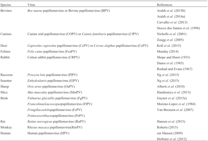
for epithelial and mucous tissue (Cotchin, 1962). These vi-ruses have genomes between 6,953 bp (CmPV1 -Chelonia mydaspapillomavirus type 1) to 8,607 bp (CRPV1 -Cotton rabbit papillomavirus type 1), divided in three regions: early (E), late (L) and long codon region (LCR), as showed in Figure 1 (van Doorslaer, 2013). The E region codifies replication proteins (E1, E2, E4), and the oncoproteins E5, E6 and E7 (Bocanetiet al., 2014). The L region codifies capsid proteins (L1 and L2) (van Doorslaer, 2013). The LCR does not codify any protein, but has the origin of repli-cation (ori) (van Doorslaer, 2013). Currently, more than 280 different types of PVs are described. More than 200 types infect humans (Munday, 2014), which are classified in 49 genera according to the International Committee on Taxonomy of Viruses (ICTV) (Araldiet al., 2015b). Phylo-genetic classification of PVs are based on the L1 open read-ing frame (ORF) sequence homology, since this is the most conserved ORF among the different PV types (de Villierset al., 2004, de Villiers, 2013; Meloet al., 2014; Mundayet al., 2015). According to this system, differences over 10% on L1 ORF sequence determine a novel virus type, while differences between 2-10%, a novel virus subtype (de Vil-liers, 2013).
Table 1- Papillomaviruses identified in different vertebrates
Species Virus References
Bovines Bos tauruspapillomavirus or Bovine papillomavirus (BPV) Araldiet al.(2015b) Araldiet al.(2014a) Carvalhoet al.(2013) Stocco dos Santoset al.(1998) Canines Canine oral papillomavirus (COPV) orCannis familiarispapillomavirus (CfPV) Nichollset al.(2001)
Zaugget al.(2005) Deer Capreolus capreoluspapillomavirus (CcPV) orCervus elaphuspapillomavirus (CePV) Králet al.(2015) Felines Felis catuspapillomavirus (FcaPV) Munday (2014) Rabbit Cotton rabbit papillomavirus (CRPV) Shope and Hurst (1933)
Danoset al.(1985) Rashad and Evans (1967) Raccoon Procyon lotopapillomavirus (PlPV) Nget al.(2015) Seaotter Enhydralutrispapillomavirus (ElPV) Nget al.(2015) Sheep Ovis ariespapillomavirus (OaPV) Albertiet al.(2010)
Mice Mus musculuspapillomavirus (MmPV) Handisuryaet al.(2013)
Birds Fulmarus glaciallispapillomavirus (FgPV) Gaynoret al.(2015a)
Francolinusleucoscepuspapillomavirus (FlPV) Moreno-Lopezet al.(1984)
Fringillacoelebspapillomavirus (FcPV) Van Bressemet al.(2007)
Psittacuserithacuspapillomavirus (PePV)
Rat Rattus norvegicuspapillomavirus (RnPV) Hansenet al.(2015)
Monkey Rhesus macacapapillomavirus(RmPV) Roberts (2015) Human Human papillomavirus (HPV) zur Hausen (2009)
Evolutionary history of papillomaviruses
Although the virus origin is still uncertain (Bernard, 1994; Holland and Domingo, 1998), studies about PVs sug-gest that they arose concomitantly with tetrapods in the Carboniferous period of the Paleozoic era (330 million years ago) (Rector and Van Ranst, 2013). This makes the PVs one of the oldest and largest known virus family (Cubie, 2013; Rector and Van Ranst, 2013).
Studies based on molecular phylogeny suggest that these viruses originated in Africa, from where they dissem-inated to all continents (Bernard, 1994). It was not a pan-demic dissemination, occurring over one million years (Bernard, 1994).
PVs genomic diversification occurred together with mammals diversification, being influenced by multiple evolutionary forces (Bravoet al., 2010), such as the addi-tion of sequence boxes, previously present in their host (García-Vallvéet al., 2005). Thus, PVs co-evolved with their respective host (Gottschlinget al., 2007). An evidence of this co-evolution is the similarity in guanine and cytosine (G + C) content; HPVs have 41-49% of G + C, and humans
40-42% (Black, 1968). Another evidence of this co-evolution is seen with the use ofin situhybridization probe for Shope papilloma virus (CRPV), which presents homo-logy with the rabbit genome sequences (Black, 1968). Moreover, replicative mechanisms of PVs and their host are similar, reinforcing co-evolution (Leatherwood, 1998). These data suggest that PVs could have originated from genomic fragments of amniotes’ common ancestor (van Doorlaer and Burk, 2010; Rector and Van Ranst, 2013; van Doorslaer, 2013).
Animal domestication favored the enzootic transmis-sion of PVs (Gottschlinget al., 2007). In this process, novel strategies of adaptation were required to guarantee the in-fection of novel hosts (Gottschlinget al., 2011). These ad-aptations favored PVs evolution, conferring specificity in terms of host to all members ofPapillomaviridaefamily, except for BPV, which is able to promote cross-infection (Bernard, 1994).
Based on this, García-Vallvéet al.(2005) described a hypothetical model to recreate PVs evolution. This model supports the existence of a proto-papillomavirus,
prised by URR-E1-E2-L2-L1 genomic regions, able to in-fect primitive amniotes. Along mammals divergence (150 million years ago), this proto-papillomavirus was added with E6 and E7 ORFs. From this moment, PVs’ interaction with their host became more specific, resulting in a co-evolution. This process also resulted in the addition of E5 ORF in the hot spot between E2 and L2 ORFs (Bravo and Felez-Sanchez, 2015). Phylogenetic analyses point out that PVs only acquired oncogenic potential after they infected humans (García-Vallvé et al., 2005). This suggests that BPV could have originated from HPV transmission to bo-vines, as a consequence of animal domestication (García-Vallvéet al., 2005)., which would justify the similarities between these viruses, making the BPV a useful model to HPV studies (Campo, 2006; Borzacchiello and Roperto, 2008; Munday, 2014).
Non-oncogenic early (E) proteins expressed by
PVs: E1, E2 and E4
E1 and E2 ORFs are expressed after PV infection, once their genetic products are essential to virus replication (Ferraroet al., 2011). The E1 ORF is the second most con-served sequence among the PVs (Forslund et al., 1999; Enemarket al., 2000). and codifies the E1 protein, which has three functional domains: (1) N-terminal, that induces Cdk2 phosphorylation, (2) C-terminal, that acts as ATP-dependent helicase and (3) central, that binds to E2 protein, resulting in E1-E2 complex (García-Vallvé et al., 2005; Wallace and Galloway, 2014). The E1-E2 complex binds to viral replication origin (ori) present in LCR (Enemark et al., 2000; Schuck and Stenlund, 2015). Next, the E1 protein forms the di-hexameric complex in ori (Gauson et al., 2016), and recruits replication proteins such as topoiso-merase I, DNA polytopoiso-merase a and replication protein A (RPA), that need to replicate (Schuck and Stenlund, 2015). Due to its helicase activity, the E1 protein can induce sim-ple strand breaks (SSBs) and double strand breaks (DSBs) in host DNA (Schuck and Stenlund, 2015).
E2 is a modular protein, composed by two domains: (1) C-terminal and (2) transactivation N-terminal (Wallace and Galloway, 2014). The E2 C-terminal binds to Brd4 pro-tein (Gausonet al., 2016). The E2-Brd4 complex interacts with lysine residues in acetylated histones (Schweiger et al., 2006, 2007), resulting in an equitable distribution of vi-rus copies after cytokinesis (Campo, 2006; Wallace and Galloway, 2014).
The E2 protein also acts as an E6 and E7 transcrip-tional regulator (García-Vallvéet al., 2005; Bogaertet al., 2008; Cai et al., 2013). In high levels, E2 binds to 5’-ACCG(N)4CGGT-3’ palindromic sequence present in
four E2 binding sites (E2BSs) in E6 and E7 promoter (P97) (Caiet al., 2013). This inhibits RNA polymerase II binding, repressing E6 and E7 transcription (García-Vallvéet al., 2005). However, under low expression levels, E2 recruits
transcription factors necessary to form the open transcription complex (Helfer et al., 2014; Jang et al., 2014). In addition, E2 is an important epigenetic regulator, since the protein interacts with the p300/CBP-p/CAF global transcription activator (Wallace and Galloway, 2014). This interaction leads toTP53hypo-acetylation, re-ducing p53 expression levels (Wallace and Galloway, 2014).
The E4 ORF codifies a family of proteins produced by splicing followed by post-translational modifications (Campo, 1997a). The E4 protein is the most expressed pro-tein of PVs (Doorbar, 2013). For this reason, E4 is easily detected in suprabasal and granulosum layers of epidermis (Rampiaset al., 2013), being recognized as important hall-mark of PVs pathogenic activity (Doorbar, 2013). The E4 protein interacts with cytokeratin filamentous, contributing to viral replication (Campo, 1997a). Moreover, E4 is asso-ciated to virus maturation and extracellular matrix (MEC) remodeling (Ferraroet al., 2011).
Late proteins: L1 and L2
The L1 ORF is the most conserved among PVs (Ber-nardet al., 2006; Hagaet al., 2013), and for this reason it is employed in virus classification (Haga et al., 2013; de Villiers, 2013). The L1 protein has 55 kDa (Bucket al., 2,013). and is able to self-organize in pentameric structures that compose the viral capsid (Ribeiro-Müller and Müller, 2014). It has a central role in viral infection mechanism, al-lowing the capsid anchorage to heparin sulfate receptors present in cell membrane (Florinet al., 2012). Considering that L1 is a late protein, it is expressed in the most differen-tiated epithelium layers (Bucket al., 2004). Therefore, L1 immunodetection has been considered the main evidence of productive infection (Nasir and Reid, 1999; Costa and Medeiros, 2014; Meloet al., 2015; Araldi et al., 2015b), which is characterized by the virus assembly (Green, 1972).
The L2 protein has 64-78 kDa (Wang and Roden, 2013). The molecular weight variation is a consequence of post-translational modifications (Wang and Roden, 2013). During the assembly of PV particles, 2 binds to viral DNA, contributing to encapsidation and then to viral release (Gar-cía-Vallvéet al., 2005; Campo, 2006).
A third structural protein (L3) has been described as present exclusively in BPV-4 (Catroxoet al., 2013). How-ever, its function remains unclear.
Oncogenic proteins expressed by PVs: E5, E6
and E7
E5 oncoprotein
vitrotransformation (Campo, 2006; Silvestreet al., 2009; Rampiaset al., 2013; DiMaio, 2014). Furthermore, it is re-sponsible for the fibrotropism verified in fibropapillomas and equine sarcoids (Chambers et al., 2003a; DiMaio, 2014).
PV-1 E5 is a transmembrane protein (Costa and Me-deiros, 2014), with 43-44 amino acids, characterized by the presence of a central hydrophobic region, which acts as a transmembrane domain (Burkhardtet al., 1989; Tomitaet al., 2007; DiMaio, 2014). E5 also presents two cysteine res-idues in the C-terminal region that confers stability for the homodimer composed by two E5 monomers (DiMaio, 2014).
The E5 oncoprotein induces cell membrane composi-tion and dynamic changes, affecting the Golgi complex (Burkhardtet al., 1989; García-Vallvéet al., 2005; Balcos
et al., 2008). This occurs because E5 is able to bind to the H+-ATPase vacuolar subunit (Burnettet al., 1992), promot-ing Golgi inner membrane alkalinization (Krawczyket al., 2010). This leads to major histocompatibility complex II (MHC-II) heavy chain sequestration, conferring an evolu-tionary mechanism of immune evasion (Venuti et al., 2011). In addition, the E5 oncoprotein inhibits the expres-sion of MHC-I and cyclooxygenase (COX) (Borzacchiello, 2007; Tomita et al., 2007; Venuti et al., 2011). These mechanisms contribute to viral infection persistence.
The E5 oncoprotein can bind to ductin, a conexon component, inhibiting the gap junction formation and therefore cell communication (Campo, 1997a; Borzac-chiello and Roperto, 2008). E5 also contributes to focal ad-hesion loss, affecting cell differentiation (Rampiaset al., 2013).
The E5 oncoprotein is able to bind to platelet-derived growth factor receptor beta (PDGFbR) (Chamberset al., 2003a; DiMaio, 2014; Costa and Medeiros, 2014). PDGFbR is a tyrosine-kinase receptor present in the cell surface (Nicolas et al., 2013). Under normal conditions, PDGFbR is activated by PDGF, which promotes receptor dimerization (DiMaio, 2014) and activation of different kinases, such as: A-cdk2, MAPK, JNK, PI3K and C-Src (Chamberset al., 2003a; Borzacchiello, 2007). However, E5 binding to PDGFbR can lead to phosphatidylinositol-3-kinase (PI3K)-mediated Akt pathway activation (Costa and Medeiros, 2014). This activates the D3 cyclin, promot-ing cell cycle deregulation (Venutiet al., 2011; Costa and Medeiros, 2014). The PI3K/Akt pathway activation also in-duces metabolic deregulation in the host cell (Vivanco and Sawyers, 2002; Hennessyet al., 2005). PDGFbR activation also recruits pericytes, stimulating angiogenesis (Pietras and Ostman, 2010).
E6 oncoprotein
E6 is a small oncoprotein with 151-158 (HPV) (Ris-trianiet al., 2000; Bouletet al., 2007) or 137 amino acids (BPV-1) (Tonget al., 1998; Mazzuchelli-de-Souza et al.,
2013), without enzymatic activity (Tanet al., 2012a). The E6 oncoprotein is characterized by the presence of a class I PDZ domain (PSD-95/Dlg/ZO-1), which is located in the C-terminal, and four PVs conserved motifs (Cys-X-X-Cys) (Chenet al., 1997; Tonget al., 1998; Nominéet al., 2006; Boon et al., 2015). The PDZ domain is found in BPV (Mischo et al., 2013) and high-risk HPVs (Boon et al., 2015) and can interact with different proteins, promoting loss of intercellular contact and cell polarity (Boonet al., 2015).
The HPV E6 oncoprotein can form a trimeric com-plex with E6AP ubiquitin ligase and p53 (Zanieret al., 2005, 2007; Tanet al., 2012a). This complex is responsible for the proteolytic degradation of p53 (Zanieret al., 2005; Liu and Baleja, 2008), which directs the p53 to 26S protea-some (Caiet al., 2013). The HPV-5 and 8 as well as BPV E6 oncoprotein interact with the CBP/p300 complex (Wer-ness et al., 1990; Zimmermann et al., 2000; Liu et al., 2005), promoting p53 downregulation (Zimmermannet al., 1999). The loss of p53 contributes to genomic instability verified in cells infected by PVs (Araldi et al., 2013, 2015b), as well as in cells treated with BPV-1 E6 recombi-nant oncoprotein (Araldiet al., 2015a).
The overexpression of p53 represents a threat to viral replication (Shamannaet al., 2013). For this reason,
differ-ent oncogenic viruses express proteins able to promote p53 deregulation. Among these proteins are antigen T of SV40, adenovirus E1B, hepatitis B virus (HBV) HBx and PVs E6 (Shamannaet al., 2013; Cuninghameet al., 2014). Thus,
p53 deregulation is a characteristic shared by oncogenic vi-ruses.
The E6 protein can interact with paxillin (Turner, 2000; Zanieret al., 2005) and AP-1 gamma subunit clathrin adapter (Tonget al., 1998). These interactions can lead to cytoskeleton alterations, affecting the vesicular traffic. E6 can also interact with fibulin 1, contributing to invasiveness phenotype (Moody and Laimins, 2010).
Studies have also demonstrated that E6 oncoprotein can induce cell transformation (Liuet al., 2002) and im-mortalization (Boonet al., 2015) due to the up-regulation of telomerases (Cuninghameet al., 2014). This occurs be-cause E6 promotes FOXM1 overexpression, resulting in cyclin B1, D1 and cdc25 expression and, therefore, cell proliferation (Chen and Lee, 2015). In addition, E6 is able to bind to LXXL motif ofMAML1transcriptional regulator, inhibiting theNotchsignaling (Tanet al., 2012a; White and Howley, 2013). The E6 oncoprotein of both HPV and BPV also promotes energy metabolic deregulation, contributing to cell transformation (Araldiet al., 2016).
cyto-genetic damages (Wallace and Galloway, 2014) and stimulates neosis (Araldiet al., 2015a).
E7 oncoprotein
The E7 oncoprotein has 127 amino acids (Borzac-chiello, 2007), and is able to bind to LXCXE conserved motif of pRb tumor suppressor protein (Moody and Lai-mins, 2010; White and Howley, 2013). This binding results in pRb phosphorylation, leading to E2F factor translocation to the nucleus (Longo, 2013). The E2F factor recruits dif-ferent chromatin modifiers, including histone deacetylases (HDAC) (Moody and Laimins, 2010). Thus, the E7 onco-protein induces the constitutive expression of E2F-respon-sive genes, such as cyclin A and E (Moody and Laimins, 2010), leading to S and G2/M cell cycle phase increase (Saccoet al., 2003; Ferraroet al., 2011). The interaction
be-tween viral oncoproteins and pRb is not exclusive of PVs, being also observed with adenovirus E1A and antigen T of SV40 (Whiteet al., 2015).
Studies have demonstrated that E7 induces DNA breaks, contributing to cell cycle deregulation (Parket al., 2014; Araldi 2015; Araldiet al., 2015b). In addition, the E7 oncoprotein increases theSirt1deacetylase expression lev-els, modulatingg-H2AX activity (Parket al., 2014), a
pro-tein that participates in DSB repair (Sedelnikova and Pilch, 2003; Chowdhuryet al., 2005). Thus, the E7 oncoprotein inhibits DSB repair, resulting in genomic instability.
Bovine papillomavirus (BPV): first reports and
questions
In 1986 in an intriguing paper by Campo and Jarret (1986) reported the description of six different BPV types (BPV-1 to BPV-6) classified into two subgroups: subgroup A, that promotes fibropapillomas, and subgroup B, which leads to true epithelial papillomas. The report also showed that the BPV-4, a member of subgroup B, was the etiologi-cal agent of papillomas of the upper digestive tract, which could become carcinomas in animals feeding on a specific bracken fern pasture (Pteridium aquilinum). Later,
Wal-ter-Mouraet al. (1988) verified the increase of
chromo-some aberrations in cells obtained from short term peripheral lymphocytes collected from bovines afflicted with bovine enzootic hematuria (BHE) that were exposed
to pastures with bracken fern. These studies were reinforced by following reports, demonstrating that carcinogenesis is associated to the interaction between BPV and carcinogens present in the fern, such as quercetin and ptaquilosides (Pennie and Campo, 1992; Shahinet al., 1999; Potter and Baird, 2000; Benistonet al., 2001; Lealet al., 2003; Lioi et al., 2004). These studies brought new questions to be answered: how can the co-factor act in syn-ergism with the virus? Why can these effects be detected in peripheral blood, considering that the virus is epithelial? Or, could the effect on chromosomes be only related to bracken fern compounds?
BPV and bovine papillomatosis (BP)
BPV is a cosmopolitan virus, worldwide distributed, independently of the level of expertise on livestock explo-ration (Heet al., 2014). It is estimated that 60% of Brazilian cattle herd is infected by BPV (Stocco dos Santos,et al., 1998). However, that rate can be higher, once virus infec-tion can be asymptomatic (Araldiet al., 2013; Silvaet al., 2013a). Furthermore, the absence of epidemiological stud-ies about BPV distribution could underestimate the real percentage of infected animals, representing a notorious difficulty in attempting to develop vaccines, once we do not know the prevalent virus types in each country.
BPV infection is endemic in both dairy and beef cattle breeding (Clauset al., 2009; Araldi, 2015,; Araldiet al.,
2015b). However, BPV presents a predilection for dairy cattle (Araldi,2015). The virus is the causal agent of bovine
papillomatosis (BP) (Muroet al., 2008), an infectious,
con-tagious and neoplastic disease, characterized by the pres-ence of multiple benign tumors (papillomas) (Figure 2) that can regress spontaneously or progress to malignant neo-plasms (Campo, 2002; Turket al., 2005; Yaguiu et al.,
2006; Borzacchiello and Roperto, 2008; Araldi et al.,
2014b; Bocanetiet al., 2014). Although BP affects
prefera-bly young cattle, the disease can occur at all ages (Börküet al., 2007). The persistence of papillomas can lead to
feed-ing and breathfeed-ing difficulties, requirfeed-ing the animal’s eutha-nasia (Campo, 2002). Moreover, BP decreases growth rate and induces weight loss (Munday, 2014). The disease also predisposes bovines to opportunist bacterial infections (Munday, 2014), mainly in breast and mammary glands,
which can result in mastitis, causing pain, reducing milk production (Campo, 2002; Muroet al., 2008; Lindseyet al., 2009; Santos et al., 2014) and causing depreciation of leather value (Monteiroet al., 2008).
Cutaneous papillomas are proliferative benign tu-mors, with complex pathogenesis (Börkü et al., 2007). These neoplasms commonly arise in vascularized areas ex-posed to physical attrition (Özsoyet al., 2011). Histolo-gically, papillomas are characterized by epithelium hyper-plasia, showing an enlargement of interpapillary ridge that extend above dermis (Monteiro et al., 2008). Morpho-logically, they are classified in: (1) typical - exophytic masses, with “cauliflower” aspect, presenting a wide or narrow insertion base, (2) pedunculated - exophytic masses connected by a narrow base with a peduncle shape, (3) atypical or flat - dense and flat exophytic masses entirely connected with the tissue, (4) filamentous - exophytic mas-ses with highly keratinized surface and a thin base, present in mammary glands and (5) rice-form - small papillomas with rice-like shape (Monteiroet al., 2008).
Currently, 15 types of BPV are described (Mundayet al., 2015), and classified in four genera:
Deltapapillomavirus (BPV-1, 2, 13 and 14),
Epsilonpapillomavirus (BPV-5 and 8), Xipapillomavirus
(BPV-3, 4, 6, 9, 10, 11, 12 and 15) and
Dyoxipapillomavirus (BPV-7) (Melo et al., 2014; Grindattoet al., 2015; Mundayet al., 2015; Silvaet al., 2016). Delta and Epsilonpapillomavirus are associated with both papillomas and fibropapillomas, while
Xipapillomavirus, only to squamous papillomas (Tomitaet al., 2007; Yuanet al., 2007; Tanet al., 2012b; Araldi, 2015; Araldiet al., 2015b).
Although there is no epidemiological study that al-lows the definition of BPV distribution, BPV-1 and 2 seem to be the most frequently identified virus types (Pathaniaet al., 2012; Meloet al., 2014; Santoset al., 2014; Alcântara
et al., 2015; Araldi, 2015; Cotaet al., 2015; Araldiet al., 2015b). Moreover, these viruses are associated with both benign and malignant neoplasms (Gopalkrishna et al., 1995; Nasir and Campo, 2008; Cotaet al., 2015). However, the global distribution of BPV is not homogenous (Santos
et al., 2014). BPV-1 and 2 have closely related serotypes (Shafti-Keramatet al., 2009), associated with urinary blad-der malignant neoplasms (Wosiackiet al., 2005; Balcoset al., 2008; Ropertoet al., 2008; Maiolinoet al., 2013; Cota
et al., 2015). BPV-4 infection is an important cause of up-per digestive tract cancer development (Tsirimonakiet al., 2003; Nasir and Campo, 2008; Lucenaet al., 2011). Studies also show that BPV-13 is associated to urothelial carcino-mas (Ropertoet al., 2015).
Diagnostic methods
Studies about BPV diversity and prevalence are man-datory to develop novel therapeutic methods (Silvaet al.,
2013b), since immunity is species-specific (Claus et al., 2007). Therefore, diagnosis is crucial.
Different methods have been implemented to identify PVs, such as: Southern blot (Leto et al., 2011), immu-nohistochemistry (IHC) (Elzeinet al., 1991; Araldiet al., 2015b), chromogenicin situhybridization (CISH) (Meloet al..2015), electron microscopy (Araldiet al., 2014b) and PCR using specific and/or degenerate primers (Stocco dos Santoset al., 1998; Araldiet al., 2013, 2014a, 2015b; Melo
et al., 2014). Among these techniques, PCR is the most used due to its high sensitivity (Letoet al., 2011). More-over, using the restriction fragment length polymorphism of PCR products (PCR-RFLP) allows to identify BPV type (Carvalhoet al., 2013), since this method shows a correla-tion of 95% with the results obtained using DNA sequenc-ing (Martenset al., 2001a; Carvalhoet al., 2013; Kawauchi
et al., 2015).
Although real-time PCR (qRT-PCR) allows to deter-mine the number of viral copies, this method has the lowest reproducibility (Guoet al., 2012). For this reason, PCR fol-lowed by DNA sequencing represents the most common method to identify and typify BPVs (Meloet al., 2014; Araldiet al., 2014b). However, there is no “gold-standard” primer employed in PVs identification (Antonssonet al., 2010).
On the one hand, although the specific primers have higher sensitivity than degenerate primers, they cannot identify the 14 BPV types simultaneously (Araldi et al., 2014b). In addition, specific primers do not allow to iden-tify novel virus types and subtypes (Araldiet al., 2014b). Moreover, evidence indicates that a specific primer for BPV-1 can anneal to BPV-2 (Hagaet al., 2013), once these virus types are considered serotypes-like (Shafti-Keramat
et al., 2009). However, in a comparative study using com-plete genomes of BPV-1-6 in 2014 we demonstrated the BPV-1 primer specificity (Araldiet al., 2014b).
Among the degenerate primers described in the litera-ture, FAP59/64 (Forslund et al., 1999) is the most em-ployed in both BPV and HPV identification. This primer was designed based on the L1 ORF homology of HPV (Forslundet al., 1999) and later optimized to identify BPV DNA sequences (Ogawaet al., 2004). Furthermore, the use of FAP59/64 primer already allowed to identity novel BPV types, including BPV-9 and 10 (Hatamaet al., 2008). How-ever, despite of these advantages of degenerate primers, there are several reports showing their low sensitivity. For instance, MY09/11, another degenerate primer frequently used in HPV diagnosis, was unable to identify BPV and HPV sequences in clinical samples (Martelli-Marzagãoet al., 2010; Zhuet al., 2014) or copies of complete cloned ge-nome (Araldiet al., 2014b).
demonstrate the physical state of these viruses (Black, 1968; Munday, 2014; Meloet al., 2015). Another addi-tional technique to identify PVs is L1 immunodetection (Nasir and Reid, 1999; Longworth and Laimins. 2004; Roperto et al., 2011; Munday, 2014; Melo et al., 2015; Araldiet al., 2015b)., which not only allows identification of the viral presence, but also provides an important evi-dence of productive infection (Nasir and Reid, 1999; Ro-pertoet al., 2011; Meloet al., 2015).
Histopathological analysis of BPV-infected lesions is a differential diagnosis method, which allows to identify intra-epithelial neoplasms with oncogenic potential (Mon-teiroet al., 2008; Araldiet al., 2015b).
BPV infection pathway and histopathological
alterations
BPV transmission can occur by direct (animal-ani-mal) or indirect contact with contaminated surfaces (Muro
et al., 2008; Cubie, 2013). Studies also show that the virus can be transmitted by flies (Finlayet al., 2009) and ticks (Muroet al., 2008).
Studies of PV infection are based on the prototype BPV-1 (Florinet al., 2012). According to the literature, the viral infection requires a tissue micro-injury, exposing hep-arin sulfate proteoglycan receptors (Ljubojevic and Sker-lev, 2014; Greber, 2016), which are needed for BPV L1 anchorage (Hartlet al., 2011; Bucket al., 2013)., This is confirmed by treatment with heparinase preventing viral infection (Kineset al., 2016). The micro-injury is necessary for the virus to access basal keratinocytes (Liu and Baleja, 2008), where the virus cycle begins (Campo, 1997a). Cur-rent studies also show that integrina6 (CD49f) and integrin 332 (laminin 5) are targets for L1 binding (Sibbetet al., 2000; Florinet al., 2012).
The L1 binding to heparin sulfate leads to confor-mational changes in capsid icosaedric structure (Buck et al., 2013). This exposes the L2 N-terminal to be cleaved by furin protein, present in the cell membrane (Bucket al., 2013). This cleavage induces a second capsid conforma-tional change, allowing L2 to bind to different receptors, such as integrina2b4 (Bucket al., 2013). Next, the virions are internalized by an clathrin-dependent endocytose mechanism, resulting in cytoplasmic vesicles that associate to lysosomes (Dayet al., 2003). The lysosomal acid content release promotes pH alterations in capsid proteins, result-ing in viral DNA release (Dayet al., 2003). The BPV ge-nome is found in epissomal form (Campo, 2002; Munday, 2014; Cotaet al., 2015), while HPV can integrate in fragile sites of the host genome (Monte and Peixoto, 2010; Moody and Laimins, 2010; Munday, 2014). A current study based on qRT-PCR, showed that cutaneous papillomas have about 2.2104viral copies (Cota
et al., 2015).
PVs do not codify polymerase (Moody and Laimins, 2010). For this reason, these viruses induce the S-phase
en-try, which was verified in BPV-infected cells (Potockiet al., 2014), in a process known as amplification (Moody and Laimins, 2010). Due to the stimulation of cell proliferation, BPV induces mitotic stress (Potockiet al., 2014), resulting in cytogenetic aberrations (Stocco dos Santoset al., 1998; Meloet al., 2011; Araldiet al., 2013; Camposet al., 2013). Moreover, the viral hyperproliferative action leads to the exophytic mass development as a consequence of acan-thosis (Cubie, 2013; Munday, 2014; Araldiet al., 2015b). As occurs with epithelium differentiation and virus assem-bly, a keratinization process is verified (Ferraro et al., 2011). This process is histologically characterized by the increase of keratin granules in the granular layer (Ferraroet al., 2011; Araldiet al., 2015b).
Virus assembly is observed in the most differentiated epithelium layers (Munday, 2014), where the virion release occurs by cell degeneration (Brobst and Hinsman, 1966; Bucket al., 2013). This process results in koilocyte forma-tion. The term koilocyte comes from the Greek word
koillos, which means “cavity” (Ferraro et al., 2011). Koilocyte formation results from the cytopathic effect of E5 and E6 oncoproteins, although the molecular mecha-nism that results in cell vacuolization remains unclear (Krawczyket al., 2008). However, the cytoplasmic vacuol-ization contributes to cell fragility and virion release (Krawczyket al., 2008; Wang and Kieff, 2013). In this sense, koilocytes are cells destined to apoptosis, which emerges as a consequence of DNA replication and macro-molecule synthesis inhibition.
After viral assembly, virions are released in the cor-neum layer, allowing infiltration in the keratin matrix (Brobst and Hinsman, 1966; Bucket al., 2013). This mech-anism confers an immune evasion, since the icosaedric morphology of PVs is immunoreactive (Bucket al., 2013). In addition, keratin confers physical protection for virions, as they are non-enveloped.
BPV as a model for HPV
Equine sarcoid: an example of cross-infection
Although considered epithelial- and mucous-tropic, some BPV types, especiallyDeltapapillomavirus, can in-fect fibroblasts causing sarcoma-like lesions, known as equine sarcoid (Chamberset al., 2003b; Geisshüsleret al., 2016). Equine sarcoid was first described by Jackson (1936), being considered a biphasic neoplasia, since it af-fects both epidermis and dermis. Equine sarcoid is the most frequent benign skin neoplasia observed in horses that are 1-6 years old (Jackson, 1936; Ottenet al., 1993; Nasir and Reid, 1999; Martenset al., 2000; Bergvall, 2013; Mosseriet al., 2014), affecting 11.5% of all horses (Knottenbelt, 2005). Differently from papillomas, sarcoids rarely present a spontaneous regression (Angeloset al., 1991).
Equine sarcoid is an invasive but not metastatic neo-plasia (Nasir and Reid, 1999), causing substantial morbid-ity and economic loss due to aesthetic and functional impairment (Angeloset al., 1991; Ottenet al., 1993). Six different sarcoid morphotypes are known: occult, verru-cous, nodular, fibroblastic, mixed and malignant (Knot-tenbelt, 2005). However, 84% of affected equines have more than one sarcoid morphotype (Goodrichet al., 1998).
Equine sarcoids are lesions with intense fibroblastic proliferation, in which fibroblasts are disposed in fusiform bundles or spirally organized, presenting a morphology of fibropapilloma-like (Martenset al., 2000). Another charac-teristic of this neoplasia is the presence of anaplastic and pleomorphic fibroblasts, perpendiculary orientated in rela-tion to basal membrane, and being observed in the der-mo-epidermal junction (Bogaertet al., 2010). The epider-mal component is only present in verrucous and mixed sarcoids (Bogaertet al., 2010). The sarcoid invasiveness capability can be attributed to the high levels of expression of metalloproteinase. MMP1 promotes the laminin and col-lagen IV degradation, resulting in extracellular matrix re-modeling (Mosseriet al., 2014)
Equine sarcoid has a multifactorial cause (Bergvall, 2013). However, theDeltapapillomavirusis recognized as an etiological factor (Nasir and Reid, 1999; Chamberset al., 2003b; Bogaertet al., 2008b; Bergvall, 2013). The as-sociation between BPV and equine sarcoid was first de-scribed by Olson and Cook in 1951 (Brandtet al., 2008). BPV-1 and 2 DNA sequences are identified in 100% of equine sarcoids (Martens et al., 2001a; Gaynor et al., 2015b). Furthermore, DNA sequences of these virus types are identified in 2/3 of asymptomatic horses (Bravoet al., 2010). These data demonstrate that the virus can be asymp-tomatic, remaining latent in epidermis and dermis (Bogaert
et al., 2008b; Brandtet al., 2008; Bergvall, 2013).
Viral latency is a characteristic shared by BPV (Bo-gaertet al., 2008b; Araldiet al., 2013; Silvaet al., 2013a) and HPV (Maranet al., 1995; Astoriet al., 1998; Forslund
et al., 2004). However, the virus infection induces DNA damages and genomic instability (Stocco dos Santos,et al., 1998; Meloet al., 2011; Araldiet al., 2013; Camposet al.,
2013; Meloet al., 2015). Moreover, studies show that 70% of BPV-infected asymptomatic horses live in contact with cattle (Bergvall, 2013). However, although the presence of BPV-1 L1 transcripts was already verified in equine sar-coids by RT-PCR (Nasir and Reid, 1999), these neoplasias are considered abortive infection sites, since virion pres-ence was not described. (Bogaertet al., 2008b; Brandtet al., 2008). The absence of productive infection in equine sarcoids suggests that the BPV cross-infection is a conse-quence of an erratic virus cycle (Ottenet al., 1993).
As verified in PB, sarcoids are frequently seen in sites most susceptible to traumatism (Angeloset al., 1991; Otten
et al., 1993; Martenset al., 2000), confirming the need of tissue micro-injury for virus infection. In addition, as in bo-vines, it is believed that insects can contribute to BPV transmission, because BPV-1 DNA sequences were al-ready identified inMusca automnalis, Fannia carnicularis
andStomoxys calcinatransflies (Bergvall, 2013).
Studies also show that the Arabic breed is the most susceptible to sarcoid development (Knottenbelt, 2005; Bogaertet al., 2008b). The reason for this is the presence of W3 and B1 MHC-II haplotypes that facilitate BPV infec-tion persistence (Chambers et al., 2003b; Bogaert et al., 2008b).
Currently, treatments for sarcoid, as well as for PB, are almost inefficient (Bergvall, 2013). Treatment methods are: (1) surgical excision of the neoplasia, with recurrence in 50-64% of the cases within six months (Lancasteret al., 1977; Martenset al., 2001b; Bergvall, 2013; Mosseriet al., 2014), (2) laser therapy, where recurrence is 38% (Martens
et al., 2001b); cryotherapy, which is inefficient for lesions larger than 2 cm2(Carr, 2009), and chemotherapy using
cisplatin or 5’-fluouracyl (5-FU), that can cause nephro and hepatotoxicity (Stewartet al., 2006). The recurrence of dis-ease is argued to be a consequence of BPV presence in the surgical margin (Martenset al., 2001a). However, a current study showed a lack of correlation between BPV DNA in surgical margins and recurrence of equine sarcoids (Taylor
et al., 2014). Meanwhile, Brandtet al. (2008) identified BPV DNA sequences in peripheral blood of sarcoid-affected equines. These data suggest that, as verified in bo-vines (Stocco dos Santoset al., 1998; Ropertoet al., 2011; Araldiet al., 2013; Meloet al., 2015), the peripheral blood can be argued as being a vehicle of viral dissemination.
Breaking paradigms
the first evidence of vertical transmission (Freitas et al., 2003). In this study, we verified the presence of BPV DNA sequences and DNA damages in peripheral blood samples of animals from different areas of Brazil (Dinizet al., 2009; Meloet al., 2011; Araldiet al., 2013). In addition, we also demonstrated the presence of different BPV types in pe-ripheral blood and cutaneous papilloma (Araldi et al., 2014a). All these data suggest a viral activity in blood cells (Stocco dos Santoset al., 1998; Araldiet al., 2013). The presence of BPV DNA sequences in peripheral blood mononuclear cells (PBMCs) was also described in the liter-ature in both bovines (Roperto et al., 2008; Silva et al., 2013a) and equines (Brandtet al., 2008, 2011), reinforcing our results. Following studies also showed the presence of BPV transcripts and the L1 capsid protein in PBMCs, dem-onstrating the productive infection in blood cells (Roperto
et al., 2011; Meloet al., 2015). We also described the pres-ence of BPV DNA sequpres-ences in different non-epithelial tis-sues such as spermatozoa, urine and milk (Lindseyet al.,
2009). These data demonstrate the need to review the natu-ral history of papillomavirus, as currently proposed by Munday (2014).
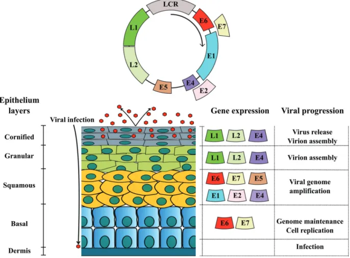
We verified that primary culture cell lines from BPV-infected cutaneous and esophageal papilloma have chromosomal aberrations similar to those verified in pe-ripheral blood (Campos et al., 2013). In addition, using BPV-4 E7 oncoprotein transformed PALF cell lines, we demonstrated the mutagenic potential of quercetin, a flavo-noid found in bracken fernP. aquilinum,which is recog-nized as a co-factor to BPV-associated upper gastric cancer (Lealet al., 2003). In a current study, we also showed that the BPV induced metabolic alteration in host cells, increas-ing reactive oxygen species and DNA damages (Araldi, 2016; Araldiet al., 2016). A summary of our main results is shown in Figure 3. These data suggest that BPV-infected cell lines are a useful model to study the pathogenic mecha-nisms that lead to cancer. Our contribution has allowed to know the BPV prevalence and distribution in Brazil (Diniz
et al., 2009; Carvalho et al., 2013; Lunardiet al., 2013; Meloet al., 2014; Araldiet al., 2014b; Cotaet al., 2015;
Gaynoret al., 2015a; Grindattoet al., 2015; Donget al., 2016; Martanoet al., 2016). These studies showed that the co-infection of at least two BPV types is frequent (Araldiet al., 2013, 2014a; Carvalhoet al., 2013), demonstrating the importance to develop multivalent vaccines.
BPV-associated malignant neoplasms
Urinary bladder carcinoma
Urinary bladder carcinoma represents 0.01% of all bovine cancers (Ropertoet al., 2015). It is estimated that urinary bladder cancer has caused economic losses of 4 million between 2000-2006 in the Azores (Costa and Medeiros, 2014).
The etiopathogenic role of BPV in urinary bladder carcinoma was first described in 1955 in Brazil and South Africa (Plowrigh, 1955). Currently, the BPV promoter ac-tion in urothelial carcinoma is well established. This is be-cause sequences of BPV-1, 2, 13 and 14, as well as the expression of E5 oncoprotein are detected in this neo-plasms (Wosiackiet al., 2006; Balcoset al., 2008; Roperto
et al., 2016; Russoet al., 2016).
Urinary bladder carcinoma is clinically characterized by bovine enzootic hematuria (BHE) (Wosiacki et al., 2002), which is verified in more than 90% of cattle with uri-nary bladder cancer (Resendeset al., 2011). BHE is charac-terized by intermittent hematuria, which can lead to anemia and weight loss (Wosiackiet al., 2002). The disease affects preferably cattle between 3-5 years old, without breed pref-erence (Wosiackiet al., 2002). BHE is associated to dietary intake of carcinogenic compounds present inP. aquilinum, P. esculetum, P. revolution, Chelanthes seiberi, Encephalartos hildebrandti, Ranunaelus montana, R. acris, Climatis vitalbi(Wosiackiet al., 2002). Among these species, P. aquilinumstands out for its wide biogeogra-phical distribution (Oliveira, 2012). Bracken fern is com-monly observed in the South of Brazil, where there is a high incidence of BEH (Diaset al., 2012).
P. aquilinum presents high levels of immunosup-pressor and carcinogenic compounds, including quercetin, ptaquilosides and shikimic acid (Shahin et al., 1999; Benistonet al., 2001; Bonadieset al., 2004). The immu-nosuppressor activity of these metabolites contributes to BPV infection persistence and represents an additional source of DNA mutation (Lealet al., 2003). According to Tokarniaet al.(2000), the diary intake of 10 kg ofPteridum
ssp.in a year can lead to BEH. Moreover, the consumption of this bracken fern can result in urinary bladder and esoph-ageal carcinoma (Masudaet al., 2011).
Therefore, BPV-1 and 2 are considered important co-factors for BEH development (Campo, 1997a; Ropertoet al., 2005; Pavelskiet al., 2014), once BPV leads to epider-mal and derepider-mal hyperproliferation at the same time that it induces DNA damages, contributing to cancer initiation (Stocco dos Santoset al., 1998; Araldiet al., 2013; Campos
et al., 2013).
Esophageal carcinoma (EC)
While the association between HPV and EC remains under discussion despite all evidences (Antonssonet al., 2016), the etiological role of BPV in EC is well established
(Borzacchielloet al., 2003). EC in cattle is a self-limiting disease, being directly associated to BPV-4 infection (Bor-zacchielloet al., 2003; Masudaet al., 2011).
Although there is no report about BPV transmission in humans, a study performed in Germany showed a high incidence of warts in veterinarians that had contact with bo-vines (Bosse and Christophers, 1964). Another study in Central Asia discussed the association between milk con-sumption and EC (Nasrollahzadeh et al., 2015). Con-sidering that BPV DNA sequences were already detected in milk (Lindseyet al., 2009) and the thermal resistance of vi-ral capsid (Módoloet al., data not published), it seems that the virus can survive to pasteurization process (zur Hausen, 2012). Therefore, more efforts are necessary to verify this possible cross-infection.
EC is the eighth most prevalent human malignancy, being considered the sixthcausa mortisfor cancer globally (Antonssonet al., 2010; Herbsteret al., 2012). Considered the third most common gastrointestinal cancer (Felinet al., 2008), EC has a high incidence in men (Bjørgeet al., 1997). In 2002, 462,000 novel cases of EC in the world were re-ported (Antonssonet al., 2010). Brazil had 10,780 novel cases of EC in 2014 and 7,636 deaths due to the disease in 2011 (INCA, 2016).; its incidence has increased in the last years (INCA, 2016). This data leads to concern, since EC has a mortality rate that is 25% higher than cervical cancer (Hanet al., 1996; Kahrilas and Hirano, 2013).
Among the clinical signs of EC in humans are pro-gressive dysphagia, weight loss, odynophagia, anorexia, fe-ver and retrosternal pain (Felin et al., 2008; Haster and
Owyang, 2013), which are similar to those verified in bo-vines (Borzacchielloet al., 2003). Scarce epidemiological
data about EC in cattle are available. In humans, EC has a variable incidence according to geographic area (Bjørgeet al., 1997; Syrjänen, 2002; Guoet al., 2012; Nasrollahzadeh et al., 2015; Antonssonet al., 2016). Among countries with
high EC incidence are China, Singapore, Iran, South Africa and Brazil (Beyet al., 1976; Hanet al., 1996; Syrjänen,
2002; Guoet al., 2012). Due to the high number of EC
cases in Central Asia (100/100,000) in relation to North America and Western Europe (5-10/100,000), the region is known as the Asian Esophageal Cancer Belt (Nasrol-lahzadeh et al., 2015). Although the reason for EC
inci-dence variation is unknown (Syrjänen, 2002; Nasrollah-zadeh et al., 2015), studies indicate the contribution of
environmental factors, in addition to infectious agents, in the oncogenic process (Changet al., 1992; Antonssonet al., 2010).
evi-dence shows the etiopathogenic contribution of different infectious agents in EC, such as cytomegalovirus (CMV), Epstein-Barr virus (EBV), herpes simplex and HPV (Syrjä-nen, 2002).
Although the association between HPV and pre-malignant cervical lesions is known since the 1970s (Syr-jänen, 2002), the participation of this virus in EC had its history marked by controversial reports in the literature (Lavergne and de Villiers, 1999). The association between HPV and EC was first proposed by Syrjänen (1982). On the one hand, HPV protein expression was observed in samples by IHC (Winkleret al., 1985; Hilleet al., 1986; Kulskiet al., 1986) and viral DNA sequences were identified by CISH in EC (Changet al., 1993; Togawaet al., 1994; Coo-peret al., 1995),. On the other hand, serological studies contested these results (Dillner et al., 1995; Han et al., 1996; Lagergrenet al., 1999). However, despite these con-troversial results, the association between HPV and EC is currently accepted, being discussed in the 18thedition of Harrison’s Principles of Internal Medicine (Kahrilas and Hirano ,2013). Thus, the absence of HPV detection by serological methods reflects a problem still observed nowa-days: the use of antibodies against late (L1) proteins (Dil-lner et al., 1995; Lagergren et al., 1999). However, considering that EC does not present an HPV productive in-fection, the late protein expression is not expected. This oc-curs because HPV presents a “hit and run” mechanism (Hanet al., 1996; Bjørgeet al., 1997).
Not only serological studies are controversial. Stu-dies based on PCR also show a low correlation between HPV and EC, mainly in clinical samples from Australia (Antonssonet al., 2010; Antonssonet al., 2016). These re-sults can be attributed to the origin of clinical samples, since Australia is not considered a high-risk area for HPV-associated EC. In addition, problems involving primer sen-sitivity are extensively discussed in the literature of both BPV and HPV (Lavergne and de Villiers, 1999; Silvaet al., 2013b; de Villiers, 2013; Araldiet al., 2014b). However, evidence of PVs etiopathogenic action in EC has accumu-lated along the last 20 years. Among these are: (1) the pres-ence of koilocytes in EC biopsies (Bjørge et al., 1997; Syrjänen, 2002; Vieiraet al., 2013), (2) loss or mutation in p53 as a consequence of the PV E6 oncoprotein (Changet al., 1994, 1995; Herbster et al., 2012), (3) increase in telomerase activity (Syrjänen, 2002), and detection of DNA sequences of HPV-6, 11, 18, 31 and 33 in EC (Vieira
et al., 2013).
Prophylactic and therapeutic vaccines against
BPV and HPV
Currently, there are few forms of treatment against BP available (Muroet al., 2008). Among the possibilities is the papilloma surgical excision (Muro et al., 2008). Al-though frequently employed, this method is inefficient in
cattle with high incidence of BPV, because it is impracticable to perform the excision of papillomas of all cattle. Another strategy frequently employed is self-hemo-therapy (Letoet al., 2011). This method consists in the re-moval and intramuscular reinjection of a volume of 10 mL of venous blood, inducing a nonspecific immune stimulus that can promote the “shedding of the warts” (Letoet al., 2011). However, this technique does not avoid BP re-occurrence, thus being a palliative method. Another possi-bility to reduce the incidence of BP is to control ecto-parasite populations, since it was demonstrated that the biological control of ticks reduces the incidence of BPV (Williamet al., 1992).
Few medical interventions proposed in the last cen-tury can match the effects that immunization exerts on lon-gevity (Schuchat and Jackson, 2013). For this reason, vac-cination is considered the best form of prevention, control and eradication of viral etiology diseases (Ribeiro-Müller and Müller, 2014). Moreover, immunization reduces both transmission and dissemination of the infectious agent (Schuchat and Jackson, 2013).
Two prophylactic vaccines against HPV are available in the market since 2006: (1) Cervarix, produced by Gla-xo-Smith Klein (GSK) and (2) Gardasil, produced by Merck (Ribeiro-Müller and Müller, 2014). These vaccines are based on virus-like particles (VLPs) of the L1 structural protein (Mariglianiet al., 2012). Cervarix is a bivalent vac-cine able to confer protection against HPV-16 and 18, em-ploying L1 VLPs produced in Baculovirus inTrichoplusia niinsect cells, using aluminum hydrophosphate as adjuvant (Ribeiro-Müller and Müller, 2014). Gardasil is able to con-fer protection against two high-risk HPV types (HPV-16 and 18) and two low-risk ones (HPV-6 and 11), associated to genital warts. This vaccine is composed by of L1 VLPs produced inSaccharomyces cerevisae, employing lipid A 3-O-diacilete-44-monophosphoryl (ASO4) as adjuvant. . Both vaccines are considered safe and well tolerated (Ri-beiro-Müller and Müller, 2014). For these reasons, more than 30 countries, including Brazil, adopted immunization programs against HPV based on these vaccines.
Australia, the first country to adopt the vaccination against HPV, observed a reduction of 70% in HPV-6, 11, 16 and 18 infection incidence. Similar results were also ver-ified in Denmark, Finland and Sweden (Ribeiro-Müller and Müller, 2014). However, available vaccines are not able to protect against all HPV types, since there are more than 200 described. Moreover, these vaccines have a high cost of production. Thus, it is necessary to invest in novel multi-valent vaccines, with lower production cost. Vaccines based on recombinant protein expressed inEscherichia coli
have demonstrated to be a useful alternative, because they have a lower cost, and not requiring the L1 VLPs, they are more stable (Ribeiro-Müller and Müller, 2014).
developing a vaccine to combat BPV began with the infec-tion of Shope papillomas extract in the 1940 decade (Sho-pe, 1937). Since then, different vaccine models were proposed (Jarretet al., 1991; Gaukrogeret al., 1996; Góes
et al., 2008; Loveet al., 2012; Mazzuchelli-de-Souzaet al., 2013). However, none of them became a commercial prod-uct. This denotes the notorious difficulty to obtain a safe and efficient vaccine, and reflects the reduced number of research groups dedicated to develop a vaccine against BPV.
Studies have demonstrated that BPV early proteins (E6 and E7) show a therapeutic action, while later proteins (L1 and L2) have a prophylactic action (Campo, 1997b). Vaccines based on E6 and E7 have also been discussed against HPV (Borysiewiczet al., 1996; Heet al., 2000; Yao
et al., 2013), reinforcing the usefulness of the BPV model not only for comprehending the pathogenic mechanisms of HPV, but also for vaccine biotechnology. However, in a current study based on BPV-1 E6 recombinant onco-protein, we demonstrated that this oncoprotein is able to in-duce clastogenesis and neosisper se(Araldiet al., 2015a). This data emphasizes the necessity of anin silicoanalysis of E6 and E7 oncoproteins, aiming to obtain novel variants that are more antigenic and less mutagenic. In order to guar-antee the immunization and safety of products, our group is now focusing on the development of a prophylactic vaccine based on L1 recombinant protein (Módoloet al., data not published).
Conclusions
Papillomaviridae comprises the most extensive known family of viruses, able to infect all vertebrates in-cluding humans, in which it is responsible for 27-30% of all infectious agent-associated incident cancer cases. More-over, PVs represent an important problem in the veterinary field, inducing papillomas in dogs, felines and cattle. Also, BPV infects equines, resulting in sarcoids. Although novel discoveries contributed to the understanding of the PVs oncogenic role, some carcinogenic mechanisms remain un-known, especially those following cancer initiation (Araldi
et al., 2016). In addition, current studies have collected evi-dence of BPV productive infection in sites earlier consid-ered as not permissive, such as PBMCs (Ropertoet al., 2011; Meloet al., 2015), placenta (Ropertoet al,.2012) and primary cell cultures (Camposet al., 2008; Camposet al., 2013). Similar results have also been described for HPV (Chiouet al., 2003; Foresta et al., 2013; Pessoa, 2014). However, despite of these advances, the natural history of PVs remains dependent on cell differentiation, emphasiz-ing the need to review the replication cycle of these viruses (White and Howley, 2013; Munday, 2014). In addition, the available vaccines against HPV do not confer protection against all virus types, providing only a limited protection. The veterinary field lives a most dramatic scene, since there is no vaccine available against BPV. Over the last 30 years,
our group has dedicated efforts to elucidate the pathogenic mechanisms of PVs, focusing on BPV, once the virus is considered a prototype for HPV studies. Although our con-tributions brought important advances, more studies are necessary to propose efficacious and safe prophylactic and therapeutic measures.
Dedication
This paper is dedicated to Dr. Maria Luiza Beçak on occasion of her 80thbirthday. She and her husband were the
founders of the Laboratory of Genetics of the Instituto Butantan, in 1961. They were Brazil’s pioneers in introduc-ing human and vertebrate cytogenetics and the first to use cytogenetics as an important tool in human genetic coun-seling. Dr. Beçak’s contribution in classic and recent papers in the genetic literature is very important and re-markable.
Acknowledgments
The authors thank the Fundação de Amparo à Pesqui-sa do Estado de São Paulo (FAPESP, process 2014/20617-5) and Fundação Butantan for the financial support.
References
Alberti A, Pirino S, Pintore F, Addis MF, Chessa B, Cacciotto C, Cubeddu T, Anfossi A, Benenati G, Coradduzza E, et al.
(2010) Ovis aries Papillomavirus 3: A prototype of a novel genus in the family Papillomaviridae associated with ovine squamous cell carcinoma. Virology 407:352-359.
Alcântara B, Alfieri A, Headley S, Rodrigues W, Otonel R, Lunardi M and Alfieri A (2015) Molecular characterization of bovine Deltapapillomavirus (BPV-1, 2, and 13) DNA in equine sarcoids. Pesqui Veterinária Bras 35:431-436. Angelos J, Marti E, Lazary S and Carmichael L (1991)
Character-ization of BPV-like DNA in equine sarcoids. Arch Virol 119:95-109.
Antonsson A, Knight L and Whiteman D (2016) Human papil-lomavirus not detected in esophageal adenocarcinoma tu-mor specimens. Cancer Epidemiol 41:96-98.
Antonsson A, Nancarrow D, Brown I, Green A, Drew P, Watson D, Hayward N and Whiteman D (2010) High-risk human papillomavirus in esophageal squamous cell carcinoma. Cancer Epidemiol Biomarkers Prev 19:2080-2087. Araldi R (2015) Bovine papillomavirus: What We Know and
What We Should Know. Lambert Academic Publishing, Germany, 124 p.
Araldi R (2016) Papillomaviruses: From mutation to metastasis. 6th Euro Virology Congress and Expo, p 4172.
Araldi R, Melo T, Diniz N, Carvalho R, Beçak W and Stocco R (2013) Bovine papillomavirus clastogenic effect analyzed in comet assay. Biomed Res Int 2013:1-7.
Araldi R, Giovanni D, Melo T, Diniz N, Mazzuchelli-de-Souza J, Sant’Ana T, Carvalho R, Beçak W and Stocco R (2014b) Bovine papillomavirus isolation by ultracentrifugation. J Virol Methods 208:119-124.
Araldi R, Mazzuchelli-de-Souza J, Modolo D, Souza E, Melo T, Spadacci-Morena D, Magnelli R, Rocha M, De-Sá-Júnior P, Carvalho R,et al.(2015a) Mutagenic potential ofBos taurus
papillomavirus type 1 E6 recombinant protein: First descrip-tion. Biomed Res Int 2015:1-15.
Araldi R, Melo T, Neves A, Spadacci-Morena D, Magnelli R, Módulo D, De-Sá-Júnio P, Mazzuchelli-de-Souza J, Car-valho R, Beçak W,et al.(2015b) Hyperproliferative action of bovine papillomavirus (BPV): Genetics and histopatho-logical aspects. Genet Mol Res 14:12942-12954.
Araldi R, Módolo D, De-Sá-Júnior P, Consonni S, Carvalho R, Roperto F, Beçak W and Stocco R (2016) Genetics and met-abolic deregulation following cancer initiation: A world to explore. Biomed Pharmacother 82:449-458.
Astori G, Lavergne D, Benton C, Hockmayr B, Egawa K, Garbe C and de Villiers E (1998) Human papillomavirus are com-monly found in normal skin of immunocompetent hosts. J Invest Dermatol 110:752-55.
Balcos L, Borzacchiello G, Russo V, Popescu O, Roperto S and Roperto F (2008) Association of bovine papillomavirus type-2 and urinary bladder tumours in cattle from Romania. Res Vet Sci 85:145-148.
Beniston RG, Morgan IM, O’Brien V and Campo MS (2001) Quercetin, E7 and p53 in papillomavirus oncogenic cell transformation. Carcinogenesis 22:1069-1076.
Bergvall K (2013) Sarcoids. Vet Clin Equine 29:657-671. Bernard H (1994) Coevolution of papillomaviruses with human
populations. Trends Microbiol 2:18-21.
Bernard H, Calleja-Macias I and Dunn S (2006) Genome varia-tion of human papillomavirus types: Phylogenetic and medi-cal implications. Int J Cancer 118:1071-1076.
Bey E, Alexander J, Whitcutt J, Hunt J and Gear J (1976) Carci-noma of the esophagus in Africans: Establishment of a con-tinuously growing cell line from a tumor specimen. In Vitro 12:107-114.
Bjørge T, Hakulinen T, Engeland A, Jellum E, Koskel P, Lehtinen M, Luostarinen T, Paavonen J, Sapp M, Schiller J, et al.
(1997) A prospective, seroepidemiological study of the role of human papillomavirus in esophageal cancer in Norway. Cancer Res 57:3989-3992.
Black P (1968) The oncogenic DNA viruses: A review of in vitro transformation studies. Annu Rev Microbiol 22:391-426. Bocaneti F, Altamura G, Corteggio A, Velescu E, Roperto F and
Borzacchiello G (2014) Bovine papillomavirus: New in-sights into an old disease. Transbound Emerg Dis 63:1-10. Bogaert L, Martens A, Van Poucke M, Ducatelle R, De Cock H,
Dewulf J, De Baere C, Peelman L and Gasthuys F (2008a) High prevalence of bovine papillomaviral DNA in the nor-mal skin of equine sarcoid-affected and healthy horses. Vet Microbiol 129:58-68.
Bogaert L, Martens A, Kast W, Van Marck E and De Cock H (2010) Bovine papillomavirus DNA can be detected in kera-tinocytes of equine sarcoid tumors. Vet Microbiol 146:269-275.
Bogaert L, Martens A, Van Poucke M, Ducatelle R, De Cock H, Dewulf J, De Baere C, Peelman L and Gasthuys F (2008b) High prevalence of bovine papillomaviral DNA in the
nor-mal skin of equine sarcoid-affected and healthy horses. Vet Microbiol 129:58-68.
Bonadies F, Borzacchiello G, Dezzi S, Nicoletti R and Roperto S (2004) Mass spectrometric analysis of ptaquiloside, the toxic sesquiterpene from bracken fern. Rapid Commun Mass Spectometry 18:825-828.
Boon S, Tomaic V, Thomas M, Roberts S and Banks L (2015) Cancer-causing human papillomavirus E6 proteins display major differences in the phospho-regulation of their PDZ in-teractions. J Virol 89:1579-1586.
Börkü M, Atalay O, Kibar M, Cam Y and Atasever A (2007) Ivermectin is an effective treatment for bovine cutaneous papillomatosis. Res Vet Sci Sci 83:360-363.
Borysiewicz L, Fiander A, Nimako M, Man S, Wilkinson G, Westmoreland D, Evans A, Adams M, Stacey S, Boursnell M,et al.(1996) A recombinant vaccinia virus encoding hu-man papillomavirus types 16 and 18, E6 and E7 proteins as immunotherapy for cervical cancer. Lancet 347:1523-1527. Borzacchiello G (2007) Bovine papillomavirus infections in
ani-mals. Commun Curr Res Educ Top Trends Appl Microbiol 673-679.
Borzacchiello G, Ambrosio V, Roperto S, Poggiali F, Tsirimo-nakis E, Venuti A, Campo M and Roperto F (2003) Bovine papillomavirus type 4 in oesophageal papillomas of cattle from the South of Italy. J Comp Pathol 128:203-206. Borzacchiello G and Roperto F (2008) Bovine papillomaviruses,
papillomas and cancer in cattle. Vet Res 39:45.
Bosse K and Christophers E (1964) Beitrag zur Epidemiologie der Warzen. Hautarzt 15:80.
Boulet G, Horvath C, Vanden B, Sahebali S and Bogers J (2007) Human papillomavirus: E6 and E7 oncogenes. Int J Bio-chem Cell Biol 39:2006-2011.
Brandt S, Haralambus R, Schoster A, Kirnbauer R and Stanek C (2008) Peripheral blood mononuclear cells represent a reser-voir of bovine papillomavirus DNA in sarcoid-affected equines. J Gen Virol 89:1390-1395.
Brandt S, Schoster A, Tober R, Kainzbauer C, Burgstaller JP, Haralambus R, Steinborn R, Hinterhofer C and Stanek C (2011) Consistent detection of bovine papillomavirus in le-sions, intact skin and peripheral blood mononuclear cells of horses affected by hoof canker. Equine Vet J 43:202-209. Bravo I and Felez-Sanchez M (2015) Papillomaviruses: Viral
evolution, cancer and evolutionary medicine. Evol Med Pu-blic Health 2015:32-51.
Bravo I, Sanjosé S and Gottschling M (2010) The clinical impor-tance of understanding the evolution of papillomaviruses. Trends Microbiol 18:432-438.
Brobst D and Hinsman E (1966) Electron microscopy of the bo-vine cutaneous papilloma. Vet Pathol 3:196-207.
Brücher B and Jamall I (2014) Epistemology of the origin of can-cer: A new paradigm. BMC Cancer 15:1-15.
Buck C, Day P and Trus B (2013) The papillomavirus major capsid protein L1. Virology 445:169-174.
Buck C, Pastrana D, Lowy D and Schiller J (2004) Efficient intracellular assembly of papillomaviral vectors. J Virol 78:751.