495 495 495 495 495 Mem Inst Oswaldo Cruz, Rio de Janeiro, Vol. 95(4): 495-501, Jul./Aug. 2000
A GBP 130 Derived Peptide from
Plasmodium falciparum
Binds to Human Erythrocytes and Inhibits Merozoite
Invasion
in Vitro
Jorge E Suarez, Mauricio Urquiza, Hernando Curtidor, Luis E Rodriguez, Marisol
Ocampo, Elizabeth Torres, Fanny Guzman, Manuel Elkin Patarroyo
+Instituto de Inmunología, Hospital San Juan de Dios, Universidad Nacional de Colombia, AA 44709, Bogotá, Colombia
The malarial GBP 130 protein binds weakly to intact human erythrocytes; the binding sites seem to be located in the repeat region and this region’s antibodies block the merozoite invasion. A peptide from this region (residues from 701 to 720) which binds to human erythrocytes was identified. This peptide named 2220 did not bind to sialic acid; the binding site on human erythrocyte was affected by treatment with trypsin but not by chymotrypsin. The peptide was able to inhibit Plasmodium falciparum
merozoite invasion of erythrocytes. The residues F701, K703, L705, T706, E713 (FYKILTNTDPNDEVERDNAD) were found to be critical for peptide binding to erythrocytes.
Key words: Plasmodium falciparum - GBP 130 - binding assay
Interactions between Plasmodium falciparum
merozoite and erythrocyte proteins are essential for the parasite’s survival. Inhibition of these inter-actions is one of the main strategies for malaria control. Merozoite proteins involved in erythrocyte recognition, binding and invasion are specific tar-gets for the development of effective weapons against malaria (Camus & Hadley 1985, Kochan et al. 1986, Perkins & Rocco 1988).
One of these erythrocyte binding proteins is the glycophorin binding protein (GBP 130) (Perkins 1984), which contains 11 highly conserved 50-resi-due-long repeats and a charged 225-resi50-resi-due-long N-terminal region. GBP 130 binds weakly intact human erythrocytes and this binding is not signifi-cantly modified by erythrocyte neuraminidase treat-ment (Perkins 1988). GBP 130 binds to glycophorin coupled to an acrylamide matrix (Perkins 1984). Al-though the specificity of its binding has been ques-tioned (Van Scharavendijk et al. 1987), the binding sites seem to be located in the repeat region, since this region alone is able to bind to glycophorin (Kochan et al. 1986). Antibodies raised against a
This research project was supported by the Presidency of the Republic of Colombia, the Ministry of Public Health, Colciencias and the German Leprosy Relief As-sociation.
+Corresponding author. Fax: +57-1-280.3999. E-mail:
jorgeess@hotmail.com Received 13 September 1999 Accepted 22 March 2000
recombinant fusion protein containing 4.5 of the 50-amino acid repetitive units from GBP 130 inhibit
in vitro merozoite invasion into erythrocytes (Kochan et al. 1986). Furthermore, immunization of splenectomized Saimiri monkeys with parasite pro-teins induces a protective immune response against
P. falciparum (Dubois et al. 1984), in which the main antigen is a 96 kDa thermostable protein that was first described in 1984 as GBP 130 (Bonnefoy et al. 1988, 1994). GBP 130 is also recognized by antibod-ies involved in the formation of “Immune Clusters of Merozoites” when the P. falciparum parasites are cultured in the presence of immune sera from
Aotus monkeys (Lyon et al. 1986, 1989, Chulay et al. 1987). On the contrary, a three repeat (GBP3R) re-combinant protein was not recognized by these an-tibodies; also immunization of Aotus monkeys with this protein provided no protection whatsoever (Aronson et al. 1991).
496 496 496 496
496 GBP 130 Peptide Binds to Erythrocytes Jorge E Suarez et al.
MATERIALS AND METHODS
Peptide synthesis - Peptides were synthesized by the solid phase method (Merrifield 1963, Houghten 1985) with 100 mg p-methyl benzy-hydrylamine resin (substitution 0.74 meq g-1). Stan-dard N α t-Boc protected amino acids were em-ployed (Bachem). Peptides were cleaved by the Low-High HF technique (Tam et al. 1983), purified by RP-HPLC and freeze-dried.
Amino acid analysis was performed on all pep-tides and amino acid sequence on those having high binding activity. In order to allow radiolabel-ing of peptides which did not contain a Tyr resi-due, the underlined residue was replaced with Tyr (Fig. 1); replacements are based on volumetric and charge analysis.
Radiolabeling - Peptides were labeled with 125I by the chloramine T method (Urquiza et al. 1996); 3.2 µl Na125I (17.2 mCi µg-1) was oxidized with 28 µg chloramine T and added to 5 µg of peptide. The reaction was stopped by addition of 14 µg so-dium bisulfite in isotonic PBS (pH 7.4). The radiola-beled peptide was purified by passage through a sephadex G-10 column.
Screening assay - Increasing concentrations of radiolabeled peptide (16-100 nM) were added to a human erythrocyte suspension (3*108 cells), in the absence (total binding) or presence (non-specific binding) of unlabeled peptide (1.25 µM), and incu-bated for 1 h at room temperature (Urquiza et al. 1996). After incubation, cells were washed five times with isotonic PBS. The specific binding was calculated as the difference between total and non-specific ing. For each peptide, the slope of the specific bind-ing curve (specific bound CPM/ total added CPM) was calculated (Fig. 1). Based on previous studies, peptides with slope values greater than or equal to 2%, were considered as being high specific binding activity (HSBA) (Urquiza et al. 1996).
The binding assay with erythrocytes from other species (Aotus monkeys, rabbits, horses, goats, and chickens) was performed under the same experi-mental conditions. For saturation assays, 3*107 cells were incubated in 140 µl and radiolabeled pep-tide concentration ranged from 20 to 400 nM.
Treatment of erythrocytes with neuraminidase
- Erythrocytes (20% hematocrit, in buffer contain-ing 5 mM sodium acetate, 140 mM NaCl, 10 mM CaCl2, and 0.1 mM PMSF, pH 5.1) were treated with 250 µU neuraminidase (ICN 9001-67-6) per ml of packed cells at 37oC for 1 h. Finally the cells were washed with TBS (Camus & Hadley 1985) and used in binding assay.
Treatment of erythrocytes with trypsin and chy-motrypsin - Erythrocytes [20% hematocrit in TBS buffer (5 mM Tris-HCl, 140 mM NaCl, pH 7.4)] were
treated with trypsin (Sigma T-1005) or chymotrypsin (Sigma C-4129) at a final concentration of 100 µg ml-1 at 25oC for 1 h. They were then washed with TBS buffer containing 0.1 mM EDTA and 0.1 mM PMSF (Camus & Hadley 1985). These erythrocytes were used in binding assay with 125I-labeled pep-tide 2220.
Competition binding assay - In order to iden-tify the residues involved in peptide binding to erythrocytes, a competition binding assay was car-ried out with peptide 2220 glycine analogues (Fig. 4). Human erythrocytes (1*108 ) in 100 µl were in-cubated with 20 nM of 125I-labeled peptide 2220 and with 0, 100 and 800 nM of each non-radiola-beled analogue peptide for 1 h at room tempera-ture. The erythrocytes were then washed five times with 25 volume of PBS. The assay was done in triplicate. It was considered that a critical amino acid replacement had taken place in any peptide analogue which was not able to inhibit at least 50% of the 125I-labeled 2220 peptide binding.
Invasion and development inhibition assays -The peptides were tested for their ability to inhibit erythrocyte invasion by merozoites. For this pur-pose in vitroP. falciparum FCB-2 strain cultures were used. A synchronized culture, in schizont stage (Trager & Jensen 1976), was seeded in 96-well plates with non-infected erythrocytes and with the peptide (200 µM final concentration, a final hemat-ocrit of 1.5% and a final parasitemia of 1.2%). After 18 h, the culture supernatant was removed and com-plete media was added with 1.33 µCi ml-1 of 3 H-hypoxanthine. Plates were then incubated for 30 h and harvested in fiberglas filters. To determine the development inhibition capacity, a simultaneous assay was performed under similar conditions, but peptides were added at the ring-stage after syn-chronization. Schizonts were harvested after 30 h of development inhibition test incubation. Two cri-teria were used to assess invasion and develop-ment: (1) the amount of newly formed rings was determined by measuring the incorporation of 3 H-hypoxanthine (metabolic labeling of rings); (2) al-ternatively, the number of newly formed rings was determined by microscopic examination in Giemsa stained thin smears. Routinely, 10,000 erythrocytes were examined and the percentage was calculated.
RESULTS
497 497497 497 497 Mem Inst Oswaldo Cruz, Rio de Janeiro, Vol. 95(4), Jul./Aug. 2000
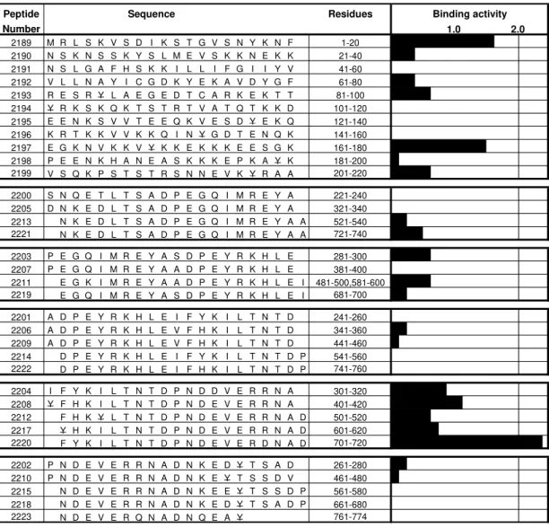
Peptide Sequence Residues Binding activity
Number 1.0 2.0
2189 M R L S K V S D I K S T G V S N Y K N F 1-20 2190 N S K N S S K Y S L M E V S K K N E K K 21-40 2191 N S L G A F H S K K I L L I F G I I Y V 41-60 2192 V L L N A Y I C G D K Y E K A V D Y G F 61-80 2193 R E S R Y L A E G E D T C A R K E K T T 81-100 2194 Y R K S K Q K T S T R T V A T Q T K K D 101-120 2195 E E N K S V V T E E Q K V E S D Y E K Q 121-140 2196 K R T K K V V K K Q I N Y G D T E N Q K 141-160 2197 E G K N V K K V Y K K E K K K E E S G K 161-180 2198 P E E N K H A N E A S K K K E P K A Y K 181-200 2199 V S Q K P S T S T R S N N E V K Y R A A 201-220
2200 S N Q E T L T S A D P E G Q I M R E Y A 221-240 2205 D N K E D L T S A D P E G Q I M R E Y A 321-340 2213 N K E D L T S A D P E G Q I M R E Y A A 521-540 2221 N K E D L T S A D P E G Q I M R E Y A A 721-740
2203 P E G Q I M R E Y A S D P E Y R K H L E 281-300 2207 P E G Q I M R E Y A A D P E Y R K H L E 381-400 2211 E G K I M R E Y A A D P E Y R K H L E I 481-500,581-600 2219 E G Q I M R E Y A S D P E Y R K H L E I 681-700
2201 A D P E Y R K H L E I F Y K I L T N T D 241-260 2206 A D P E Y R K H L E V F H K I L T N T D 341-360 2209 A D P E Y R K H L E V F H K I L T N T D 441-460 2214 D P E Y R K H L E I F Y K I L T N T D P 541-560 2222 D P E Y R K H L E I F H K I L T N T D P 741-760
2204 I F Y K I L T N T D P N D D V E R R N A 301-320 2208 Y F H K I L T N T D P N D E V E R R N A 401-420 2212 F H K Y L T N T D P N D E V E R R N A D 501-520 2217 Y H K I L T N T D P N D E V E R R N A D 601-620 2220 F Y K I L T N T D P N D E V E R D N A D 701-720
2202 P N D E V E R R N A D N K E D Y T S A D 261-280 2210 P N D E V E R R N A D N K E Y T S S D V 461-480 2215 N D E V E R R N A D N K E E Y T S S D P 561-580 2218 N D E V E R R N A D N K E D Y T S A D P 661-680 2223 N D E V E R Q N A D N Q E A Y 761-774
Fig. 1: GBP 130 synthetic peptides’ specific binding activity to human erythrocytes. Amino acid sequences are given in the left column. Binding activity: is the slope value of specific binding curve. Underlined amino acids were substituted with Tyr to allow radiolabeling. Each one of the black bars represents the slope of the specific binding curve, which is named high specific binding activity (HSBA). Peptides with HSBA ≥ 2% were considered as high specific binding peptides to erythrocytes, since these peptides recognize more than 200 specific binding sites per cell at low concentrations of radiolabeled peptide (200 nM).
activity has already been reported (Kochan et al. 1986). Some other peptides (2204, 2208, 2212, 2217), with very similar sequences to peptide 2220 (RDN
is substituted by RRN), show lower binding
activi-ties than peptide 2220. Peptides which only share the N-terminal (2201, 2206, 2209, 2214, 2222) or the
C-terminal (2202, 2210, 2215, 2218, 2223) sequence with peptide 2220 showed no specific binding to erythrocytes.
In order to investigate whether the peptide la-beling, which introduces an iodine atom into a ty-rosine residue, affected its interaction with human erythrocytes, an additional assay was carried out
with peptide 2220 labeled with non-radioactive NaI. Fig. 2 shows that both iodinated and non-iodinated peptides identically displaced the radiolabeled ligand. Therefore, iodinated tyrosine can be ex-cluded as significantly altering the erythrocyte-peptide interaction.
498 498 498 498
498 GBP 130 Peptide Binds to Erythrocytes Jorge E Suarez et al.
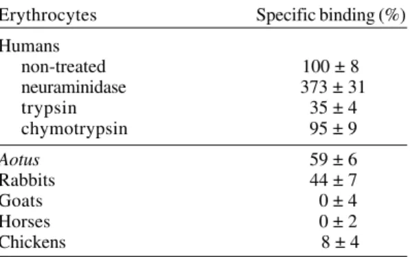
Peptide 2220 and interaction with enzyme treated erythrocytes - In order to determine whether the peptide 2220 interacts with sialic acid or sialoglycoproteins, binding assays were performed with enzyme treated erythrocytes. Table I shows the percentages of specific binding compared to peptide binding of normal human erythrocytes.
0 50 100 150 200
0 1 2 3 4
Added peptide (CPM*10-6)
Bound peptide (CPM*10
-3)
Fig. 2: peptide 125I-2220 binding to human erythrocyte.
Total binding: binding to erythrocyte in absence of compe-tition (%). Non specific-binding: binding in presence of non-labeled peptide (o). Non specific binding in presence of peptide iodinate with non-radioactive NaI (!).
Fig. 3: saturation curve for peptide 2220. The graph shows the total (%) binding, non-specific (!) binding and spe-cific (o) binding of the radiolabeled peptide to human erythrocytes. In the Hill plot (inset graph) the axis are: the abscissa is log F, and the ordinate is log (B/Bmax-B). B: fmol bound peptide; Bmax: maximum fmol bound peptide; F: nM free peptide
Human erythrocytes treated with neuraminidase showed a 373% increase in specific binding for peptide 2220. However, human erythrocytes treated with trypsin showed markedly lower affinity for peptide 2220, with a reduction to 35%. It was also noted that, when human erythrocytes were treated with chymotrypsin, there was no effect on peptide 2220 specific binding.
Peptide 2220 bound mainly to human eryth-rocytes - Peptide 2220 was assayed with erythro-cytes of different species with the aim of determin-ing if the binddetermin-ing is species specific (Table I). Pep-tide 2220 possesses a higher specific binding to human erythrocytes than to erythrocytes from other species. Aotus and rabbit erythrocytes showed specific binding values of 59% and 44% respec-tively, compared to human erythrocytes. No bind-ing was observed to goat, horse or chicken eryth-rocytes.
TABLE I
Binding of 2220 peptide to enzyme treated human erythrocyte and non-human erythrocytes
Erythrocytes Specific binding (%)
Humans
non-treated 100 ± 8
neuraminidase 373 ± 31
trypsin 35 ± 4
chymotrypsin 95 ± 9
Aotus 59 ± 6
Rabbits 44 ± 7
Goats 0 ± 4
Horses 0 ± 2
Chickens 8 ± 4
Critical residues for peptide-erythrocyte inter-action - The importance of each residue in the bind-ing of peptide 2220 to erythrocytes was determined by analyzing the ability of glycine analogs to in-hibit the binding of the unmodified peptide (com-petition binding assay, Fig. 4). Residues F701, K703, L705, T706 and E713 were critical, since the replacement of any of these amino acids dramati-cally reduced their binding affinity (Fig. 4).
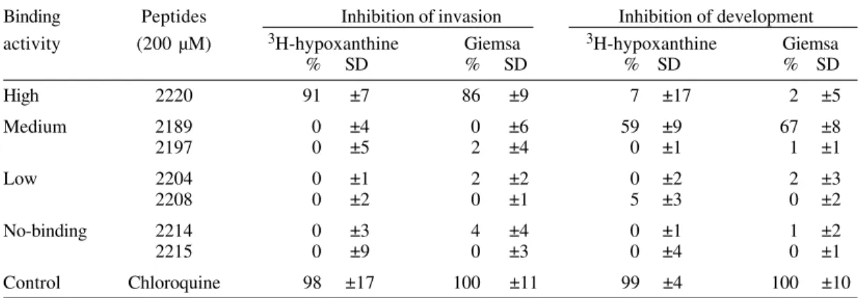
Effect on in vitro peptide 2220 P. falciparum cultures - To determine whether the peptide 2220 is biologically relevant, in vitro invasion and devel-opment inhibition assays were performed. Both assays were performed in triplicate and six pep-tides were used as controls: peppep-tides 2189 and 2197 (medium specific-binding); peptides 2204 and 2208 (low specific-binding); and peptides 2214 and 2215 (non-binding). As shown in Table II, the invasion process is inhibited only by peptide 2220 (91% in-hibition). The slides of this assay, staining with
0 200 400 600 800
0 100 200 300 400
-1,7
-0,4
0,9
1,4 2 2,6
Free peptide (nM)
499 499499 499 499 Mem Inst Oswaldo Cruz, Rio de Janeiro, Vol. 95(4), Jul./Aug. 2000
Giemsa show that the parasites in the slide of pep-tide 2220 has the same morphology of the control, but the number of parasited erythrocytes was di-minished. None of the control peptides showed any effect on P. falciparum parasite invasion. One con-trol peptide (2189) markedly affected the develop-ment of the parasite (59%), but did not inhibit inva-sion. This suggests that it may have a toxic effect on the intracellular parasite, but is not capable of interacting with erythrocyte proteins to block the invasion process.
DISCUSSION
Merozoite surface proteins play an important role in erythrocyte invasion, since they determine the first contact between erythrocyte and merozo-ite. Among these, GBP 130 has been implicated as being an erythrocyte binding protein in merozoites (Perkins 1984, 1988). Peptide 2220 was identified as being a part of the GBP 130 sequence which binds specifically to erythrocytes. This peptide belongs to the repeat region which binds to human erythro-cytes; antibodies against this region inhibited in vitro erythrocyte invasion by merozoites (Kochan et al. 1986). Peptides 2204, 2208, 2212, 2217 and 2220 have a very similar sequence, including the resi-dues critical for binding to erythrocytes. However, the 2220 peptide has the highest binding affinity. The change of R for D in position 717 increases the affinity of peptide 2220 to erythrocytes.
The interaction between peptide 2220 and erythrocytes is charge-independent, since peptides 2202 and 2205, which have a similar charge, did not bind specifically to erythrocytes. Consequently, the amino acid sequence is the determinant factor in the binding. Peptide 2220, iodinated with non-ra-dioactive NaI, displaced the radiolabeled ligand identically to displacement produced by the non-labeled peptide (Fig. 2), showing that the modifica-tion caused by iodinamodifica-tion of the peptide does not affect this peptide’s interaction with erythrocytes. A 150 nM affinity constant shows that the interac-tion between peptide 2220 and erythrocytes is very strong. This interaction could be mediated by salt-bridges, since peptide 2220 has a high proportion of charged amino acids (8/20) and the E713 amino acid is critical to binding. A Hill coefficient of 2 suggests positive cooperativity.
Residue
100 nM
0 50 100 150 200 250
F Y K I L T N T D P N D E V E R D N A D
2220
800 nM
0 50 100 150
F Y K I L T N T D P N D E V E R D N A D
2220
TABLE II
Inhibition of parasite invasion and development to erythrocyte by GBP 130 peptides
Binding Peptides Inhibition of invasion Inhibition of development
activity (200 µM) 3H-hypoxanthine Giemsa 3H-hypoxanthine Giemsa
% SD % SD % SD % SD
High 2220 91 ±7 86 ±9 7 ±17 2 ±5
Medium 2189 0 ±4 0 ±6 59 ±9 67 ±8
2197 0 ±5 2 ±4 0 ±1 1 ±1
Low 2204 0 ±1 2 ±2 0 ±2 2 ±3
2208 0 ±2 0 ±1 5 ±3 0 ±2
No-binding 2214 0 ±3 4 ±4 0 ±1 1 ±2
2215 0 ±9 0 ±3 0 ±4 0 ±1
Control Chloroquine 98 ±17 100 ±11 99 ±4 100 ±10
Fig. 4: specific binding to erythrocytes of peptide 2220 in competition assays with glycine analog peptides. The height of the black bars is proportional to the binding inhibition of the original radiolabeled peptide (20 nM) by the original or peptide unlabeled analogues (100 and 800 nM). The letter at the bottom represents the amino acid changed by glycine.
500 500 500 500
500 GBP 130 Peptide Binds to Erythrocytes Jorge E Suarez et al.
Peptide 2220 shows a high increase in specific binding to neuraminidase treated erythrocytes (Table I), indicating that the interaction does not involve sialic acid molecules. This agrees with pre-vious observations, which demonstrate that GBP 130 receptors are not dependent on sialic acid (Perkins 1988). The binding of peptide 2220 to eryth-rocytes was affected by the treatment with trypsin and not by chymotrypsin. As trypsin cleaves the external proteic part of glycophorin A and C (Pasvol 1984), it is possible that glycophorin A, C or other related molecules act as a receptor for this peptide. However, as the binding was not completely abro-gated by trypsin treatment, other proteins (such as glycophorin B) could be involved in the binding. The membrane receptor of peptide 2220 has not been identified, although it is known that its bind-ing activity is sensitive to trypsin treatment.
Binding competition assays with peptide 2220 glycine analogues show that some amino acids (FYKILTNTDPNDEVERDNAD) were critical in the binding; probably these residues are not only in-volved in direct binding interactions (salt-bridges and hydrophobic), but could also be essential for the induction of the tridimensional structure re-quired for binding. In some cases the replacement of 2220 residues by glycine led to peptides with higher binding affinity (I704, P710, N711, V714). Probably these residues’ side chains hinder opti-mal 2220 peptide-receptor interactions. Substitu-tion of two different threonine residues (T706 and T708) has a very different effect on peptide bind-ing (Fig. 4). This further emphasizes that peptide 2220 binding is highly dependent on its primary structure, and not only on a particular amino acid composition.
The binding assays with peptide 2220 and erythrocytes from different species (Table I) showed that this peptide binds to human erythrocytes with higher affinity than to erythrocytes from other spe-cies. However, it also interacts with Aotus mon-keys’ erythrocytes, albeit to a lesser extent. This might explain why both species are susceptible to
P. falciparum infection. Peptide 2220 also binds sig-nificantly to rabbit erythrocytes. It has been dem-onstrated that some proteins involved in merozoite invasion are able to bind to rabbit erythrocytes (Orlandi et al. 1990), but P. falciparum merozoites do not invade these erythrocytes (Breuer et al. 1983). Probably the binding domain of the peptide receptor on the erythrocyte is different in the stud-ied species, which will change the affinity constant of this interaction.
Two assays were carried out in order to test the peptide effect on in vitro cultures of P. falciparum. In one assay, peptide was added before merozoites were released at the schizont stage. In this assay,
peptide 2220 inhibited invasion by 91%. The pep-tide may affect merozoite release or erythrocyte in-vasion. However, we did not see delay in parasite growth or death parasites. This could be indicating that peptide 2220 may compete for the receptor sites on the erythrocyte and block the merozoite-inva-sion. In another assay, when peptide was added after invasion, ring stage peptide 2220 did not af-fect parasite development. However, the difference in membrane permeability between ring stage and schizont stage restricts the interpretation of this results. The peptide concentration needed for in-vasion-inhibition was higher than the affinity con-stant value; this may be due to higher GBP 130 affinity than peptide 2220, or because the effective concentration is diminished by proteolytic degra-dation. Interestingly, peptide 2220 belongs to a re-peat region which binds to glycophorin and elicits antibodies able to block merozoite invasion of eryth-rocytes (Kochan et al. 1986).
REFERENCES
Aronson NE, Silverman C, Wasserman GF, Kochan J, Hall T, Esser K, Young JE, Chulay JD 1991. Immu-nization of owl monkeys with a recombinant pro-tein containing repeated epitopes of a Plasmodium falciparum glycophorin-binding protein. Am J Trop Med Hyg 45: 548-559.
Bonnefoy S, Gysin J, Blisnick T, Gillotte M, Carcy B, Pereira Da Silva L, Mercereau-Puijalon O 1994. Im-munogenicity and antigenicity of a Plasmodium falciparum protein fraction (90-110 kDa) able to protect squirrel monkeys against asexual blood stages.
Vaccine12: 32-40.
Bonnefoy S, Mattei D, Dubremetz JF, Guillotte M, Jouin H, Ozaki LS, Sibilli L, Mercereau-Puijalon O 1988.
Plasmodium falciparum: molecular analysis of a putative protective antigen, the thermostable 96-kDa Protein. Exp Parasitol65: 69-83.
Breuer WV, Ginsburg H, Cabantchik ZI 1983. An assay of malaria parasite invasion into human erythrocytes: the effects of chemical and enzimatic modification of erythrocyte membrane components. Biochim Biophys Acta755: 263-271.
Camus D, Hadley TJ 1985. A Plasmodium falciparum
antigen that binds to host erythrocytes and mero-zoites. Science230: 553-556.
Chulay JD, Lyon JA, Haynes JD, Meierovics AI, Atkinson CT, Aikawa M 1987. Monoclonal anti-body characterization of Plasmodium falciparum
antigens in immune complexes formed when sch-izonts rupture in the presence of immune serum. J Immunol139: 2768-2774.
Dubois P, Dedet JP, Fandeur T, Rousslhon C, Jendoubi M, Pauillac S, Mercereau-Puijalon O, Pereira Da Silva L 1984. Protective immunization of the squirrel monkey against asexual blood stages of Plasmodium falciparum by use of parasite protein fractions. Proc Natl Acad Sci USA81: 229-232.
solid-501 501501 501 501 Mem Inst Oswaldo Cruz, Rio de Janeiro, Vol. 95(4), Jul./Aug. 2000
phase synthesis of large numbers of peptides: speci-ficity of antigen-antibody interaction at the level of individual amino acids. Proc Natl Acad Sci USA82: 5131-5135.
Kochan J, Perkins ME, Ravetch JV 1986. A tandemly repeated sequence determines the binding domain for an erythrocyte receptor-binding protein of Plas-modium falciparum. Cell44: 689-696.
Lyon JA, Haynes JD, Diggs CL, Chulay JD, Pratt-Rossiter JM 1986. Plasmodium falciparum antigens synthesized by schizonts and stabilized at the mero-zoite surface by antibodies when schizonts mature in the presence of growth inhibitory immune serum.
J Immunol136: 2252-2258.
Lyon JA, Thomas AW, Hall T, Chulay JD 1989. Speci-ficities of antibodies that inhibit merozoite dispersal from malaria-infected erythrocytes. Mol Biochem Parasitol36: 77-86.
Merrifield RB 1963. Solid phase peptide synthesis. l. The synthesis of a tetrapeptide. J Am Chem Soc85: 2149-2154.
Orlandi PA, Sim BK, Chulay JD, Haynes JD 1990. Char-acterization of the 175-kilodalton erythrocyte bind-ing antigen of Plasmodium falciparum.Mol Biochem Parasitol40: 285-294.
Pasvol G 1984. Receptors on red cells for Plasmodium falciparum and their interaction with merozoites. Phil
Trans R Soc Lond B307: 189-200.
Perkins ME 1984. Surface proteins of Plasmodium falciparum merozoites binding to the erythrocyte receptor, glycophorin. J Exp Med160: 788-798. Perkins ME 1988. Stage dependent processing and
lo-calization of a Plasmodium falciparum protein of 130,000 molecular weight. Exp Parasitol65: 61-68. Perkins ME, Rocco LJ 1988. Sialic acid-dependent bind-ing of Plasmodium falciparum merozoite surface antigen, Pf200, to human erythrocytes. J Immunol 141: 3190-3196.
Tam JP, Heath WF, Merrifield RB 1983. SN2 deprotection of synthetic peptides with a low con-centration of HF in dimethyl sulfide: evidence and application in peptide synthesis. J Am Chem Soc 105: 6442-6455.
Trager W, Jensen J 1976. Human malaria parasites in continuous culture. Science193: 673-675.
Urquiza M, Rodriguez LE, Suárez JE, Guzman F, Ocampo M, Curtidor H, Segura C, Truillo E, Patarroyo ME 1996. Identification of Plasmodium falciparum MSP-1 peptides able to bind to human red blood cells.
Parasite Immunol18: 515-526.