J Bras Pneumol. 2011;37(2):277-280
respectively. Single-photon emission CT (SPECT) scans, in conjunction with standard CT scans, revealed an irregular mass (approximately 8 × 8 cm in size) in the anterior mediastinum, varying from 40 to 54 HU in density (Figure 1a). Bronchoscopy revealed hyperemic, edematous bronchial mucosa with hemorrhagic stippling and traces of blood in the bilateral basal segments, although there was no active bleeding. There was no invasion of the lumen or ulceration of the bronchial wall. A CT-guided biopsy of the mediastinal mass was performed. The histopathological study of the biopsy sample showed necrosis, cell detritus, and large pleomorphic cells, in which the cytoplasms were clear, the nuclei contained coarse chromatin, and the nucleoli were evident, with a neoplastic appearance. Immunohistochemical staining showed a positive response for cytokeratin AE1/ AE3, chorionic gonadotropin hormone, and alpha fetoprotein, although the responses to synaptophysin and the leukocyte antigen, which are common in neoplastic cells, were negative. The anatomopathological diagnosis was of a seminomatous GCT, a diagnosis that was underscored by the findings of normal serum alpha fetoprotein and moderately elevated serum Beta-hCG. To evaluate the extent of the tumor, we carried out SPECT/CT scans of the abdominal cavity, pelvic cavity, brain, and liver but failed to identify any abnormalities. The TM was identified through ultrasound (Figure 1b). Vascularity was found to be adequate in both testicles. The patient received six cycles of chemotherapy with cisplatin, etoposide, and bleomycin. After the treatment, the hemoptysis resolved. In addition, post-treatment SPECT/ CT fusion imaging showed a reduction in the size of the lesion and the absence of any
To the editor:
Primary mediastinal tumors comprise a heterogeneous group of abnormalities composed of neoplastic, congenital and inflammatory lesions. The probability of malignancy in mediastinal tumors is associated with three factors: tumor location, patient age at the time of diagnosis, and the presence of symptoms. In general, the tumors that are the most likely to become malignant are those that are found in the anterior compartment and those that are accompanied by symptoms.(1)
We recently treated a patient diagnosed with a primary seminomatous germ cell tumor (GCT) accompanied by testicular microlithiasis (TM). To our knowledge, there are only six published cases documenting an association between primary seminoma of the mediastinum and TM.(2-7) Another aspect that made this case
exceptional was that the initial symptom was hemoptysis, a feature that has been reported in less than 1% of patients with extragonadal GCTs affecting the mediastinum.(8)
The patient was a 27-year-old, nonsmoking male construction worker with no family history of cancer. He sought treatment at our hospital, complaining of a two-month history of intermittent hemoptysis (≈30-60 mL/24 h), weight loss, and dyspnea (Modified Medical Research Council grade 2). The results of the physical examination, biochemical tests, and hematological studies were all normal. A chest X-ray taken at admission revealed an evident widening of the mediastinum. On the basis of these findings, we then quantified the serum tumor markers alpha fetoprotein and beta-human chorionic gonadotropin (Beta-hCG), obtaining values of 2.59 ng/mL (reference values, 0.67-3.2 ng/mL) and 13.5 mIU/mL (reference value for males, < 2.5 mIU/mL),
Hemoptysis in a patient with testicular microlithiasis
and a germ cell tumor: a rare combination
Hemoptise em um paciente com microlitíase testicular e tumor de células germinativas: uma combinação rara
278
J Bras Pneumol. 2011;37(2):277-280
metabolic activity within the lesion, a condition that persisted at one year after the end of the treatment (Figure 1c).
Although most GCTs originate in the gonads, 2-5% are extragonadal.(8) Of all mediastinal
masses, extragonadal GCTs account for 10-15% and seminomas account for 2-4%.(9)
In individuals with mediastinal seminomas, symptoms appear when the tumor reaches a given size and tend to be secondary to the compression of the adjacent structures. Bokemeyer et al.(8) studied 104 patients with
mediastinal seminomas and identified only one case of hemoptysis. In the case presented here, hemoptysis was the initial symptom. Although we were unsuccessful in identifying the exact cause of the hemoptysis, we documented the hyperemic character and hemorrhagic stippling of the bronchial mucosa. Neither the biopsy of the mucosa nor the examination of the BAL fluid revealed identifiable tumor cells.
Typically identified through ultrasonography, TM consists of multiple, randomly distributed, echogenic micronodules, with no acoustic shadow, measuring 1-3 mm in diameter. Although the clinical significance of TM remains unclear, studies of the prevalence of in situ cancer in the presence of TM have been conducted. The frequency of in situ cancer in the general population of patients with TM is 1.2%, compared with 10.8% in the population of those referred to fertility specialists.(10)
Cast et al.(11) analyzed 4,892 scrotal ultrasounds
in 4,819 patients and identified 33 patients with TM, which translates to a prevalence of 0.68%. In addition, the authors found that 7 patients with TM also had concurrent tumors. The relative risk of concurrent tumor was found to be 21.6 times greater for individuals with TM than for those without.
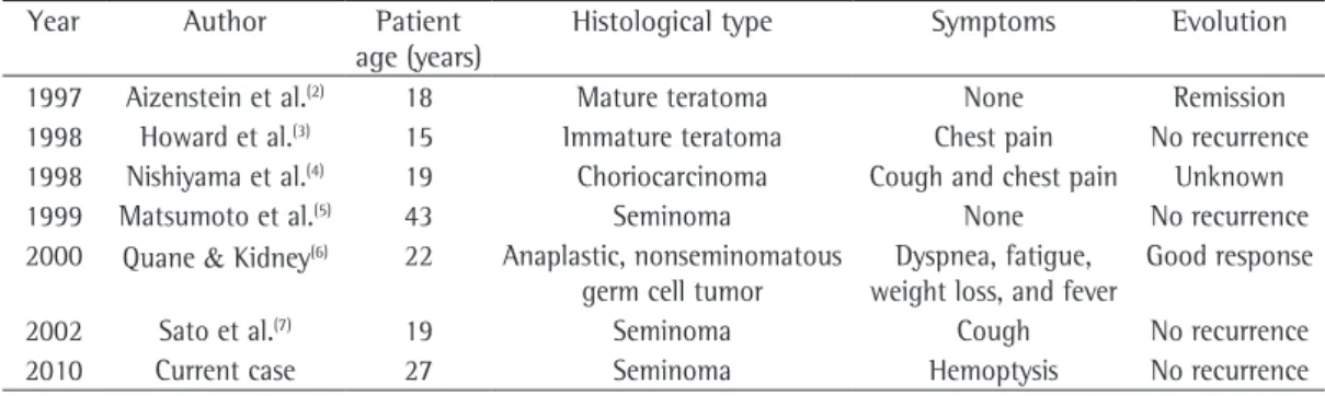
The combination of an extragonadal GCT and TM was clearly documented in our patient. This combination is quite rare. As previously mentioned, only six such cases have been published to date. The first was described by Aizenstein et al. in 1997.(2) The additional cases
were reported between 1998 and 2002.(3-7) The
characteristics of all seven cases (those six cases
J Bras Pneumol. 2011;37(2):277-280
279
Daniel Mendoza Posada Physician. Oncology Department, Dr. Ismael Cosío Villegas National
Institute of Respiratory Diseases, Mexico City, Mexico
Luis Torre Bouscoulet Physician. Department of Clinical Pulmonology, Dr. Ismael Cosío Villegas
National Institute of Respiratory Diseases, Mexico City, Mexico
References
1. McKenney JK, Heerema-McKenney A, Rouse RV. Extragonadal germ cell tumors: a review with emphasis on pathologic features, clinical prognostic variables, and differential diagnostic considerations. Adv Anat Pathol. 2007;14(2):69-92.
2. Aizenstein RI, Hibbeln JF, Sagireddy B, Wilbur AC, O’Neil HK. Klinefelter’s syndrome associated with testicular microlithiasis and mediastinal germ-cell neoplasm. J Clin Ultrasound. 1997;25(9):508-10.
3. Howard RG, Roebuck DJ, Metreweli C. The association of mediastinal germ cell tumour and testicular microlithiasis. Pediatr Radiol. 1998;28(12):998.
4. Nishiyama T, Terunuma M, Iwashima A, Souma T, Hirahara H. Testicular microlithiasis with mediastinal choriocarcinoma: a case report. Int J Urol. 1998;5(3):301-2.
5. Matsumoto K, Iwamura M, Katsuta M, Satoh T, Ohori M, Egawa S, et al. Extragonadal seminoma with testicular microlithiasis: a case report [Article in Japanese]. Hinyokika Kiyo. 1999;45(10):725-7.
6. Quane LK, Kidney DD. Testicular microlithiasis in a patient with a mediastinal germ cell tumour. Clin Radiol. 2000;55(8):642-4.
7. Sato K, Komatsu K, Maeda Y, Ueno S, Koshida K, Namiki M. Case of mediastinal seminoma with testicular microlithiasis. Int J Urol. 2002;9(2):114-6.
8. Bokemeyer C, Nichols CR, Droz JP, Schmoll HJ, Horwich A, Gerl A, et al. Extragonadal germ cell tumors of the
plus the case described here) are summarized in Table 1.
Seminomas respond well to chemotherapy, and the five-year survival rate is over 88%. Although TM has yet to be formally recognized as a premalignant lesion, patients with TM should be monitored and it should be borne in mind that the mediastinum is a potential site for GCT development. The case presented here is interesting not only because the TM-extragonadal GCT combination is rare but also because the patient sought medical attention due to hemoptysis, which is seen in less than 1% of individuals with primary mediastinal GCTs. This case underscores the importance of broadening the differential diagnosis of hemoptysis and of performing scrotal ultrasound in patients with extragonadal GCTs located in the mediastinum.
Acknowledgments
The authors would like to thank Paul Kersey (Language Laboratory, The College of Michoacán) for correcting errors in language usage.y
Arturo Cortés Télles Physician. Department of Clinical Pulmonology, Dr. Ismael Cosío Villegas
National Institute of Respiratory Diseases, Mexico City, Mexico
José de Jesús López Luna Physician. Pathology Department, Dr. Ismael Cosío Villegas National
Institute of Respiratory Diseases, Mexico City, Mexico
Table 1 - Reported cases of extragonadal germ cell tumors accompanied by testicular microlithiasis.
Year Author Patient
age (years)
Histological type Symptoms Evolution
1997 Aizenstein et al.(2) 18 Mature teratoma None Remission
1998 Howard et al.(3) 15 Immature teratoma Chest pain No recurrence
1998 Nishiyama et al.(4) 19 Choriocarcinoma Cough and chest pain Unknown
1999 Matsumoto et al.(5) 43 Seminoma None No recurrence
2000 Quane & Kidney(6) 22 Anaplastic, nonseminomatous
germ cell tumor
Dyspnea, fatigue, weight loss, and fever
Good response
2002 Sato et al.(7) 19 Seminoma Cough No recurrence
280
J Bras Pneumol. 2011;37(2):277-280
10. Meissner A, Mamoulakis C, de la Rosette JJ, Pes MP. Clinical update on testicular microlithiasis. Curr Opin Urol. 2009;19(6):615-8.
11. Cast JE, Nelson WM, Early AS, Biyani S, Cooksey G, Warnock NG, et al. Testicular microlithiasis: prevalence and tumor risk in a population referred for scrotal sonography. AJR Am J Roentgenol. 2000;175(6):1703-6. mediastinum and retroperitoneum: results from an
international analysis. J Clin Oncol. 2002;20(7):1864-73. 9. Moran CA, Suster S, Przygodzki RM, Koss MN.