Ureteroscopic Pneumatic Lithotripsy of Impacted Ureteral
Calculi
Artur H. Brito, Anuar I. Mitre, Miguel Srougi
Division of Urology, School of Medicine, University of Sao Paulo, Sao Paulo, SP, Brazil
ABSTRACT
Introduction: This work evaluates the results of ureteroscopic treatment of impacted ureteral stones with a pneumatic lithotripter.
Materials and Methods: From March 1997 to May 2002, 42 patients with impacted ureteral stones were treated by retro-grade ureteroscopic pneumatic lithotripsy. Twenty-eight patients were female and 14 were male. The stone size ranged from 5 to 20 mm. The ureteral sites of the stones were distal in 21, middle in 12 and proximal in 9.
Results: Considering stones with distal location in the ureter, 1 patient had ureteral perforation and developed a stricture in the follow-up (4.7%). As for stones in the middle ureter, 2 perforations and 1 stricture were observed (8.3%) and regarding stones located in the proximal ureter, 5 perforations and 4 strictures occurred (44%). In the mid ureter, 1 ureteral avulsion was verified. In 34 patients without ureteral perforation, only 1 developed a stricture (2.9%). Of 8 patients who had perfo-ration, 6 developed strictures. The overall incidence of stricture following treatment of impacted ureteral calculi was 14.2%.
Conclusions: Ureteroscopy for impacted ureteral calculi is associated with a higher incidence of ureteral perforation and stricture. Ureteroscopy of proximal ureteral calculi is associated with a high risk of perforation, when compared to mid or distal ureteral calculi. Ureteral perforation at the site of the stone seems to be the primary risk factor for stricture formation in these cases.
Key words: ureteroscopy; ureteral calculi; lithotripsy; injury; stenosis
Int Braz J Urol. 2006; 32: 295-9
INTRODUCTION
Impacted ureteral stone is commonly con-sidered as a condition where a stone remains at the same site for more than 2 months (1). Impacted ure-teral stone are the most difficult to treat, because there is a severe ureteral inflammation. This fact results in an increased risk of ureteral injury by in-struments during endoscopic procedure (2). Extra-corporeal shockwave lithotripsy (SWL) is widely used for treatment of ureteral stones as well as
Harmon et al. reported the rate of stricture formation after ureteroscopy to be 0.5% in 1992 compared to 1.5% 10 years earlier (8). In the present study, we assessed prospectively the effectiveness of ureteroscopic pneumatic lithotripsy for treatment of impacted ureteral stones.
MATERIALS AND METHODS
From March 1997 to May 2002, 42 patients with impacted ureteral stones were treated by ureteroscopic pneumatic lithotripsy. Twenty-eight patients were female and 14 were male. The age ranged from 23 to 72 years old. The stone size ranged between 5 and 20 mm. One patient had a 5 mm calcu-lus, 11 had calculi between 6 and 10 mm, 18 had culi between 11 and 15 mm and 12 patients had cal-culi between 16 and 20 mm. Stones were located in the distal ureter in 21 patients, in the mid ureter in 12 and in the proximal ureter in 9.
The access to the calculi was retrograde in all patients. The procedures were carried out with the
patients in the lithotomy position under lumbar anes-thesia. Ureteral orifice dilatation was necessary in 3 patients.
A retrograde ureteropyelogram was per-formed in all cases to study the ureteral anatomy and a guide wire (Teflon or hydrophilic) was inserted prior to the introduction of the ureteroscope. The hydro-philic guide wire was used in the cases where it was impossible to pass the Teflon guide wire.
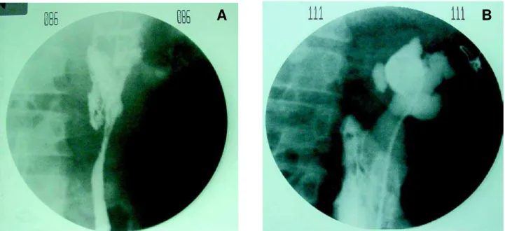
Ureteroscopy was performed with 8.5F semi rigid ureteroscope (Wolf Medical Instruments, Vernon Hills, Illinois, USA). The ureteroscope was introduced just under the stone and confirmation of its attach-ment to the edematous and hyperemic ureteral mu-cosa was obtained. The fragmentation of the stones was done with a pneumatic lithotripter (Electro Medi-cal Systems, Kaufering, Germany). The fragments were removed with a grasping forceps or a Dormia basket. At the end of the procedure, a retrograde ureteropyelogram was performed to verify whether there was perforation and a double-J catheter was introduced in all the patients and left in place for 3 weeks (Figure-1). All the procedures were performed
A
B
under fluoroscopic and visual guidance. All patients were submitted to an intravenous pyelogram after 2 months to verify ureteral patency.
RESULTS
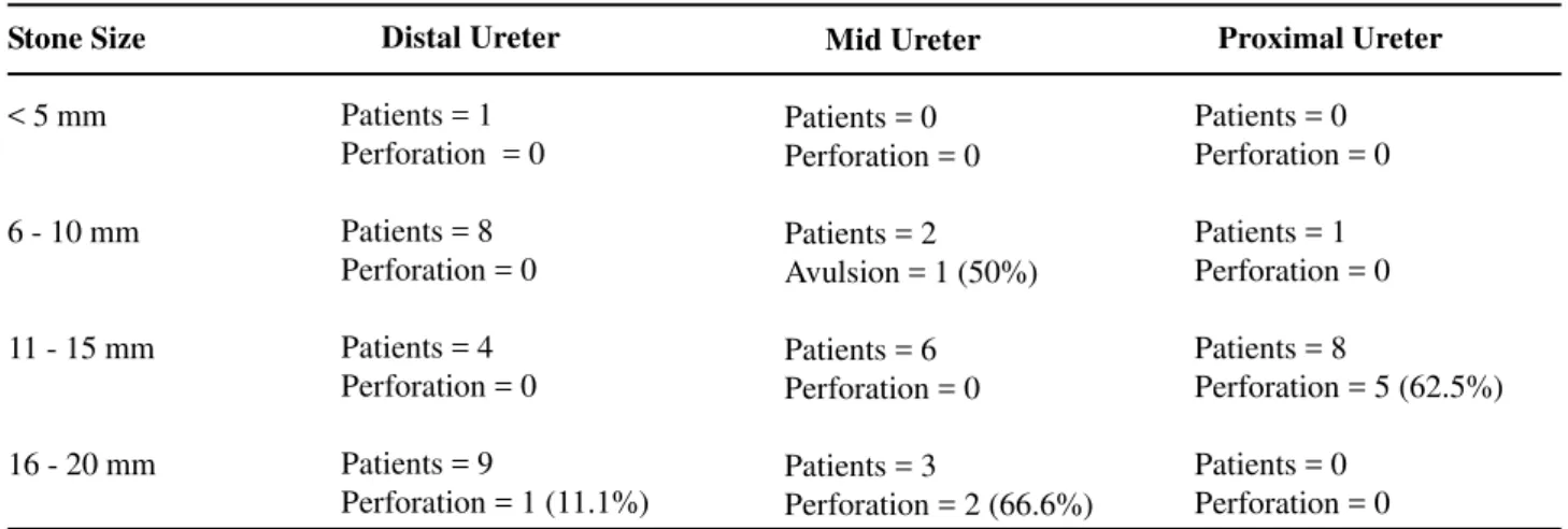
No complication was observed for stone smaller than 5 mm. For the 11 stones between 6 and 10 mm, there was 1 ureteral avulsion with the Dormia basket. This complication was treated by ureteral reimplantation with a Boari bladder flap. Postopera-tive evaluation at 6 months, by intravenous pyelo-gram, showed a good result. For the 18 calculi be-tween 11 and 15 mm there were 5 ureteral perfora-tions and for the 12 calculi between 16 and 20 mm, there were 3 cases of ureteral perforation (Table-1).
The relation of the calculi site and perfora-tion showed that in 21 distal calculi there were only 1
perforation and 1 stricture (4.7%). In the 12 mid ure-teral calculi, there were 1 ureure-teral avulsion, 2 perfo-rations and 1 stricture (8.3%), and in the 9 proximal ureteral calculi, there were 5 perforations and 4 stric-tures (44%). In 3 of these 4 patients that developed strictures, it was not possible to withdraw all frag-ments, because during the procedure there has been a mild hemorrhage and it was stopped (Table-2).
The overall incidence of strictures was 14.2%. For the treatment of ureteral strictures, the renal function was also available in 4 patients that presented large pelvicaliceal dilation. In these pa-tients, a nephrostomy tube was placed and a renal scintigraphy was done. In 3 patients, renal scintigra-phy shows a renal function lower than 20%, being indicated nephrectomy for these cases and in 1 pa-tient the renal function was higher than 20% being indicated incision of the stenosis with the Acucise®
catheter (Applied Urology, Rancho Santa Margarita,
Table 2 – Complications of impacted ureteral calculus.
Calculus Site
Patients
Complications Perforation Avulsion
Stricture
Distal Ureter
21
01 (4.7%)
00
0
01 (4.7%)
Mid Ureter
12
02 (16.6%)
01 (11.1%)
01 ( 8.3%)
Proximal Ureter
9
5 (55.5%) 0
4 (44%)
Table 1 – Stone size and incidence (percentage) of ureteral perforation.
Stone Size
< 5 mm
6 - 10 mm
11 - 15 mm
16 - 20 mm
Distal Ureter
Patients = 1 Perforation = 0
Patients = 8 Perforation = 0
Patients = 4 Perforation = 0
Patients = 9
Perforation = 1 (11.1%)
Mid Ureter
Patients = 0 Perforation = 0
Patients = 2 Avulsion = 1 (50%)
Patients = 6 Perforation = 0
Patients = 3
Perforation = 2 (66.6%)
Proximal Ureter
Patients = 0 Perforation = 0
Patients = 1 Perforation = 0
Patients = 8
Perforation = 5 (62.5%)
California, USA). Three patients had ureteral steno-sis with good renal function and an incision of the stenosis with the Acucise® catheter was indicated.
Evaluation by intravenous pyelogram, at 6 months, of the 4 patients in that was did incision with Acucise®
catheter, showed good results in all.
Analysis of all cases of ureteral stricture showed that in 34 patients without ureteral perfora-tion only one presented late stricture (2.9%), while in 8 patients that had perforation, 6 presented stric-tures (75%).
The overall stone free rate was 92.9%.
DISCUSSION
SWL can be a modality treatment for most upper urinary tract stones, because of its simplicity, noninvasiveness and minimal morbidity. However, some stones are difficult to fragment by SWL or the fragments may remain in the urinary tract even after successful fragmentation of the stone. Since residual stones can cause hydronephrosis followed by a de-crease in renal function or urinary tract infection, re-sidual fragments should be removed even if they are less than 4 mm in diameter (9). Impacted ureteral calculi are more difficult to fragment with SWL than stones lying in the renal pelvis, because of the lack of natural expansion space for stones in ureter (10,11). In the aforementioned situations, ureterolithotripsy is the best treatment option (12).
Four sources of energy for intracorporeal lithotripsy are now available, that is, electrohydrau-lic, ultrasound, pneumatic and Holmium laser. With electrohydraulic and ultrasonic energy, there is more risk of complication, as for example ureteral perfora-tion. The pneumatic energy is strong enough for frag-menting all types of stones and is cheaper than Hol-mium laser. However, with the pneumatic lithotripter, there is more retrograde migration of the ureteral stone during its fragmentation.
Impacted ureteral stone, is considered a con-dition where a stone remains at the same site for more than 2 months (1). All cases in our series fulfilled this criterion and were treated by ureteroscopic pneu-matic lithotripsy.
Migration of stone fragments to the kidney is an unfavorable aspect that may occur during pneu-matic lithotripsy requiring special care during stone fragmentation (13). Removal of all the stone frag-ments is important to prevent additional chronic mu-cosal inflammation leading to stricture formation (14,15).
Ureteral stricture formation is a recognized complication of ureteral instrumentation and stone removal. The mechanism of stricture formation has not yet been completely elucidated and it is likely to be multi-factorial. However, direct mechanical trauma (perforation or avulsion), relative ischemia from the use of large diameter ureteral instruments and ther-mal injury have been implicated as contributing fac-tors in stricture formation (1).
Patients with chronically impacted stones, show inflammation and edema of the ureteral wall, and these changes may spread to the surrounding tis-sues. Histological studies have revealed chronic in-flammation, interstitial fibrosis and urothelial hyper-trophy at the site of impacted stones. Ureteral edema and fibrosis may arise from ischemia secondary to chronic pressure or from an immunological reaction to the stone material (2,15).
Dretler & Young identified residual stones as an etiological factor of ureteral stricture. They found foreign body reaction around calcium oxalate crys-tals, at the site of the stricture, in patients who under-went stone fragmentation before extraction. This find-ing suggests that fragments of calculi embedded in the ureteral mucosa may stimulate inflammation that may result in stricture formation (15).
After this study, we believe that large im-pacted ureteral stones, in the proximal ureter, should be treated by retroperitoneoscopy or flexible ureteroscope with Holmium laser stone-fragmenta-tion and not with semi rigid ureteroscopic. In this study, we work only with semi rigid ureteroscopic and stone fragmentation with pneumatic lithotripter because we did not have flexible ureteroscope and Holmium laser by that time.
avoided with meticulous technique, the luminal patho-logical changes increase odds of the injury and stric-ture formation.
CONCLUSIONS
Ureteroscopic pneumatic lithotripsy is a useful treatment modality of impacted ureteral calculi, but it is associated with a higher incidence of strictures. The treatment of proximal ureteral calculi has an increased risk of perforation, when compared to distal ureteral calculi, and ureteral perforation increases the risk of stricture formation.
CONFLICT OF INTEREST
None declared.
REFERENCES
1. Roberts WW, Cadeddu JA, Micali S, Kavoussi LR, Moore RG: Ureteral stricture formation after removal of impacted calculi. J Urol. 1998; 159: 723-6. 2. Morgentaler A, Bridge SS, Dretler SP: Management
of the impacted ureteral calculus. J Urol. 1990; 143: 263-6.
3. Streem SB: Contemporary clinical practice of shock wave lithotripsy: a reevaluation of contraindications. J Urol. 1997; 157: 1197-203.
4. Erhard M, Salwen J, Bagley DH: Ureteroscopic re-moval of mid and proximal ureteral calculi. J Urol. 1996; 155: 38-42. Erratum in: J Urol 1996; 155: 1039. 5. Dretler SP, Keating MA, Riley J: An algorithm for the
management of ureteral calculi. J Urol. 1986; 136: 1190-3.
6. Lingeman JE, Shirrell WL, Newman DM, Mosbaugh PG, Steele RE, Woods JR: Management of upper ure-teral calculi with extracorporeal shock wave lithotripsy. J Urol. 1987; 138: 720-3.
7. Kramolowsky EV: Ureteral perforation during ureterorenoscopy: treatment and management. J Urol. 1987; 138: 36-8.
8. Harmon WJ, Sershon PD, Blute ML, Patterson DE, Segura JW: Ureteroscopy: current practice and long-term complications. J Urol. 1997; 157: 28-32. 9. Yagisawa T, Kobayashi C, Ishikawa N, Kobayashi H,
Toma H: Benefits of ureteroscopic pneumatic lithot-ripsy for the treatment of impacted ureteral stones. J Endourol. 2001; 15: 697-9.
10. Mueller SC, Wilbert D, Thueroff JW, Alken P: Extra-corporeal shock wave lithotripsy of ureteral stones: clinical experience and experimental findings. J Urol. 1986; 135: 831-4.
11. Chaussy CG, Fuchs GJ: Current state and future de-velopments of noninvasive treatment of human urinary stones with extracorporeal shock wave lithotripsy. J Urol. 1989; 141: 782-9.
12. Biri H, Kupeli B, Isen K, Sinik Z, Karaoglan U, Bozkirli I: Treatment of lower ureteral stones: extra-corporeal shockwave lithotripsy or intraextra-corporeal lithotripsy? J Endourol. 1999; 13: 77-81.
13. Mitre AI, Chambo JL, Arap S: Ureteroscopy. In: Glenn’s Urologic Surgery. Philadelphia, Lippincott-Raven Publishers. 1998; pp. 942-5.
14. Chambo JL, Mitre AI, El Hayek OR, Brito AH, Arap S: Experiencia com um novo tipo de litotridor intracorporeo: litotridor balistico-pneumatico Swiss Litoclast. J Bras Urol. 1996; 22: 68-70.
15. Dretler SP, Young RH: Stone granuloma: a cause of ureteral stricture. J Urol. 1993; 150: 1800-2.
Accepted after revision: March 7, 2006
Correspondence address:
Dr. Artur Henrique Brito Rua Barata Ribeiro, 414 / 36 São Paulo, SP, 01308-000, Brazil Fax: + 55 11 3255-1044