Copyright © 2008 by Sociedade Brasileira de Pediatria
O
RIGINALA
RTICLEPhototherapy causes DNA damage in peripheral
mononuclear leukocytes in term infants
Ali Aycicek,1 Abdurrahim Kocyigit,2 Ozcan Erel,2 Hakan Senturk3
Abstract
Objective:Our aim was to determine whether endogenous mononuclear leukocyte DNA strand is a target of
phototherapy.
Methods:The study included 65 term infants aged between 3-10 days that had been exposed to intensive (n = 23)
or conventional (n = 23) phototherapy for at least 48 hours due to neonatal jaundice, and a control group (n = 19). DNA
damage was assayed by single-cell alkaline gel electrophoresis (comet assay). Plasma total antioxidant capacity and
total oxidant status levels were also measured, and correlation between DNA damage and oxidative stress was
investigated.
Results:Mean values of DNA damage scores in both the intensive and conventional phototherapy groups were
significantly higher than those in the control group (p < 0.001). Mean values and standard deviation were 32 (9), 28
(9), 21 (7) arbitrary unit, respectively. Total oxidant status levels in both the intensive and conventional phototherapy
groups were significantly higher than those in the control group (p = 0.005). Mean (standard deviation) values were
18.1 (4.2), 16.9 (4.4), 13.5 (4.2) µmol H2O2equivalent/L, respectively. Similarly, oxidative stress index levels in both
the intensive and conventional phototherapy groups were significantly higher than those in the control group (p = 0.041).
Plasma total antioxidant capacity and total bilirubin levels did not differ between the groups (p > 0.05). There were no
significant correlations between DNA damage scores and bilirubin, total oxidant status and oxidative stress levels in
either phototherapy group (p > 0.05).
Conclusions:Both conventional phototherapy and intensive phototherapy cause endogenous mononuclear
leukocyte DNA damage in jaundiced term infants.
J Pediatr (Rio J). 2008;84(2):141-146:DNA damage, comet assay, hyperbilirubinemia, oxidative stress, phototherapy.
Introduction
Phototherapy is the most widely used form of therapy for newborn infants with hyperbilirubinemia in order to decrease the body burden of neurotoxic bilirubin.1-3Wide clinical expe-rience suggests that long-term adverse biological effects of phototherapy are absent, minimal, or unrecognized. How-ever, those using phototherapy should remain alert to these possibilities and avoid any unnecessary use because unto-ward effects on DNA have been demonstratedin vitro.4
Peroxidases can also catalytically generate nitrogen diox-ide (NO2) using H2O2and nitrite as substrates.5Excess
amounts of reactive oxygen and nitrogen species (ROS and RNS) can cause injury to host cells and may induce DNA strand breaks. Accumulation of DNA damage with time can lead to gene modifications in cells that may be mutagenic or carcino-genic.6As yet, although there are some studies on oxidative status in phototherapy treated infants,7-10there is no report available on DNA damage in these patients. We previously reported that phototherapy has a negative impact on
numer-ous parts of the oxidant/antioxidant defense system in new-born hyperbilirubinemic infants and exposes them to potent
oxidative stress.11In this study, we measured endogenous
1. MD. Pediatrics Department, Medical Faculty, Harran University, Sanliurfa, Turkey.
2. MD. Clinical Biochemistry Department, Medical Faculty, Harran University, Sanliurfa, Turkey. 3. MD. Pediatrics Department, Sanliurfa Children’s Hospital, Sanliurfa, Turkey.
No conflicts of interest declared concerning the publication of this article.
Suggested citation:Aycicek A, Kocyigit A, Erel O, Senturk H. Phototherapy causes DNA damage in peripheral mononuclear leukocytes in term infants. J Pediatr (Rio J). 2008;84(2):141-146.
Manuscript received Oct 22 2007, accepted for publication Dec 19 2007.
doi:10.2223/JPED.1765
mononuclear leukocyte DNA strand breaks, and investigated
the correlation between DNA damage score and plasma total antioxidant capacity (TAC), total oxidant status (TOS), and
oxidative stress index (OSI) in jaundiced term infants treated with conventional and intensive phototherapy.
Methods
Subjects
Sixty-five term newborn (38-41 weeks) 3- to 10-day-old infants who were delivered vaginally and admitted to
Sanli-urfa Children’s Hospital because of clinically significant
indi-rect hyperbilirubinemia comprised the subjects of this study. All the infants were being breastfed and had no etiological
fac-tor for hyperbilirubinemia. Infants with severe congenital mal-formation, prematurity or postmaturity, maternal diabetes,
birth asphyxia, sepsis or hemolytic-type hyperbilirubinemia due to blood group (Rh or ABO) incompatibility, those that had
received conventional or intensive phototherapy before blood drawing, those in which within the first 24 hours of birth the
total plasma bilirubin level had risen by more than 5 mg/dL
per day or was higher than 24 mg/dL; and those with signs and symptoms suggestive of serious illness were excluded
from the study. Clinically significant indirect hyperbilirubine-mia was defined as being present in infants with a plasma total
bilirubin concentration of more than 13 mg/dL.12
Conventional phototherapy systems consisted of six white fluorescent tubes (Philips TL 52/20W) placed 40 cm above the
infant. Intensive phototherapy systems consisted of 12 white fluorescent tubes (Philips TL03) placed within 20 cm under
and above the infant’s front and back. The infants were placed naked, except for a diaper and eye patches, in an incubator or
cradle, or intensive phototherapy unit (Bilicrystal,
Medes-time). The light energy of the phototherapy units was mea-sured using a standard photometer (Light Meter VF, Minolta,
Japan), conventional phototherapy units were 12-16 µW/cm2/nm, and intensive phototherapy units were 30-34
µW/cm2/nm. For bilirubin levels greater than 22 mg/dL, inten-sive phototherapy was applied; otherwise, lower than 22
mg/dL, randomized conventional or intensive phototherapy was applied. Intensive and conventional phototherapy were
exposed to continuous at least for 48 hours (maximum 72
hours, intensive mean = 58 hours, conventional = 61 hours), except during feeding, cleaning, and sampling. The control
group comprised 19 infants whose plasma total bilirubin con-centrations were 12.9-19.1 mg/dL but who did not receive
phototherapy (before phototherapy). DNA damage score and serum total antioxidants/oxidants levels were measured in
samples drawn from hyperbilirubinemic infants after treated with phototherapy (intensive or conventional groups) and
before phototherapy (controls). Maximum 2.5 mL blood
samples were taken per infant for all parameters. This time was chosen to allow sampling simultaneous routine bilirubin
tests, thus avoiding a blood-taking procedure solely for the
purpose of the study. Controls were not included in
photo-therapy groups. All babies were breast-fed. Local ethics com-mittee approved this study. The parents gave their consent
for the newborns’ involvement in the study.
Sample preparation
Blood samples were collected from a peripheral vein into
heparinized tubes, stored at 10 ºC in the dark to prevent fur-ther DNA damage, and were processed within 2 hours.
Mono-nuclear leukocyte isolation for the comet assay was performed by use of Histopaque 1077 (Sigma); 1 mL of heparinized whole
blood was carefully layered over 1 mL of Histopaque and
cen-trifuged for 35 min at 500 x g and 25 ºC. The interface band containing mononuclear leukocytes was washed with
phosphate-buffered saline (PBS) and then collected after 15 min centrifugation at 400 x g. The resulting pellets were
resus-pended in PBS and the cells were counted in a Neubauer cham-ber. Membrane integrity was assessed by means of the trypan
blue exclusion method. The remaining blood was centrifuged
at 1,500 x g for 10 min to obtain the plasma. The separated plasma was divided into two parts, one of which was used to
measure total and direct bilirubin, while the other one was stored at -80 ºC until further analysis of TAC and TOS.
DNA damage determination by alkaline comet assay
Endogenous DNA damage in peripheral mononuclear leu-kocytes was analyzed by alkaline comet assay according to
Singh et al.13with minor modification.14All of the analysis steps were conducted under red light or without direct light to
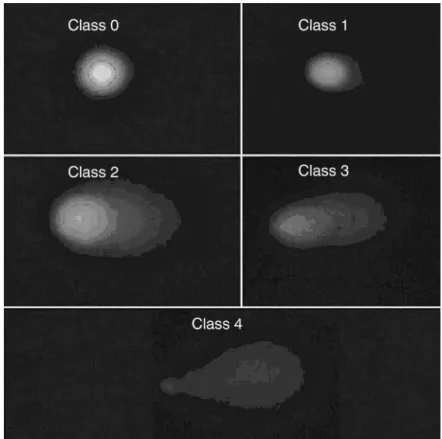
prevent additional DNA damage. The images of 100 ran-domly chosen nuclei (50 nuclei from each of two replicate
slides) were analyzed visually for each subject. Each image was classified according to the intensity of the fluorescence in
the comet tail and was given a value of 0, 1, 2, 3, or 4 (from
undamaged, class 0, to maximally damaged, class 4) (Figure 1), so that the total score of a slide could be between 0 and
400 arbitrary units (AU). Photomicrographs of representa-tive samples were depicted in Figures 2 and 3. The same
bio-chemistry staff completed all procedures and a single observer unaware of the subject’s group determined DNA damage
score.
TAC and TOS levels were measured by Erel’s meth-ods.15,16The percentage of TOS level to TAC level was
regarded as the OSI.17,18To perform the calculation, the result unit of TAC, mmol Trolox equivalents per liter, was changed to
µmol Trolox equivalent per liter, and the OSI value was
calcu-lated as follows: OSI = [(TOS, µmol per liter/(TAC, µmol Trolox equivalent per liter)/100].
Statistical analysis
Testing for normality of variances was accomplished using
Levene statistical tests. Variances in this assay were homo-geneous. The data were compared using one-way analysis of
comparison test. Sex ratio was compared using a chi-square test. Bivariate associations between variables were assessed by Pearson’s correlation test. The data were expressed as mean ± standard deviation and differences were considered statistically significant at p < 0.05. Statistical analysis was
conducted using SPSS for Windows Release 11.5 (SPSS, Chi-cago, IL, USA).
Results
Intensive phototherapy group’s mean age was 7±3 days,
mean height was 50±3.2 cm, mean body weight was 3.1±1.6
Figure 1 - Alkaline comet assay. Images were classified according to fluores-cence intensity in the comet tail and were given a value of 0-4 (from undamaged class 0 to maximally damaged class 4)
Figure 2 - Photomicrograph of representative sample inducing DNA damage in intensive phototherapy-treated infant. Cells were processed by alkaline single-cell gel electro-phoresis assay. Peripheral blood mononuclear leuko-cytes exposed to photototherapy exhibit comet tails indicative of damaged DNA
kg, mean duration of phototherapy was hours 54±6, and sex
ratio was 12/11 (M/F). Conventional phototherapy group’s mean age was 7±4 days, mean height was 50±3.6 cm, mean
body weight was 3.2±1.2 kg, mean duration of phototherapy was hours 61±10, and sex ratio was 13/10 (M/F). Control
group’s mean age was 5±2 days, mean height was 50±2.9
cm, mean body weight was 3±1.1 kg, and sex ratio was 10/9 (M/F). We found no significant differences between the three
groups in terms of age, length, weight, duration of photo-therapy (between intensive and conventional), gestational
age (all groups were 39±1) or male/female distribution (p > 0.05).
DNA damage scores, and TAC, TOS, OSI, and total biliru-bin levels are shown in Table 1. DNA damage scores were
32±9 in the intensive group, 28±9 in the conventional group, and 21±7 in the control group (p < 0.001). TOS and OSI
lev-els were significantly higher in the intensive and conventional groups than they were in the control group (p < 0.05). DNA
damage scores and TOS and OSI levels did not differ
signifi-cantly between the conventional and intensive phototherapy groups. Plasma TAC and total bilirubin levels did not differ
between the groups (p >0.05). There were no significant cor-relations between DNA damage scores and bilirubin, TOS and
OSI levels in the conventional or in the intensive photo-therapy groups (p > 0.05).
Discussion
The major results of the present study are that endog-enous mononuclear leukocyte DNA strand breaks, which are
a well-known type of DNA damage, were significantly increased in both conventional and intensive
phototherapy-treated infants when compared to the
con-trols. Interestingly, there was no significant correlation between DNA damage scores and TOS levels in the intensive
or conventional phototherapy groups. This is the first report showing an association between mononuclear leukocytes DNA
damage in intensive or conventional phototherapy-treated
jaundiced term infants.
Human peripheral mononuclear leukocytes have been
widely used to monitor environmentally induced genetic dam-age by a variety of methods, such as micronucleus,
chromo-some aberration, and sister-chromatid exchange assays.19 Among the various assays for measuring DNA damage, the
single cell gel electrophoresis (comet) assay is a sensitive and powerful method for determining DNA strand breaks.20,21It
has also been reported that strand breaks arise from DNA
damage generated by oxidative stress.14,22Measuring DNA damage with the comet assay in the human epidermis from
phototherapy-treated infants is not possible due to the diffi-culty in obtaining viable single cells from this tissue in infants.
We therefore used this method to measure DNA damage in circulating mononuclear leukocytes. However, future
assess-ments of DNA oxidation in the epidermis may shed further light on the role of oxidative stress in phototherapy-treated
infants.
Although we have demonstrated presence of DNA
dam-age in the form of direct strand breaks, we cannot rule out base oxidation. The repair of strand-break ligation (halftime
of approximately 30 min) is a much faster process than that of oxidized base lesions by base excision repair (halftime of
approximately 3 hours).23Thus, the repair procedures already existed for phototherapy 48 hours later. Similarly, the partial
inhibition of repair induced by the DNA polymerase inhibitor after phototherapy may represent interference with the
rejoining of direct strand breaks. Although presence of
com-ets after phototherapy may also indicate that strand breaks were induced as primary lesions, strand breaks measured by
the comet assay immediately after phototherapy could just as well represent nicks arising between the incision and
rejoin-ing stages of excision repair processes initiated durrejoin-ing solar irradiation.24
Table 1- Comparison of DNA damage, oxidative and antioxidative parameters of phototherapy-treated jaundiced newborns and controls
Intensive
phototherapy (n = 23)
Conventional
phototherapy (n = 23)
Control
(n = 19) p*
DNA damage (arbitrary unit) 32±9 28±9 21±10 < 0.001†‡
TAC (mmol Trolox equiv/L) 0.72±0.26 0.75±0.27 0.87±0.39 0.051
TOS (µmol H2O2equiv/L) 18.1±4.3 16.9±4.4 13.5±4.2 0.005†‡
Total bilirubin (mg/dL) 12.2±2.2 15.1±4.1 18.2±3.4 < 0.001†§
OSI (arbitrary unit) 2.9±1.5 2.6±1.5 1.9±1.3 0.041†‡
OSI = oxidative stress index; TAC = total antioxidant capacity; TOS = total oxidant status. Data are given as mean±standard deviation.
* One-way ANOVA with Tukey’s honestly significant difference multiple comparison test. †The difference was between phototherapy groups and controls.
Photoreactions are able to induce mutagenic
photoprod-ucts or lesions in DNA among adjacent pyrimidines in the form of dimmers.25Although wavelength from 245 to 290 nm is
absorbed maximally by DNA,26as seen in this study, at the wavelength 460 nm DNA damage resulted. This result may
be caused by the greater wavelength penetrating a deep layer
of the tissue, especially in the newborn’s vulnerable soft skin.27It appears that under phototherapy rate of damage
exceeds repair capacity. This study showed that mono-nuclear leukocyte DNA damages are greater in both
conven-tional phototherapy and intensive phototherapy-treated infants than in not treated phototherapy term jaundiced
infants. However, in this study, the main limitation is not using the same patients as their own control (before and during
pho-totherapy). In alkaline comet assay is very complex and
samples waiting are not possible.14Also, laboratory could accept blood samples for study of comet assay only at
par-ticular times of the week. These difficulties caused lost of cases and not using the same patients as their own control (before
and during phototherapy). We cope with these difficulties owing to using different patients treated with phototherapy
for at least 48 hours and controls (not exposure
photo-therapy). We think that new studies are needed to determine whether this changes in similar cases.
Free radicals can adversely alter lipids, proteins, and DNA.28,29Reactions of bilirubin involving free radicals or toxic
oxygen reduction products have been well documented: unconjugated bilirubin scavenges singlet oxygen with high
efficiency, reacts with superoxide anions and peroxyl radi-cals, and serves as a reducing substrate for peroxidases in
the presence of hydrogen peroxide or organic
hydroperox-ides.30,31This deterioration is especially evident in the pres-ence of oxidative stress such as phototherapy.7Bohles et al.
reported a significant decrease in antioxidants during photo-therapy.32However, our study showed that TAC levels were
not altered significantly by phototherapy. The oxidative/ antioxidative balance shifted significantly to the oxidative
side, because other indicators of oxidative status, TOS and OSI level, were significantly increased in infants exposed to
conventional and intensive phototherapy. We also found that
there were no significant correlations between DNA damage ratios and TOS and OSI levels (p>0.05). In this study, these
results indicate that DNA damage was not only caused by oxi-dative stress but also by other factors such as direct
interac-tion of phototherapy light with DNA. Interestingly, intensive and conventional phototherapy cause similar degrees of DNA
damage and oxidative stress. There may be a threshold effect
of phototherapy on these parameters.
It is stated that phototherapy’s noninvasive nature, easy
availability, low cost, and few side effects reported initially have almost led to the assumption that it is innocuous.33
How-ever, those using phototherapy should remain alert to these possibilities and avoid any unnecessary use because
unto-ward effects on DNA have been demonstratedin vitro.3,4,11
Our study showed that both intensive and conventional
pho-totherapy cause unfortunate effects on DNAin vivo.
In conclusion, both conventional phototherapy and
inten-sive phototherapy cause mononuclear leukocyte DNA dam-age in jaundiced term infants.
Acknowledgements
We are most grateful to the nurses of the Neonatal Ser-vice at Sanliurfa Children’s Hospital and Harran University
Medical School Biochemistry Department for their assistance in conducting this study.
References
1. Porter ML, Dennis BL.Hyperbilirubinemia in the term newborn.
Am Fam Physician. 2002;65:599-606.
2. Tan KL.Phototherapy for neonatal jaundice.Acta Paediatr. 1996; 85:277-9.
3. Lamola AA, Blumberg WE, McClead R, Fanaroff A.
Photoisomerized bilirubin in blood from infants receiving phototherapy.Proc Natl Acad Sci U S A. 1981;78:1882-6.
4. Babara JS, Robert MK. Jaundice and hyperbilirubinemia in the newborn. In: Behrman RE, Kliegman RM, Jenson HB, editors. Nelson Textbook of Pediatrics. 17th ed. Philadelphia, PA: Saunders; 2004. p. 592 7.
5. Brennan ML, Wu W, Fu X, Shen Z, Song W, Frost H, et al.A tale of two controversies: defining both the role of peroxidases in nitrotyrosine formation in vivo using eosinophil peroxidase and myeloperoxidase-deficient mice, and the nature of peroxidase-generated reactive nitrogen species.J Biol Chem. 2002;277:17415-27.
6. Valko M, Izakovic M, Mazur M, Rhodes CJ, Telser J.Role of oxygen radicals in DNA damage and cancer incidence.Mol Cell Biochem. 2004;266:37-56.
7. Gathwala G, Sharma S.Oxidative stress, phototherapy and the neonate.Indian J Pediatr. 2000;67:805-8.
8. Turgut M, Basaran O, Cekmen M, Karatas F, Kurt A, Aygun AD.
Oxidant and antioxidant levels in preterm newborns with idiopathic hyperbilirubinaemia.J Paediatr Child Health. 2004; 40:633-7.
9. Akisu M, Yilmaz D, Tuzun S, Kultursay N.Antioxidant defense systems in newborns undergoing phototherapy.Indian J Pediatr. 1999;66:651-5.
10. McDonagh AF. The role of singlet oxygen in bilirubin photo-oxidation.Biochem Biophys Res Commun. 1971;44: 1306-11.
11. Aycicek A, Erel O.Total oxidant/antioxidant status in jaundiced newborns before and after phototherapy.J Pediatr (Rio J). 2007; 83:319-22.
12. Newman TB, Maisels MJ.Evaluation and treatment of jaundice in the term newborn: A kinder, gentler approach.Pediatrics. 1992;89:809-18.
13. Singh NP, McCoy MT, Tice RR, Schneider EL.A simple technique for quantitation of low levels of DNA damage in individual cells.
14. Kocyigit A, Keles H, Selek S, Guzel S, Celik H, Erel O.Increased DNA damage and oxidative stress in patients with cutaneous leishmaniasis.Mutat Res. 2005;585:71-8.
15. Erel O.A novel automated direct measurement method for total antioxidant capacity using a new generation, more stable ABTS radical cation.Clin Biochem. 2004;37:277-85.
16. Erel O.A new automated colorimetric method for measuring total oxidant status.Clin Biochem. 2005;38:1103-11.
17. Aycicek A, Erel O, Kocyigit A.Increased oxidative stress in infants exposed to passive smoking.Eur J Pediatr. 2005;164: 775-8.
18. Harma M, Harma M, Erel O.Oxidative stress in women with preeclampsia.Am J Obstet Gynecol. 2005;192:656-7.
19. Cole J, Skopek TR.International Commission for Protection Against Environmental Mutagens and Carcinogens. Working paper no. 3. Somatic mutant frequency, mutation rates and mutational spectra in the human population in vivo. Mutat Res. 1994;304:33-105.
20. Moller P, Knudsen LE, Loft S, Wallin H.The comet assay as a rapid test in biomonitoring occupational exposure to DNA-damaging agents and effect of confounding factors.Cancer Epidemiol Biomarkers Prev. 2000;9:1005-15.
21. Garcia O, Mandina T, Lamadrid AI, Diaz A, Remigio A, Gonzalez Y, et al.Sensitivity and variability of visual scoring in the comet assay. Results of an inter-laboratory scoring exercise with the use of silver staining.Mutat Res. 2004;556:25-34.
22. Rojas E, Lopez MC, Valverde M.Single cell gel electrophoresis assay: methodology and applications.J Chromotogr B Biomed Sci Appl. 1999;722:225-54.
23. Sparrow JR, Zhou J, Cai B.DNA is a target of the photodynamic effects elicited in A2E-laden RPE by blue-light illumination.Invest Ophthalmol Vis Sci. 2003;44:2245-51.
24. Arlett CF, Lowe JE, Harcourt SA, Waugh AP, Cole J, Roza L, et al.Hypersensitivity of human lymphocytes to UV-B and solar irradiation.Cancer Res. 1993;53:609-14.
25. Matsumura Y, Ananthaswamy HN.Toxic effects of ultraviolet radiation on the skin.Toxicol Appl Pharmacol. 2004;195: 298-308.
26. Tornaletti S, Pfeifer GP.UV damage and repair mechanisms in mammalian cells. Bioessays 1996;18:221-8.
27. Meulemans CC, Werner M. Skin penetration depth optical radiation.http://www.solarcsystems.com/pdfs/philips_light_ sources_for_phototherapy.pdf. Access: 19/9/2007.
28. Warner BB, Wispe JRFree radical mediated diseases in pediatrics.
Semin Perinatol. 1992;16:47-57.
29. Halliwell B.Free radicals, antioxidants, and human disease: curiosity, cause, or consequence?Lancet. 1994;344:721-4.
30. Stocker R, Ames BN.Potential role of conjugated bilirubin and copper in the metabolism of lipid peroxides in bile.Proc Natl Acad Sci USA. 1987; 84:8130-4.
31. Stocker R, Glazer AN, Ames BN.Antioxidant activity of albumin bound bilirubin.Proc Natl Acad Sci USA. 1987;84:5918-22.
32. Bohles H, Schnall B.The effect of phototherapy on serum uric acid.Klin Padiatr. 1981;193:308-10.
33. Tan KL.Phototherapy for neonatal jaundice.Clin Perinatol. 1991; 18:423-39.
Correspondence: Ali Aycicek
Pediatrics Department, Medical Faculty, Harran University 63300 - Sanlıurfa - Turkey
Tel.: +90 (414) 314.8410 Fax: +90 (414) 313.9615