Int. Arch. . vol.19 número2
Texto
Imagem

Documentos relacionados
The results showed that the higher effi ciency in light energy uptake, paired with better photochemical performance and better CO 2 fi xation in plants under direct sunlight
In ragione di queste evidenze, il BIM - come richiesto dalla disciplina comunitaria - consente di rispondere in maniera assolutamente adeguata alla necessità di innovazione dell’intera
It is estimated that patients undergoing treatment for haematologic malignancies, such as leukaemia, have a mortality rate of 35% due to systemic fungal infections (Bhatt et
The length in 3D MRI images of the same cochlea were checked against each temporal bone (Table 2), using predefined measurements from the vestibule to the apex of the cochlea
The probability of attending school four our group of interest in this region increased by 6.5 percentage points after the expansion of the Bolsa Família program in 2007 and
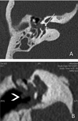
High resolution CT scan of temporal bone and head revealed a large soft tissue mass showed post contrast enhancement involving middle ear cavity and external auditory canal
Computed tomography (CT) findings showed the mandible fracture and complete opacification of the right maxillary sinus, with expansion and focal destruction of the cortical
Coronal CT with bone algoritm (axial plane) and MRI axial T2WI: inner table osteoma in the right temporal bone.... CT - coronal with bone algoritm and MRI coronal T1WI: outer
The CT scan showed a complex depressed skull fracture in the left temporal bone; two depressed skull fractures in the occipital bone and signs of pneumocephalus.. The 3D CT
