UNIVERSIDADE FEDERAL DA PARAÍBA CENTRO DE CIÊNCIAS DA SAÚDE
PROGRAMA DE PÓS-GRADUAÇÃO EM ODONTOLOGIA
EFEITO IN VITRO DE VERNIZES FLUORETADOS
CONTENDO NAF E TIF
4NAS PROPRIEDADES
NANOMECÂNICAS E DE SUPERFÍCIE DO
ESMALTE DENTÁRIO SUBMETIDO À EROSÃO
Maria Isabel Dantas de Medeiros
i MARIA ISABEL DANTAS DE MEDEIROS
EFEITO IN VITRO DE VERNIZES FLUORETADOS CONTENDO
NAF E TIF
4NAS PROPRIEDADES NANOMECÂNICAS E DE
SUPERFÍCIE DO ESMALTE DENTÁRIO SUBMETIDO À EROSÃO
Dissertação apresentada ao Programa de Pós-Graduação em Odontologia, da Universidade Federal da Paraíba, como parte dos requisitos para obtenção do título de Mestre em Odontologia – Área de Concentração em Odontologia Preventiva e Infantil.
Orientador: Profa. Dra. Fabíola Galbiatti de Carvalho Carlo Co-orientador: Prof. Dr. Hugo Lemes Carlo
ii
M488e Medeiros, Maria Isabel Dantas de.
Efeito in vitro de vernizes fluoretados contendo NaF e TiF4
nas propriedades nanomecânicas e de superfície do esmalte dentário submetido à erosão / Maria Isabel Dantas de
Medeiros.-- João Pessoa, 2014. 44f. : il.
Orientadora: Fabíola Galbiatti de Carvalho Carlo Coorientador: Hugo Lemes Carlo
Dissertação (Mestrado) - UFPB/CCS
1. Odontologia. 2. Odontologia preventiva e infantil. 3.Erosão dentária. 4. Módulo de elasticidade. 5. Fluoretos. 6.Microscopia de força atômica.
iii MARIA ISABEL DANTAS DE MEDEIROS
EFEITO IN VITRO DE VERNIZES FLUORETADOS CONTENDO
NAF E TIF
4NAS PROPRIEDADES NANOMECÂNICAS E DE
SUPERFÍCIE DO ESMALTE DENTÁRIO SUBMETIDO À EROSÃO
Banca Examinadora
_____________________________________________
Profa. Dra. Fabíola Galbiatti de Carvalho Carlo Orientadora - UFPB
_____________________________________________
Prof. Dr. Hugo Lemes Carvalho Carlo Co-orientador - UFPB
______________________________________________
Prof. Dr. Rogério Lacerda dos Santos Examinador - UFPB
______________________________________________
iv DEDICATÓRIA
Dedico essa minha dissertação de mestrado, bem como todos os meus esforços para ser uma boa profissional, a minha irmã Maria Hortência Dantas de Medeiros, que está viva em meu coração, e aos muitos pacientes que assim como ela tanto necessitaram e necessitam dos cuidados odontológicos.
As pessoas por muitas vezes precisam de um exemplo de vida para se espelhar e vencer as adversidades da vida, e Hortência é esse exemplo para mim, por ter sido uma pessoa feliz e alegre mesmo nos momentos mais difíceis, quando as limitações e a dor se faziam presentes.
v AGRADECIMENTOS
A Deus por ter me reservado esta oportunidade de aumentar meus conhecimentos, e colocar as pessoas certas no meu caminho durante esta jornada.
Aos meus pais, Maria do Céu e Ademar Sales, pelo incentivo e apoio durante a realização deste mestrado, assim como pelo cuidado e dedicação a mim dispensados, por toda minha vida, para construção da minha carreira profissional. “Mainha” obrigada por está ao meu lado, sempre.
Ao meu esposo Paulo André pelo incentivo e compreensão nesta etapa de minha vida. Obrigada pela cumplicidade e apoio que tem me dado independente das minhas escolhas. A sua atenção e carinho foram fundamentais neste processo.
A minhas irmãs, Luiza e Cecília, pelo apoio e atenção durante este mestrado.
Ao meu tio Antônio Ivo pela atenção e colaboração com meus estudos, e pelo exemplo que você deixou como profissional da saúde.
À Profa. Dra. Fabíola Galbiatti de Carvalhopela excelente orientação, pelos ensinamentos, dedicação e por acreditar no meu trabalho. Minha profunda admiração e respeito.
Ao Prof. Dr. Hugo Lemes Carlo, Coordenador do Mestrado Acadêmico em Odontologia da UFPB, pelo apoio e colaboração com o presente estudo.
Aos Profs. Wilton Padilha e Ana Maria Valença, que me ensinaram a gostar de pesquisa e me despertaram o interesse pela vida acadêmica.
Aos Profs. Frederico e Severino Jackson pela atenção e disponibilização dos laboratórios onde foi realizado o nosso trabalho.
vi À Universidade Federal da Paraíba-UFPB, instituição onde tive a oportunidade de dar um importante passo rumo ao crescimento científico e profissional.
A todos os funcionários desta instituição que contribuíram para a realização do meu trabalho durante este período.
Aos colegas do curso de mestrado por compartilhar comigo as dificuldades e conquistas neste curso de mestrado.
Ao doutorando Bruno, pela colaboração com o nosso estudo, sempre disponível e atencioso aos nossos questionamentos.
vii EPÍGRAFE
“Quem espera que a vida seja feita de ilusão
Pode até ficar maluco ou morrer na solidão É preciso ter cuidado pra mais tarde não sofrer É preciso saber viver...
Toda pedra do caminho você deve retirar Numa flor que tem espinhos você pode se arranhar Se o bem e o mal existem você pode escolher É preciso saber viver...”
viii RESUMO
Objetivos: Analisar o efeito dos vernizes de NaF e TiF4 na nanodureza (N) e módulo de elasticidade (E) da superfície do esmalte após um curto tempo de exposição a bebida a base de cola, simulando a ingestão de uma lata de refrigerante, e também avaliar a espessura e topografia da camada protetora formada, por meio de microscópio de força atômica (AFM).
Métodos: Trinta blocos de esmalte humano (4x4 mm) foram divididos em 3 grupos (n = 10): controle (sem verniz), verniz de NaF e verniz de TiF4. As amostras permaneceram em saliva artificial durante 24 horas, e os vernizes foram aplicados apenas uma vez. Após 6h os espécimes foram submetidos a desafio erosivo (10 ciclos: 5s em bebida a base de cola/5s em saliva artificial). A espessura da camada protetora formada e a topografia da superfície foram avaliadas por microscopia óptica e AFM. Os dados foram submetidos à ANOVA Two-Way, Tukey e T- Student (α = 0,05).
Resultados: Houve diferença estatística dos valores de N e E (GPa), antes e após o desafio erosivo, apenas para os grupos controle e TiF4. Após o desafio erosivo o grupo NaF mostrou estatisticamente maiores valores de N e E que os grupos controle e TiF4. No entanto, os valores da espessura e profundidade da indentação mostraram que os valores encontrados de N e E para o grupo TiF4 estavam relacionados com a camada protetora formada e não com o esmalte erodido. Ambos os vernizes mostraram, por meio de AFM, a formação de uma camada protetora globular, e a espessura da camada formada foi significativamente maior para o grupo TiF4 que NaF.
O verniz de NaF foi capaz de proteger as propriedades nanomecânicas de esmalte após curto desafio erosivo e o verniz de TiF4 apresentou formação de camada protetora espessa e homogênea com propriedades nanomecânicas . Conclusões: O verniz de NaF foi capaz de proteger as propriedades nanomecânicas de esmalte após curto desafio erosivo e o verniz de TiF4 apresentou formação de camada protetora espessa e homogênea com propriedades nanomecânicas.
ix ABSTRACT
Objectives: To analyze the effect of NaF and TiF4 varnishes on nanohardness (N) and elastic modulus (E) of enamel surface after very short exposure time to simulation of drinking a can of soft drink; and evaluate the thickness and topography of protective layer formed, by atomic force microscopy (AFM).
Methods: Thirty blocks of human enamel (4x4 mm) were divided into 3 groups
(n=10): control, NaF varnish and TiF4 varnish. The specimens remained in artificial saliva for 24h, and the varnishes were applied only once. After 6h, specimens were submitted to erosive challenge (10 cycles: 5s in cola drink/5s in artificial saliva). The thickness of protective layer formed and the surface topography were evaluated by optical microscope and AFM. The data were subjected to Two-Way
ANOVA, Tukey and Student’s-t(α=0.05).
Results: The values shown for N and E (GPa) before and after erosive challenge
differed statistically, only for control and TiF4 groups. After the erosive challenge, NaF group showed statistically higher values for N and E than those found for control and TiF4 groups. However, the thickness and depth of indentation values showed that N and E values found for TiF4 group were related to the protective layer formed. By AFM, both varnishes showed globular protective layer formation, and the thickness of layer was significantly higher for TiF4 than NaF group.
Conclusions: NaF varnish was able to protect the nanomechanical properties of
enamel after short erosive challenge, and TiF4 varnish showed a thick and homogeneous protective layer formation with nanomechanical properties.
x LISTA DE ABREVIATURAS E SIGLAS
NaF – Fluoreto de Sódio TiF4 – Tetrafluoreto de Titânio
AFM – Microscópio de força atômica N – Nanodureza
E – Módulo de elasticidade CaF2 – Fluoreto de cálcio H – dureza
Pmax - Pico de carga Ac - Área de contato V - coeficiente de Poisson
xi SUMÁRIO
1. INTRODUÇÃO 01
2. CAPÍTULO 1 05
3. CONSIDERAÇÕES GERAIS 23
4. CONCLUSÃO 25
REFERÊNCIAS 26
ANEXO 1 33
ANEXO 2 34
1 1. INTRODUÇÃO
Um declínio na prevalência de cárie dentária em países industrializados ou em desenvolvimento tem sido observado nas últimas décadas 9, 39, 54, e paralelamente a esta redução, outra lesão causada pela desmineralização da estrutura dentária tem recebido destaque: a erosão dentária 20.
Estudos recentes demonstram uma alta prevalência de erosão dentária 7, 21, 38, 44. Bartlett et al. 7 estudaram a prevalência de erosão dentária em uma amostra de jovens adultos europeus com idades entre 18-35 anos e relataram que 57,1% se enquadravam nos escores de 1 a 3 de BEWE (Basic Erosive Wear Examination) que vai desde perda inicial da superfície até perda de superfície maior que 50%. Murakami et al. 38 avaliaram a prevalência de desgaste dentário erosivo em 967 pré-escolares brasileiros, e constataram que estava presente em 51,6% das crianças. Na Noruega Søvik et al. 44 relataram a uma prevalência de erosão dentária de 59%. Isaksson 21 observou a prevalência de erosão dentária, constatando que 75% dos indivíduos estudados em uma coorte de 494 suecos de 20 anos de idade apresentavam erosão dentária, sendo também encontrada uma relação entre erosão dentária e estilo de vida e sobrepeso/obesidade.
A erosão dentária é definida como a perda de estrutura dentária devido a processos químicos, decorrentes de exposição ácida, sem o envolvimento bacteriano 1, 49. Os fatores etiológicos podem ser intrínsecos relacionados ao ácido gástrico em indivíduos que são acometidos por doença gástrico esofágica, doenças metabólicas e psicossomáticas 11, 28, e fatores extrínsecos advindos de ácidos provenientes da dieta e medicamentos, bem como ácidos de origem ambiental/ocupacional no qual o indivíduo pode absorver substâncias (líquidos ou vapores ácidos) como trabalhadores em indústrias de produção de baterias e fertilizantes e atletas nadadores 28, 53, 55.
2
doenças gástrico esofágicas 45. Assim, a aplicação tópica de flúor é uma maneira simples e acessível para proteger o esmalte frente à desmineralização por ácidos 12.
A ação de fluoretos é principalmente atribuída a precipitação de glóbulos de fluoreto de cálcio (CaF2) na superfície do esmalte dentário, o qual atua como uma barreira física que dificulta o contato dos ácidos com o esmalte subjacente. Esta precipitação de CaF2 forma um reservatório mineral, o qual é “atacado” pelo
desafio erosivo, liberando íons que auxiliam no tamponamento dos ácidos e na remineralização 35, 41. A formação da camada de CaF2 e seu efeito protetor contra a desmineralização dependem do pH, concentração de íons fluoreto, tipo de sal do fluoreto e veículo 35. As preparações de fluoretos mais ácidas, assim como as mais concentradas podem formar camadas mais espessas de CaF2, podendo portanto, oferecer uma maior proteção contra a erosão dentária 37.
Pesquisas mostram que compostos a base de fluoreto de sódio, o qual é comumente utilizado, previnem significativamente a progressão da erosão dentária 12, 22, 23. Entretanto, existem estudos que demonstram que outros compostos fluoretados, como o tetrafluoreto de titânio (TiF4), podem apresentar efeito protetor significativamente maior contra a erosão dentária que o fluoreto de sódio 17, 18, 30, 50, 51.
3
Wiegand et al. 51 observaram em seu estudo que a solução de TiF4 pode diminuir a perda de cálcio no esmalte e na dentina durante a erosão, sendo que o seu efeito a longo prazo é limitado e restrito a dentina. Dessa forma estes autores sugeriram a realização de novas investigações em que o veículo fluoretado tenha melhor capacidade de aderência a superfícies dentais, tais como géis ou vernizes, o que poderia aumentar seu efeito protetor.
Magalhães et al. 30, compararam in vitro o efeito do verniz experimental de TiF4 a vernizes utilizados comercialmente a base de fluoreto de sódio, e a solução de TiF4, sobre a erosão em esmalte de dente bovino, e concluíram que, nas condições do estudo, o verniz de TiF4 reduziu a erosão em esmalte bovino, mostrando melhores resultados em relação aos dois vernizes de fluoreto de sódio. Levy et al. 24 analisaram in vitro o efeito dos vernizes e soluções de TiF4 e NaF
na proteção contra a erosão do esmalte dentário com e sem abrasão. Estes autores observaram que todos os vernizes fuoretados foram capazes de reduzir significativamente a perda de esmalte, de modo que o verniz de TiF4 apresentou os menores valores de perda mineral. Foi concluído que o verniz de TiF4 pode ter um efeito promissor na redução da perda de esmalte sob condições erosivas e abrasivas in vitro.
Uma das formas de avaliação in vitro utilizada em estudos relacionados às
alterações sobre as propriedades mecânicas das estruturas dentárias é a técnica de nanoindentação 3, 6, 8, 10. Nos estágios iniciais da erosão, o teste de nanoindentação é capaz de medir pequenas alterações de estrutura fornecendo a nanodureza e o módulo de elasticidade 3, 4, 8, 10, 14, 26, 43. Esta técnica permite a aplicação de pequenas forças resultando em deformações na estrutura em escala nanométrica. Além disso, muitos estudos associam o teste de nanoindentação juntamente com a análise em microscopia de força atômica, o que possibilita correlacionar as alterações mecânicas de ultraestrutura com a topografia de superfície 4, 10, 14, 26, 27, 43.
4
O módulo de elasticidade do esmalte dentário submetido à erosão foi investigado por Barbour et al. 5, Barbour et al. 6, Beyer et al. 8 e Lippert, Parker e Jandt 26. Beyer et al. 8 observaram redução dos valores de nanodureza e módulo de elasticidade em esmalte dentário humano submetido a diferentes soluções ácidas (ácido tartárico, maleico, láctico, ascórbico, fosfórico e cítrico), bem como Barbour et al. 5, que utilizaram soluções de ácido cítrico com diferentes valores de pH pra produzir erosão. Lippert, Parker e Jandt 26 investigaram um possível reendurecimento da superfície amolecida de esmalte por meio de solução remineralizante com propriedades químicas semelhantes as da saliva humana, porém não foi observado aumento da nanodureza do esmalte após a remineralização. Porém, ainda não foi investigado o efeito da aplicação de agentes fluoretados no módulo de elasticidade e nanodureza do esmalte dentário durante o desenvolvimento inicial da erosão.
5 2. CAPÍTULO 1
O manuscrito a seguir foi submetido para publicação no periódico “Journal of Dentistry” e encontra-se em análise.
Effect of NaF and TiF4 varnishes on nanomechanical properties of enamel surface and protective layer after a short erosive challenge
Maria Isabel Dantas Medeirosa, Bruno Alessandro Silva Guedes Limab, Hugo Lemes Carloc, Rogério Lacerda dos Santosd, Fabíola Galbiatti de Carvalhoa
a Department of Clinical and Social Dentistry, Health Science Center, UFPB – Federal University of Paraíba, Brazil;
b Department of Mechanical Tecnology, Tecnological Center, UFPB - Federal University of Paraíba, Brazil;
c Department of Operative Dentistry, Health Science Center, UFPB - Federal University of Paraíba, Brazil;
d Department of Biological Science, Division of Dentistry, UFCG - Federal University of Campina Grande, Brazil
Key Words: tooth erosion; elastic modulus; fluorides; microscopy, atomic force
Corresponding Author: Prof. Fabíola Galbiatti de Carvalho - Department of Clinical and Social Dentistry - Federal University of Paraíba - Health Science Center – Campus I - João Pessoa, Paraíba, Brazil
6
Effect of NaF and TiF4 varnishes on nanomechanical properties of enamel surface and protective layer after a short erosive challenge
Abstract
Objectives: To analyze the effect of NaF and TiF4 varnishes on nanohardness (N) and elastic modulus (E) of enamel surface after very short exposure time to simulation of drinking a can of soft drink; and evaluate the thickness and topography of protective layer formed, by atomic force microscopy (AFM).
Methods: Thirty blocks of human enamel (4x4 mm) were divided into 3 groups
(n=10): control, NaF varnish and TiF4 varnish. The specimens remained in artificial saliva for 24h, and the varnishes were applied only once. After 6h, specimens were submitted to erosive challenge (10 cycles: 5s in cola drink/5s in artificial saliva). The thickness of protective layer formed and the surface topography were evaluated by optical microscope and AFM. The data were subjected to Two-Way
ANOVA, Tukey and Student’s-t(α=0.05).
Results: The values shown for N and E (GPa) before and after erosive challenge
differed statistically, only for control and TiF4 groups. After the erosive challenge, NaF group showed statistically higher values for N and E than those found for control and TiF4 groups. However, the thickness and depth of indentation values showed that N and E values found for TiF4 group were related to the protective layer formed. By AFM, both varnishes showed globular protective layer formation, and the thickness of layer was significantly higher for TiF4 than NaF group.
Conclusions: NaF varnish was able to protect the nanomechanical properties of
enamel after short erosive challenge, and TiF4 varnish showed a thick and homogeneous protective layer formation with nanomechanical properties.
Clinical Significance: After very short erosive challenge the enamel surface
7 Introduction
The strategy to control or to prevent erosive demineralization of teeth involves the topical application of conventional fluoride, such as sodium fluoride (NaF).1-3 The action of NaF fluoride is mainly attributed to precipitation of CaF2-like material on the eroded dental surface forming a protective layer.1,4 The topical application of highly concentrated NaF varnishes is often recommended to prevent erosion5 and these have demonstrated effective protection against dental erosion due to their higher fluoride concentration and capacity to adhere to enamel.6-8
As the anti-erosive effect of conventional fluorides requires a very intensive fluoridation regime,9 current studies have focused on fluoride compounds that might have higher level of efficacy, for example, compounds containing polyvalent metal ions such as titanium tetrafluoride (TiF4). The protective capacity of TiF4 is
related to increased uptake of fluoride by acidic pH and the formation of a glaze-like surface layer that might primarily act as an acid-resistant diffusion barrier.10, 11 Although some studies have shown that TiF4 varnishes had an inhibitory effect on dental erosion,3, 7, 12, 13 the results about its effect are still contradictory.14, 15
Several studies have investigated the effect of NaF and TiF4 varnishes on enamel erosion by experimental models of erosive challenge with a variety of daily cycles performed in a period from 5 to 7 days 3, 7, 12, 13 to simulate patients with dental erosion. However, little attention has been given to the initial alterations in the enamel structure caused by occasional soft drink consumption, such as drinking a can of soft drink or a single intake of soft drink during a meal, which is a very common habit in the world population. This fact is relevant because a very short time of exposure to an acidic beverage may lead to the early stages of dental erosion, which are characterized by softening of the enamel surface.16
The initial changes in enamel caused by acidic beverages have been measured by the nanoindentation test.16- 20 However, the effect of NaF and TiF4 varnishes on the nanomechanical properties of enamel after initial erosion and the thickness and topography of the protective layer formed by both varnishes have
not been reported before. Therefore, the hypotheses tested were꞉ 1) There was
8
between the thickness and topography of the protective layer formed by the two varnishes on eroded enamel, when analyzed by atomic force microscopy (AFM).
Material and method Specimen Preparation
After obtaining approval from the Research Ethics Committee, twenty-five sound human third molars were selected for this study. Teeth were stored in 0.1% thymol at 4oC and used within 1 month after extraction. From each tooth, two enamel specimens (4 x 4 x 3 mm) were cut with a flexible diamond disc (7016, KG Sorensen, Barueri, SP, Brazil) at low speed under water cooling, and fifty specimens were obtained. They were embedded in acrylic resin (Vipi Flash –
Pirassununga, SP, Brazil) and the enamel surfaces were ground flat with SiC paper discs (400, 600 and 1200 grit) and polished with 1µm alumina suspension (Erios Corp., São Paulo, SP, Brazil). Afterwards, the specimens were cleaned in an ultrasonic device (UltrasonicCleaner, model USC1400, Unique Ind. Com. Ltda, São Paulo, SP, Brazil) for 2 min and checked for the presence of cracks and fractures using a microscope (Nikon 88286, Tokyo, Japan) at 40x magnification. The baseline nanohardness of the enamel surface was determined and specimens with 4.85 GPa ± 20% of this value were selected 21 to standardize the initial hardness. Specimens without the pre-determined values were discarded. Thirty specimens were selected and allocated to three groups (n=10), according to fluoride varnish application: 1. Control group – without agent application; 2. NaF group – NaF varnish application (Duraphat, Colgate-Palmolive Ind. Com. Ltda, São Bernardo, SP, Brazil) and 3.TiF4 group – experimental TiF4 varnish application (FGM, Joinville, Santa Catarina, Brazil) (Table1). Ten specimens were selected for evaluation of thickness of the protective layer formed by varnish application.
Fluoride varnish application
9
For the NaF and TiF4 groups, 20 μL of each fluoride varnish was applied on the specimen surface and spread with microbrush. After this, the specimens were immersed in artificial saliva for 6 h, for clinical simulation of the contact time of the varnish with the tooth surface.12, 14, 15 After this period, the varnishes were carefully removed from the surface using acetone and a scalpel blade, taking care to avoid touching the enamel surface.14, 15 In the control group no product was applied and specimens were immersed in artificial saliva for 6 h. The method of producing artificial saliva used was similar to that of McKnight-Hanes and Whitford, 23 but without sorbitol (in g:L). The varnishes were applied only once before the erosive challenge. 6, 15
To evaluate the thickness of the protective layer formed by TiF4 and NaF varnishes, five specimens were prepared for each varnish group. The enamel surface was divided into two equal parts: one half of the surface was covered with 2 layers of acid-resistance nail varnish (control surface - without varnish application) and on the other half, fluoride varnish was applied according to each group. After fluoride application, the nail varnish was removed with a scalpel blade and the entire surface of the specimen was submitted to the erosive challenge.
Erosive challenge
The erosive challenge was based on simulating a short time of exposure to a cola beverage (pH 2.6; coke, Porto Real, RJ, Brazil) according to Wongkhantee et al.24: the specimens were immersed in 32.5 mL of cola drink for 5 s at room temperature, rinsed in deionized water and then immersed in 32.5 mL of artificial saliva at room temperature for 5 s. This cycle was repeated 10 times. After this, the specimens were stored in 100% humidity until the nanoindentation test was performed.
Nanoindentation test
10
nanohardness and elastic modulus. Measurements were taken at baseline and after erosive challenge. All readouts were performed by the same examiner.
The method described by Oliver and Pharr25 was used to calculate the hardness (H) and elastic modulus of enamel (E), using the formulas:
H= Pmax / Ac
1/Er= (1 - v2)/E + (1 – vi2)/Ei,
where Pmax is peak load and Ac is the contact area, and E and v are Young’s
modulus and Poisson’s ratio for the specimen and Ei and vi are the same
parameters for the indenter, respectively. Poisson’s ratio for enamel was assumed
to be 0.4.26
The depth of indentation for each specimen after varnish application and erosive challenge was given by ultramicrohardness software and it was determined as Maximum Height (nm). The depth of indentation values were compared with the thickness values of the protective layer formed by varnish application to investigate whether the indentation was made in the enamel surface or in the protective layer.
Optical Microscopy
Five specimens from each group were randomly selected for analysis of the enamel surface after varnish application and erosive challenge, by Optical Microscope (Axiotech), at 100x magnification.
Atomic Force Microscopy
Five randomized specimens of each group were analyzed using atomic force microscopy (AFM; SPM-9600, Shimadzu, Kyoto, Japan). Each specimen
was fixed to the microscope holder on a stub (2 x 3 mm). The block surface morphology was probed in ‘‘contact mode’’. Imaging was performed with standard
geometry silicon nitride Micro-Cantilever (OMCL-TR, Olympus, Tokyo, Japan) and probed with 0.15 N/m constant elastic and 24 KHz resonant frequency. Images 30 µm x 30 µm with a resolution of 256 X 256 pixels and operating point of 1.5 V were collected at a very low scan rate to obtain details of the enamel structure and to avoid damaging the tip.
11
parameters as described above. After image capture, the Height Trace tool was
used. Four parallel lines were made on each specimen. Each outline started on the control half (without fluoride application) and finished on the half to which the fluoride varnish was applied. The difference in height of each line was recorded by
the Height Trace tool and the thickness of the protective layer was measured. The
mean thickness (nm) of the protective layer was obtained for each group.
Statistical analysis
Data analysis was performed with the GraphPad Instat version 2.0 (GraphPad software, La Jolla, USA) at a level of significance of α = 0.05. Since all the variables tested satisfied the assumptions of equality and normal distribution
(Bartlett and Kolmogorov–Smirnov tests, respectively), Two-Way ANOVA and Tukey tests were carried out for statistical comparisons of enamel nanohardness and elastic modulus among the groups. To compare the baseline and after erosive challenge nanohardness and elastic modulus values in the same group the paired-T test was used.
Results
Mean values (± sd) of enamel nanohardness, elastic modulus for each group are reported in Table 2. There were no statistically significant differences among groups for baseline values of nanohardness and elastic modulus (p=0.73 and p=0.81, respectively). After erosive challenge, there were significant differences of nanohardness and elastic modulus values among groups (p=0.001). The Control Group had intermediate values of nanohardness (3.4 ± 0.4 GPa) and elastic modulus (104.0 ± 8.5 GPa) and the TiF4 varnish group statistically showed the lowest nanohardness (2.4 ± 0.8 GPa) and elastic modulus (82.2 ±14.3 GPa) values (p=0.001). The NaF varnish group showed the highest nanohardness (4.4 ± 0.5 GPa) and elastic modulus (121.7 ± 15.3 GPa) values. The statistical comparison before (baseline) and after erosion for each group showed that only the Control and TiF4 varnish groups showed significant differences for nanohardness (p=0.001 and p=0.002, respectively) and elastic modulus values (p=0.008 and p=0.001, respectively).
12
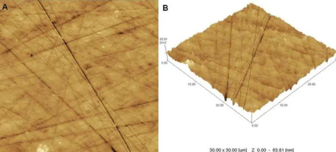
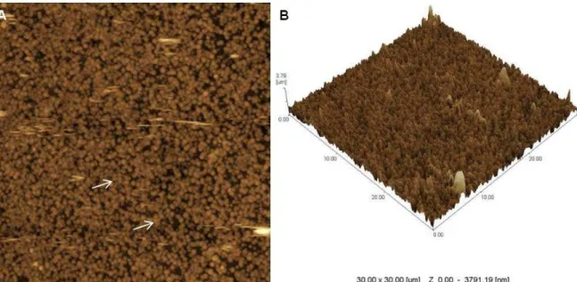
protective layer (Figs. 1A and 1B). The varnishes showed different surface topographies. The NaF varnish group showed a surface interspersed with globular deposits forming a non-homogeneous globular layer (Figs. 2A and 2B). The TiF4 varnish group showed homogeneous globular deposition forming a protective layer that completely hid the enamel surface (Figs. 3A and 3B).
Figure 4 shows the optical microscopy images after fluoride varnish application and erosive challenge. Both varnishes formed protective layer on the enamel surface (Fig 4B and 4C), however, the TiF4 varnish showed a more homogeneous layer formation, completely covering the enamel surface (Fig. 4C). The NaF varnish showed the formation of an interspersed protective layer on enamel (Fig. 4B). The Control group showed no protective layer formation (Fig. 4A).
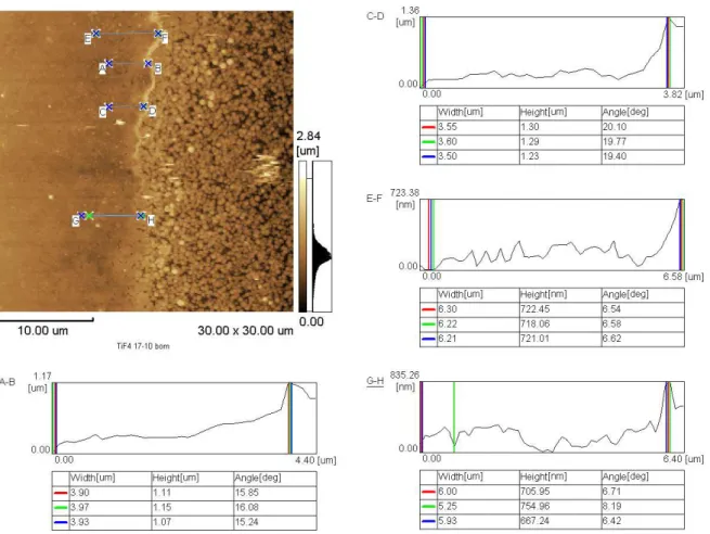
Mean thickness values (± sd) of the layer formed by the TiF4 varnish and NaF varnish groups, and mean values (± sd) of maximum indentation height for each group are reported in Table 3. The depth of indentation post-erosion was significantly higher for the TiF4 varnish group (296.9 ± 49.2 nm), followed by Control (237.5 ± 19.7 nm) and NaF varnish groups (213.7 ± 17.1). The protective layer thickness of the TiF4 varnish group also was significantly higher (953 ± 55.7 nm) than that of the NaF varnish group (53.1 ± 3.7 nm). Thus, the thickness of the protective layer formed was higher than the indentation depth for the TiF4 varnish group. Figure 5 illustrates the measurement of thickness of the protective layer formed by TiF4 varnish application after the erosive challenge, analyzed by AFM.
Discussion
The nanomechanical properties of enamel surfaces subjected to initial erosion after the application of remineralizing agents have not been extensively investigated and only solutions with or without the addition of fluoride have been tested.17, 27 Asfluoride varnish is recommended to prevent erosion,5 the innovative approach of the present study was related to the effect of NaF and TiF4 varnishes on the nanomechanical properties of enamel surfaces after a short time of exposure to a cola beverage.
13
structure overlying sound tissue.16 If the exposure to acid is prolonged or repeated at short intervals, the mineral loss continues and the outermost layer of softened tissue is completely dissolved and irreversibly lost.16 The majority of studies have investigated the effect of NaF and TiF4 varnishes on enamel erosion caused by acidic beverages for a prolonged period of time. 7, 13, 14, 22 However, it was
demonstrated by nanoindentation test that there was significant enamel softening
after 30 s exposure to soft drink,28 thus making it clinically relevant to investigate the effect of short times of exposure to acidic beverages on enamel surfaces.
The experimental model of erosive challenge used in the present study was designed to simulate an individual drinking a can of soft drink (325 mL) with the washing effect of saliva, by cyclic specimen immersion and time comparable with that taken for the intake of a single drink, as was used by Wongkhantee et al.24 and Panich and Poolthong 29. Furthermore, the nanoindentation test has a limited use for longer exposure times than the one evaluated in the present study.28, 19
The standardization of baseline nanohardness values (Table 2) made it possible to establish nanomechanical comparisons among the groups after treatment. The NaF varnish group was the only that showed no statistically significant difference before and after erosive challenge (Table 2), demonstrating that this varnish was effective in preventing the initial demineralization or softening of enamel caused by a very short time of exposure to cola beverage. This protective effect of NaF varnish has also been observed in previous studies,6, 14, 30 and results from the formation of a CaF2 layer that acts as a mineral reservoir. It also behaves as a physical barrier that prevents the acid from coming into contact with the underlying enamel, leading to decreased susceptibility to further enamel dissolution.4, 13 AFM images (Fig. 2) showed globular deposits on the surface, indicating formation of the CaF2 layer, with a thickness of 53.1 ± 3.7 nm even after the erosive challenge (Table 3).
14
nanohardness and elastic modulus of enamel. This result emphasizes the importance of prevention of dental erosion even in patients who have not yet developed the disease, but are considered a risk group, such as those who habitually consume soft drinks. Wongkhantee et al.24 and Panich and Poolthong 29 also related the reduction in enamel microhardness values after using the same erosive challenge.
Some factors have been shown to influence the efficacy of fluoride agents against demineralization, mainly the composition of fluoride salt, fluoride concentration and pH.4 In the present study, TiF4 showed higher concentration of fluoride (2.45%) and lower pH (3.4) in comparison with NaF (2.26% and 4.5, respectively). From this aspect, one would expect a better effect of TiF4 when compared with NaF, not only because of the titanium, but also due to the high concentration and low pH, which could increase the enamel fluoride uptake. Although this study found lower nanohardness and elastic modulus values for the TiF4 group, these results were not consistent with the optical and AFM images (Figs. 3 and 4). AFM and optical images (Figs.3 and 4C) showed a homogeneous globular deposition completely hiding the enamel surface, showing the protective layer formed by TiF4 varnish application. This layer probably is the glaze-like surface layer cited by others authors.10, 11 The glaze-like layer is assumed to be formed from a new compound (hydrated hydrogen titanium phosphate) or organometallic complexes by substitution of calcium by titanium ions in the apatite lattice.10, 11 Magalhães et al.7 investigated the surface layer coating by TiF4 varnish, using scanning electron microscopy and confirmed the titanium deposits with concentrations of 3.5 ± 1.0% Ti and 2.0 ± 0.8% of fluoride. This layer can be characterized as barrier acid-resistance diffusion, 7, 10, 11 which might have prevented the reduction in nanomechanical properties of enamel.
15
related to the enamel surface treated. To author´s knowledge, this is thought to be the first demonstration that the precipitates formed after the application of TiF4 produced a metal-rich layer that has a significantly higher level of nanomechanical properties and thickness than the layer formed by NaF varnish (Table 3). The nanohardness and elastic modulus values of the TiF4 layer formed (2.4 ± 0.8 and 82.2 ±14.3 GPa, respectively) were significant lower than eroded enamel values (3.4 ± 0.4 and 104.0 ± 8.5 GPa, respectively) (Table 2). However, these values may be considered high, since the numerical difference between them was 1 GPa for nanohardness and 21.8 GPa for elastic modulus. Based on this fact, it can be suggested that in addition to the metal-rich layer perhaps being able to act as barrier acid-resistance diffusion, it might also be able to support mechanical forces and protect the enamel surface against erosion and abrasion.
Although the nanomechanical properties of enamel after TiF4 varnish application could not be measured, AFM images (Figs. 3 and 5) showed the formation of a homogeneous, thick and mechanically resistant layer on the surface, indicating the protective effect of this varnish. The hypotheses tested in this study were accepted and the potential of TiF4 varnish for use as a remineralizing agent is of special interest. However, maintenance of the mechanical properties of the TiF4 layer formed need to be investigated after aggressive erosive challenge, and clinical studies are still necessary to evaluate its protective effect against erosion.
Conclusion
The NaF varnish was able to protect the nanomechanical properties of enamel surface and to create a protective layer after a very short time of exposure to cola beverage. For TiF4 varnish, the nanohardness and elastic modulus values post-erosion were related to the homogeneous, thick, mechanically resistant layer formed on surface, which demonstrated the protective effect of this varnish.
Acknowledgment
16 References
1. Magalhães AC, Wiegand A, Rios D, Honório HM, Buzalaf MA. Insights into preventive measures for dental erosion. Journal of Applied Oral Science 2009;
17:75-86.
2. Ganss C, Schlueter N, Hardt M, Schattenberg P, Klimek J. Effect of fluoride compounds on enamel erosion in vitro: a comparison of amine, sodium and stannous fluoride. Caries Research 2008;42:2-7.
3. Levy FM, Rios D, Buzalaf MAR, Magalhães AC. Efficacy of TiF4 and NaF varnish and solution: a randomized in situ study on enamel erosive–abrasive wear.
Clinical Oral Investigations 2013; DOI 10.1007/s00784-013-1096-y.
4. Saxegaard E, Rølla G. Fluoride acquisition on and in human enamel during topical application in vitro. Scandinavian Journal of Dental Research 1988;96
:523-35.
5. Mohammed A, Dusara K. What is the role of topical fluoride application in preventing dental erosion? Evidence-Based Dentistry 2013;14:59-62.
6. Murakami C, Bönecker M, Corrêa MSNP, Mendes FM, Rodrigues CRMD. Effect of fluoride varnish and gel on dental erosion in primary and permanent teeth.
Archives of Oral Biology 2009;54:997- 1001.
7. Magalhães AC, Kato MT, Rios D, Wiegand A, Attin T, Buzalaf MAR. The effect of an experimental 4% TiF4 varnish compared to NaF varnishes and 4% TiF4 solution on dental erosion in vitro. Caries Research 2008;42:269–74.
8. Vieira A, Jager DH, Ruben JL, Huysmans MC. Inhibition of Erosive Wear by Fluoride Varnish. Caries Research 2007;41:61-7.
9. Ganss C, Klimek J, Brune V, Schürmann A. Effects of two fluoridation measures on erosion progression in human enamel and dentine in situ. Caries Research
2004;38:561–6.
10. Büyükyilmaz T, Øgaard B, Rølla G. The resistance of titanium tetraf luoride-treated human enamel to strong hydrochloric acid. European Journal of Oral
Sciences 1997;105:473–7.
11. Ribeiro CC, Gibson I, Barbosa MA. The uptake of titanium ions by hydroxiapatite particles - structural changes and possible mechanisms.
17
12. Magalhães AC, Romanelli AC, Rios D, Comar LP, Navarro RS, Grizzo LT et al. Effect of a single application of TiF4 and NaF varnishes and solutions combined with Nd:YAG laser irradiation on enamel erosion in vitro. Photomedicine and Laser
Surgery 2011;29:537- 44.
13. Levy FM, Magalhães AC, Gomes MF, Comar LP, Rios D, Buzalaf MAR. The erosion and abrasion-inhibiting effect of TiF4 and NaF varnishes and solutions on enamel in vitro. International Journal of Paediatric Dentistry 2012;22:11- 6.
14. Magalhães AC, Stancari FH, Rios D, Buzalaf MAR. Effect of an experimental 4% titanium tetrafluoride varnish on dental erosion by a soft drink. Journal of
Dentistry 2007;35:858-61.
15. Magalhães AC, Levy FM, Rios D, Buzalaf MAR. Effect of a single application of TiF4 and NaF varnishes and solutions on dentin erosion in vitro. Journal of
Dentistry 2010;38:153- 7.
16. Barbour ME, Shellis RP. An investigation using atomic force microscopy nanoindentation of dental enamel demineralization as a function of undissociated acid concentration and differential buffer capacity. Physics in Medicine and Biology
2007;52:899-910.
17.Lippert F, Parker DM, Jandt KD. In vitro demineralization/remineralization cycles at human tooth enamel surfaces investigated by AFM and nanoindentation.
Journal of Colloid and Interface Science 2004;280:442- 8.
18. Cheng Z, Wang X, Cui F, Ge J, Yan J. The enamel softening and loss during early erosion studied by AFM, SEM and nanoindentation. Biomedical Materials
2009;4:1-7.
19. White AJ, Yorath C, ten Hengel V, Leary SD, Huysmans MC, Barbour ME. Human and bovine enamel erosion under 'single-drink' conditions. European
Journal of Oral Sciences 2010;118:604-9.
20. Beyer M, Reichert J, Bossert J, Sigusch BW, Watts DC, Jandt KD. Acids with an equivalent taste lead to different erosion of human dental enamel. Dental
Materials 2011;27:1017-23.
18
22. Magalhães AC, Rios D, Honório HM, Jorge AMJ, Delbem ACB, Buzalaf MAR. Effect of 4% titanium tetrafluoride solution on dental erosion by a soft drink: an in situ/ex vivo study. Archives of Oral Biology 2008;53:399 - 404.
23. Mcknight-hanes C, Whitford GM. Fluoride release from three glass ionomer materials and the effects of varnishing with or without finishing. Caries Research
1992;26:345-50.
24. Wongkhantee S, Patanapiradej V, Maneenut C, Tantbirojn D. Effect of acidic food and drinks on surface hardness of enamel, dentine, and toothcoloured filling materials. Journal of Dentistry 2006;34:214-20.
25. Oliver WC, Pharr GM. An improved technique for determining hardness and elastic modulus using load and displacement sensing indentation experiments.
Journal of Materials Research 1992;7:1564- 83.
26. Habelitz S, Marshall SJ, Marshall GW, Balooch M. Mechanical properties of human dental enamel on the nanometre scale. Archives of Oral Biology
2001;46:173 - 83.
27. Abdullah AZ, Ireland AJ, Sandy JR, Barbour ME. A nanomechanical investigation of three putative anti-erosion agents: remineralisation and protection against demineralisation. International Journal of Dentistry DOI
10.1155/2012/768126.
28. Barbour ME, Parker DM, Allen GC, Jandt KD. Human enamel erosion in constant composition citric acid solutions as a function of degree of saturation with respect to hydroxyapatite. Journal of Oral Rehabilitation 2005;32:16- 21.
29. Panich M, Poolthong S. The effect of casein phosphopeptide-amorphous calcium phosphate and a cola soft drink on in vitro enamel hardness. Journal of
the American Dental Association 2009;140:455-60.
19 Figure legends
Figure 1 - AFM image of enamel surface after erosive challenge and no-varnish
application. (A) “Straches” lines caused by polish procedure. (B) Rough surface
without protection layer.
20
Figure 3 - AFM image of enamel surface after TiF4 varnish application and erosive challenge. (A) (→) Homogeneous globular deposits on surface. (B) Enamel surface completely hidden by a layer of globular deposits.
21
Figure 5 - AFM image of TiF4 varnish group specimen with surface divided on side with varnish application + erosive challenge and side without varnish application + erosive challenge. The image shows the Height Trace tool used to obtain the
22
Table 1. Composition of fluoride varnishes tested in the study.
Material Composition (batch number)
Duraphat
(Colgate-Palmolive, Ind. Com. Ltda, São Bernardo, SP, Brazil)
2.26% sodium fluoride, alcohol, natural resins, wax, saccharine, flavor - pH 4.5
(051001)
Experimental TiF4 varnish (FGM, Joinville, SC, Brazil)
2.45% titanium tetrafluoride, ethanol, synthetic resin and natural resin – pH 3.4
(200313)
Table 2. Mean values ± standard deviation (GPa) of nanohardness and elastic modulus before and after NaF and TiF4 varnishes application and erosive challenge.
* Same lowercase letters indicate that there was no significant difference between baseline and post-erosive challenge values of each agent (paired-t- test, p>0.05).
** Same uppercase letters indicate that there was no significant difference among groups in each experimental period (baseline and post-challenge) (Two-way ANOVA and Tukey test, p>0.05).
Table 3. Mean values ± standard deviation (nm) of depth of indentation (maximum height) and thickness of protective layer formed by NaF and TiF4 varnishes application after erosive challenge.
Groups
(n=5) (Maximum Height - nm)Depth Indentation protective layer (nm) Thickness of
Control 237.5 ± 19.7 *B __
NaF 213.7± 17.1 C 53.1 ± 3.7 B
TiF4 296.9± 49.2 A 953 ± 255.7 A
* Same uppercase letters indicate that there was no significant difference among groups (Two-way ANOVA and Tukey test, p>0.05).
Nanohardness Elastic modulus Groups
(n=10) Baseline Post-erosion Baseline Post-erosion Control 4.9 ± 0.3 a*,A** 3.4 ± 0.4 b, B 124.2 ± 15.5 a, A 104.0 ± 8.5 b, B
NaF 4.8 ± 0.5 a, A 4.4 ± 0.5 a, A 127.4 ± 10.3 a, A 121.7 ± 15.3 a, A
23 3. CONSIDERAÇÕES GERAIS
De acordo com os resultados obtidos no Capítulo 1 foi constatado que o consumo eventual de bebida a base de cola é capaz de promover o desenvolvimento inicial de erosão do esmalte dentário, ressaltando dessa forma, a importância da prevenção da erosão dentária mesmo em pacientes que ainda não tenham desenvolvido a doença, mas que se apresentem em um grupo de risco, como aqueles que costumam consumir refrigerantes.
Para isso o presente estudo mostrou o efeito do verniz de NaF e do verniz experimental de TiF4. O verniz de NaF foi capaz de preservar as propriedades nanomecânicas do esmalte dentário quando da curta exposição a bebida a base de cola, com a formação e preservação da camada de CaF2. Isso demonstra que este verniz foi eficaz na prevenção da desmineralização inicial ou amolecimento do esmalte causado por um eventual consumo de bebida ácida.
Quanto ao efeito do verniz experimental de TiF4, este mostrou a formação de uma camada protetora mais espessa e homogênea comparada a do verniz de NaF, a tal ponto da análise de propriedades nanomecânicas terem sido feitas nesta camada e não no esmalte dentário.
Diante do exposto no presente estudo e em outras pesquisas 24, 25, 30, 33, em relação aos efeitos promissores do verniz experimental de TiF4, é de grande relevância clínica, para o tratamento da erosão dentária, que este verniz seja incorporado no mercado, pois existem relatos demonstrando que a eficácia dos vernizes fluoretados usados convencionalmente no tratamento da erosão dentária apresentam eficácia limitada, devido a possível dissolução da camada de CaF2 formada por estes compostos em ambiente ácido 13, 48. Entretanto para isso se faz necessário a realização de mais pesquisas que procurem investigar as propriedades do verniz de TiF4 quando armazenado por longos períodos de tempo.
Outra questão a ser investigada seria a citotoxicidade deste composto, sabendo-se que essa citotoxicidade foi avaliada apenas para a solução de TiF4 em um único estudo in vitro 42, no qual foi relatado um potencial citotóxico em
24
Dessa forma o presente estudo sugere a aplicação profissional do verniz a base de NaF para a prevenção da erosão dentária causada por eventuais consumos de bebidas ácidas, visto que entendemos que entre um eventual consumo de bebida ácida e outro, pode ocorrer desmineralização, caso a saliva e o flúor presente no ambiente bucal não consigam restabelecer o pH favorável para remineralização. O verniz de TiF4 possui potencial para ser utilizado clinicamente na prevenção e tratamento da erosão dentária 24, 25, 30, 33, porém novas pesquisas devem ser conduzidas, principalmente in situ e in vivo, com este
25 4. CONCLUSÃO
26 REFERÊNCIAS*
1 Amaechi BT, Higham SM. Dental erosion: possible approaches to prevention and control. J Dent. 2005; 33: 243-252.
2 Anusavice K J. Phillips Materiais Dentários. São Paulo: Elsevier; 2005.
3 Barbour ME, Parker DM, Allen GC, Jandt KD. Human enamel erosion in constant composition citric acid solutions as a function of degree of saturation with respect to hydroxyapatite. J Oral Rehabil. 2005; 32: 16–21.
4 Barbour ME, Shellis RP. An investigation using atomic force microscopy nanoindentation of dental enamel demineralization as a function of undissociated acid concentration and differential buffer capacity. Phys Med Biol. 2007; 52: 899-910.
5 Barbour ME, Parker DM, Allen GC, Jandt KD. Human enamel dissolution in citric
acid as a function of pH in the range 2.30 ≤ pH ≤ 6.30 – a nanoindentation study. Eur J Oral Sci. 2003; 111: 258-262.
6 Barbour ME, Finke M, Parker DM, Hughes JA, Allen GC, Addy M. The relationship between enamel softening and erosion caused by soft drinks at a range of temperatures. J Dent. 2006; 34: 207-213.
7 Bartlett DW, Lussi A, West NX, Bouchard P, Sanz M, Bourgeois D. Prevalence of tooth wear on buccal and lingual surfaces and possible risk factors in young European adults. J Dent. 2013; 41:1007-13.
_______________________
27
8 Beyer M, Reichert J, Bossert J, Sigusch BW, Watts DC, Jandt KD. Acids with an equivalent taste lead to different erosion of human dental enamel. Dent Mater. 2011; 27: 1017-1023.
9 Brasil. Ministério da Saúde. Secretaria de Atenção à Saúde. Secretaria de Vigilância em Saúde. SB Brasil 2010: Pesquisa Nacional de Saúde Bucal: resultados principais. 2012, 116 p.
10 Cheng Z, Wang X, Cui F, Ge J, Yan J. The enamel softening and loss during early erosion studied by AFM, SEM and nanoindentation. Biomed.Mater. 2009; 4: 1-7.
11 Chuajedong P, Kedjarune-Leggat U, Kertpon V, Chongsuvivatwong V, Benjakul P. Associated factors of tooth wear in southern Thailand. J Oral Rehabil. 2002; 29: 997-1002.
12 De Carvalho Filho ACB, Sanches RP, Martin AA, Do Espírito Santo AM, Soares LES. Energy Dispersive X-Ray Spectrometry Study of the Protective Effects of Fluoride Varnish and Gel on Enamel Erosion. Microsc Res Tech. 2011; 74: 839–844.
13 Ganss C, Schlueter N, Friedrich D, Klimek J: Retention of KOH-soluble fluoride on enamel and dentine under erosive conditions: A comparison of in vitro and in situ results. Arch Oral Biol 2007; 52: 9-14.
14 Ge J, Cui FZ, Wang XM, Feng HL. Property variations in the prism and the organic sheath within enamel by nanoindentation. Biomaterials. 2005; 26: 3333-3339.
28
16 Hjortsjö C, Jonski G, Young A, Saxegaard E. Effect of acidic fluoride treatments on early enamel erosion lesions—A comparison of calcium and profilometric analyses. Arch Oral Biol. 2010; 55: 229-234.
17 Hove LH, Holme B, Young A, Tveit AB. The Protective Effect of TiF4, SnF2 and NaF against Erosion-Like Lesions in situ. Caries Res. 2008; 42: 68–72.
18 Hove LH, Holme B, Young A, Tveit AB. The erosion-inhibiting effect of TiF4, SnF2, and NaF solutions on pellicle-covered enamel in vitro. Acta Odontol Scand. 2007; 65: 259-264.
19 Hove LH, Young A, Tveit AB. An in vitro Study on the Effect of TiF 4 Treatment against Erosion by Hydrochloric Acid on Pellicle-Covered Enamel. Caries Res. 2007; 41: 80–84.
20 Imfeld, T. Prevention of progression of dental erosion by professional and individual prophylactic measures. Eur J Oral Sci 1996; 104: 215–220.
21 Isaksson H. On dental caries and dental erosion in Swedish young adults. Swed Dent J Suppl. 2013; 232:1-60.
22 Jones L, Lekkas D, Hunt D, McIntyre J, Rafir W. Studies on dental erosion: An in vivo-in vitro model of endogenous dental erosion – its application to testing protection by fluoride gel application. Aust Dent J. 2002; 47: 304-308.
23 Kato MT, de Moraes Italiani F, de Araújo JJ, Garcia MD, de Carvalho Sales-Peres SH, Buzalaf MA. Preventive effect of an iron varnish on bovine enamel erosion in vitro. J Dent. 2009; 37: 233–236.
29
25 Levy FM, Rios D, Buzalaf MAR, Magalhães AC: Efficacy of TiF4 and NaF varnish and solution: a randomized in situ study on enamel erosive–abrasive wear. Clin Oral Investig 2013; DOI 10.1007/s00784-013-1096-y.
26 Lippert F, Parker DM, Jandt KD. In vitro demineralization/remineralization cycles at human tooth enamel surfaces investigated by AFM and nanoindentation. J Colloid Interface Sci. 2004; 280: 442-448.
27 Lippert F, Parker DM, Jandt KD. Susceptibility of deciduous and permanent enamel to dietary acid-induced erosion studied with atomic force microscopy nanoindentation. Eur J Oral Sci. 2004; 112: 61-66.
28 Lussi A, Jaeggi T. Erosion-Diagnosis and risk factors. Clin Oral Investig 2008; 12: 5-13.
29 Lussi A, Jaeggi T, Gerber C, Megert B. Effect of amine/sodium fluoride rinsing on toothbrush abrasion of softened enamel in situ. Caries Res. 2004; 38: 567-71.
30 Magalhães AC, Kato MT, Rios D, Wiegand A, Attin T, Buzalaf MAR. The Effect of an Experimental 4% TiF 4 Varnish Compared to NaF Varnishes and 4% TiF 4 Solution on Dental Erosion in vitro. Caries Res. 2008; 42: 269–274.
31 Magalhães AC, Levy FM, Rios D, Buzalaf MAR. Effect of a single application of TiF4 and NaF varnishes and solutions on dentin erosion in vitro. J Dent. 2010; 38: 153-157.
32 Magalhães AC, Rios D, Honório HM, Jorge Jr. AM, Delbem ACB, Buzalaf MAR. Effect of 4% titanium tetrafluoride solution on dental erosion by a soft drink: An in situ/ex vivo study. Arch Oral Biol. 2008; 53: 399-404.
30
34 Magalhães AC, Stancari FH, Rios D, Buzalaf MAR. Effect of an experimental 4% titanium tetrafluoride varnish on dental erosion by a soft drink. J Dent. 2007; 35: 858-861.
35 Magalhães AC, Wiegand A, Rios D, Honório HM, Buzalaf MA. Insights into preventive measures for dental erosion. J Appl Oral Sci. 2009; 17: 75-86.
36 Mcknight-hanes C, Whitford GM. Fluoride release from three glass ionomer materials and the effects of varnishing with or without finishing. Caries Res 1992; 26: 345 - 350.
37 Murakami C, Bönecker M, Corrêa MSNP, Mendes FM, Rodrigues CRMD. Effect of fluoride varnish and gel on dental erosion in primary and permanent teeth. Arch Oral Biol. 2009; 54: 997-1001.
38 Murakami C, Oliveira LB, Sheiham A, Corrêa MSNP, Haddad AE, Bönecker M. Risk indicators for erosive tooth wear in Brazilian preschool children. Caries Res. 2011; 45:121-9.
39 Narvai PC, Frazão P, Roncalli AG, Antunes JL. Cárie dentária no Brasil: declínio, polarização, iniquidade e exclusão social. Rev Panam Salud Publica. 2006; 19: 385-393.
40 Oliver WC, Pharr GM. An improved technique for determining hardness and elastic modulus using load and displacement sensing indentation experiments. J Mater Res 1992; 7: 1564 - 1583.
41 Saxegaard E, Rølla G. Fluoride acquisition on and in human enamel during topical application in vitro. Scand J Dent Res. 1988; 96: 523-535.
31
43 Shellis RP, Ganss C, Ren Y, Zero DT, Lussi A. Methodology and Models in Erosion Research: Discussion and Conclusions. Caries Res. 2011; 45: 69-77.
44 Søvik JB, Tveit AB, Storesund T, Mulic A. Dental erosion: a widespread condition nowadays? A cross-sectional study among a group of adolescents in Norway. Acta Odontol Scand. 2014; Doi:10.3109/00016357.2013.875588.
45 Vieira A, Jager DH, Ruben JL, Huysmans MC. Inhibition of Erosive Wear by Fluoride Varnish. Caries Res. 2007; 41: 61-67.
46 Vieira A, Lugtenborg M, Ruben JL, Huysmans MC. Brushing abrasion of eroded bovine enamel pretreated with topical fluorides. Caries Res. 2006; 40: 224-230.
47 Vieira A, Ruben, JL, Huysmans MC. Effect of titanium tetrafluoride, amine fluoride and fluoride varnish on enamel erosion in vitro. Caries Res. 2005; 25: 371-379.
48 Wiegand A, Attin T: Influence of fluoride on the prevention of erosive lesions –
a review. Oral Health Prev Dent 2003; 1: 245-253.
49 Wiegand A, Köwing L, Attin T. Impact of brushing force on abrasion of acid-softened and sound enamel. Arch Oral Biol. 2007; 52: 1043-1047.
50 Wiegand A, Waldheim E, Sener B, Magalhães AC, Attin T. Comparison of the Effects of TiF 4 and NaF Solutions at pH 1.2 and 3.5 on Enamel Erosion in vitro. Caries Res. 2009; 43: 269–277.
32
52 Wiegand A, Magalhães AC, Attin T. Is titanium tetrafluoride (TiF4) effective to prevent carious and erosive lesions? A review of the literature. Oral Health Prev Dent. 2010; 8: 159-64.
53 Wongkhantee S, Patanapiradej V, Maneenut C, Tantbirojn D. Effect of acidic food and drinks on surface hardness of enamel, dentine, and toothcoloured filling materials. J Dent. 2006; 34: 214-220.
54 World Health Organization. The world oral health report 2003. Continuous improvement of oral health in the 21st century – the approach of the WHO global oral health programme. Genebra: WHO; 2003.
33 ANEXO 1
34 ANEXO 2
35 APÊNDICE
METODOLOGIA
Preparo das amostras
Após obtenção da aprovação do Comitê de Ética da Universidade Federal da Paraiba (UFPB) (Anexo 1), 25 terceiros molares hígidos foram selecionados para este estudo (Figura 1A). Os dentes coletados foram armazenados em solução de timol a 0,1% por um período máximo de 1 mês, e congelados a 4°C até o momento de sua utilização.
Dois blocos de esmalte (4 x 4 x 3 mm) com área de 16 mm2 foram obtidos da superfície mais plana de cada dente utilizando disco diamantado dupla face (7016, KG Sorensen –Barueri, SP, Brasil) em peça de mão do micromotor, sob refrigeração (Figura 1B). Cinquenta corpos de prova foram confeccionados por meio do embutimento de cada bloco de esmalte em resina acrílica autopolimerizável (Vipi Flash – Pirassununga, SP, Brasil). Para este fim, a superfície externa do esmalte de cada bloco foi fixada em fita adesiva, aderida a uma placa de vidro. Em seguida, cada bloco de esmalte foi envolto por um tubo de PVC e a resina acrílica foi inserida no interior do mesmo (Figura 1C).
36
Figura 1 – Imagem do preparo das amostras. (A) Terceiros molares hígidos. (B) Blocos de esmalte 4 x 4 x 3 mm. (C) Embutimento dos blocos de esmalte. (D) Polimento em politriz.
Nanoindentação inicial
Na superfície de esmalte de cada corpo de prova (Figura 2A) foi realizada a análise de nanodureza e módulo de elasticidade inicial. Cinco nanoindentações no centro da superfície do esmalte de cada corpo de prova foi feito utilizando o Ultramicrodurômetro (Shimadzu – DUH-211S, Kyoto, Japão), do laboratório de Laboratório de Solidificação Rápida – Centro de tecnologia (CT)/UFPB (Figura 2B), com a ponta de diamante Berkovich, a qual possui uma geometria piramidal de três lados, com cada lado formando um ângulo de 65,3° com um plano normal à base da pirâmide. A carga de 5000 µN (5mN) e velocidade de carregamento de
A B
37
311,3µN/s foram utilizadas 4. A média dos valores das cinco indentações foi obtida e considerada como medida inicial.
O método descrito por Oliver e Pharr 37 foi utilizado para o cálculo da nanodureza (H) e do módulo de elasticidade do esmalte (E) por meio das fórmulas:
H= Pmax/Ac
Onde Pmax é o pico da carga e Ac é a área de contato. 1/Er= (1 – v2) / E + (1- vi2)/Ei
Onde E e v são o módulo de elasticidade e coeficiente de Poisson da amostra, e Ei e vi são os parâmetros do indentador, respectivamente. Coeficiente de Poisson do esmalte foi de 0.4 14.
Baseado no estudo de Lippert, Parker e Jandt 25, onde o valor médio de nanodureza, para superfície de esmalte de dente permanente humano, encontrado foi de 4,85 GPa, foram selecionadas apenas as amostras com valores de nanodureza até 20% acima ou 20% abaixo desse valor.
Figura 2 – Teste de nanoindentação. (A) corpo de prova. (B) Ultramicrodurômetro (Shimadzu – DUH-211S, Kyoto, Japão).
A
38 Tratamento com vernizes fluoretados
Foram selecionados 30 corpos de prova, e distribuídos aleatoriamente em 3 grupos (n=10), de acordo com a aplicação dos vernizes fluoretados (Tabela 1)
Grupo 1: controle – sem aplicação de agente fluoretado.
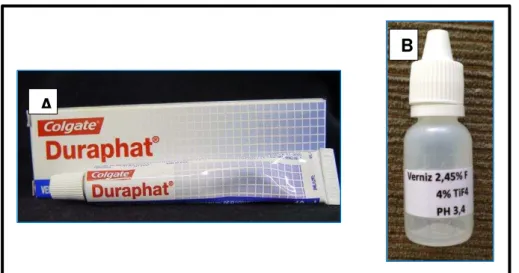
Grupo 2: Verniz Duraphat® (NaF, 2,26% F, pH 4,5, Colgate, São Bernardo, SP, Brasil) (Figura 3A);
Grupo 3: Verniz experimental de TiF4 (2,45%F, pH 3,4, FGM, Joinvile, Santa Catarina, Brasil) (Figura 3B);
Tabela 1 - Composição dos vernizes fluoretados testados neste estudo.
Material Composição (número do lote)
Duraphat(Colgate-Palmolive, Ind. Com.
Ltda, São Paulo, Brasil)
Verniz experimental de TiF4 (FGM, Joinville,
SC, Brasil)
Fluoreto de Sódio a 2,26%, álcool, resinas naturais, cera, sacarina, sabor - pH 4,5
(05 10 01)
Tetrafluoreto de titânio a 2,45%, etanol, resina sintética e resina natural – pH 3,4 (200313)
Figura 3– Vernizes fluoretados. (A) Verniz Duraphat®. (B) Verniz experimental de TiF4.
A
39
Além das amostras distribuídas nos grupos, mais dez amostras foram selecionadas para avaliação da espessura da camada protetora formada pela aplicação dos vernizes.
Os corpos de prova foram imersos em saliva artificial durante 24 horas (Figura 4A) para simular a formação da película adquirida do esmalte 30. Após, em cada amostra foi aplicado o verniz de acordo com cada grupo e seguindo as recomendações dos fabricantes. Cada produto foi inserido individualmente em seringa de insulina de 0,3 mL (BD Ultra-fine, Franklin Lakes, NJ, USA) para padronizar a quantidade do agente aplicado na superfície de esmalte.
40
Figura 4– Tratamento com vernizes fluoretados. (A) Saliva artificial por 24 horas. (B) Aplicação do verniz. (C) Saliva artificial por 6 horas. (D) e (E) Remoção do verniz.
Para avaliação da espessura da camada protetora formada pelos vernizes de TiF4 e NaF, cinco amostras foram preparadas para cada grupo do verniz (Figura 5A). A superfície do esmalte foi dividida em duas partes iguais: uma metade da superfície foi coberta com duas camadas, ácido-resistente, de esmalte de unhas (superfície controle - sem aplicação de verniz) (Figura 5B) e na outra metade foi aplicado o verniz fluoretado, tal como descrito, de acordo com cada grupo (Figura 5C). Após a aplicação de flúor, o esmalte foi removido com uma lâmina de bisturi (Figura 5D), e toda a superfície do espécime foi submetida ao desafio erosivo.
A B C
41
Figura 5 – Preparo de amostras para avaliação da espessura da camada protetora. (A) Amostra. (B) Cobertura com esmalte de unha. (C) Aplicação do verniz fluoretado. (D) Remoção da camada de esmalte de unha.
Desafio erosivo
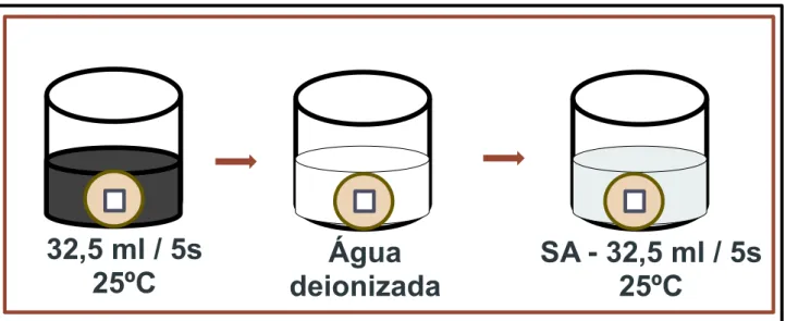
O desafio erosivo foi realizado para simular um curto tempo de exposição a refrigerante a base de cola (Coca cola®, pH 2,6, Coca-Cola Company, Porto Real, RJ, Brasil) de acordo com Wongkhantee et al. 49: as amostras foram imersas em 32,5 mL de coca cola por 5 s em temperatura ambiente (25°C), lavadas em água deionizada e, em seguida, imersas em 32,5 mL de saliva artificial em temperatura ambiente (25°C) por 5 s (Figura 6). Este ciclo foi repetido 10 vezes. As amostras foram mantidas em 100% de umidade.
A B
42
Figura 6 – Esquema do desafio erosivo utilizado
Nanoindentação final
Na área de superfície foram realizadas as medidas de nanodureza e módulo de elasticidade final (após desafio erosivo) como descrito no item de
“Nanoindentação inicial”.
A profundidade da indentação para cada amostra após aplicação do verniz e desafio erosivo foi dada pelo software do ultramicrodurômetro e determinada como altura máxima (nm). Os valores de profundidade da indentação foram comparados com os valores de espessura da camada protetora formada pela aplicação dos vernizes, para investigar se a endentação foi realizada sobre a superfície de esmalte ou na camada protetora.
Microscopia Óptica
Cinco espécimes de cada grupo foram selecionados aleatoriamente para avaliar o aspecto da camada formada após a aplicação do verniz e desafio erosivo por meio de Microscópio Óptico (Axiotech) (Figura 7), com um aumento de 100x.