The Brazilian Journal of
INFECTIOUS DISEASES
w w w . e l s e v i e r . c o m / l o c a t e / b j i d
Review article
Modulation of cerebral malaria by curcumin
as an adjunctive therapy
Kunal Jain
1,∗, Sumeet Sood
1, K. Gowthamarajan
Department of Pharmaceutics, J.S.S. College of Pharmacy, Udhagamandalam, Tamilnadu 643001, India
a r t i c l e
i n f o
Article history:
Received 12 January 2013 Accepted 21 March 2013 Available online 29 July 2013
Keywords:
Cerebral malaria Curcumin Adjunctive Cognitive deficit
a b s t r a c t
Cerebral malaria is the most severe and rapidly fatal neurological complication of Plasmo-dium falciparuminfection and responsible for more than two million deaths annually. The current therapy is inadequate in terms of reducing mortality or post-treatment symptoms such as neurological and cognitive deficits. The pathophysiology of cerebral malaria is quite complex and offers a variety of targets which remain to be exploited for better therapeutic outcome. The present review discusses on the pathophysiology of cerebral malaria with par-ticular emphasis on scope and promises of curcumin as an adjunctive therapy to improve survival and overcome neurological deficits.
© 2013 Elsevier Editora Ltda. All rights reserved.
Introduction
Parasitic diseases have grown to become a health burden as around 30% of the world’s population experience parasitic infections.1Among various parasitic infections, malaria is the
most life-threatening disease and accounts for 225 million clinical cases and 781,000 deaths per year worldwide of which 91% are in the African region. Beside this background, it is reported that each year about 85% of deaths globally are in children under five years of age, mainly due to the immunolog-ical factors.2A total of US$ 38–45 billion will be spent from 2006
to 2015 for the diagnosis and treatment of malaria, mainly in underdeveloped countries which are the most affected by this deadly disease.3Cerebral malaria (CM) is the most severe
and rapidly fatal neurological complication ofPlasmodium fal-ciparuminfection and responsible for more than two million
∗ Corresponding authors.
E-mail addresses: kunaljain [email protected] (K. Jain), [email protected] (K. Gowthamarajan).
1 These authors contributed equally to this work.
deaths annually in non-immune individuals characterized by impaired consciousness.4 This represents an enormous
burden of disease, due to the high prevalence of infection. According to the World Health Organization (WHO), CM is defined as a clinical syndrome characterized by coma (inabil-ity to localize a painful stimulus) at least 1 h after termination of a seizure or correction of hypoglycaemia, presence of asex-ual forms ofP. falciparummalaria parasites on peripheral blood smears and exclusion of other causes of encephalopathy.5
Till now, no effective vaccine is available against malaria because of the antigenic variation and complexity of parasite biology. The current therapy for management of CM is based on parenteral administration of either quinine or artemisinin derivatives such as artemether and artesunate. However, the emergence of drug resistance ofP. falciparumto anti-malarials poses a serious challenge to malaria control. In addition, there are several non-parasitic events that contribute to
pathogenesis of CM. The current therapy is aimed at reducing parasite burden and is inadequate in terms of reducing mor-tality or post-treatment symptoms such as neurological and cognitive deficits. These facts necessitate novel interventions for better therapeutic outcome in CM. Therefore, there is an urgent need to develop new antimalarial drugs, drug combinations or alternative strategies for the management of CM. The present review discusses on the pathophysiology of CM with particular emphasis on the use of curcumin, a natural polyphenol, as an adjunctive therapy.
Clinical manifestations of cerebral malaria
The clinical hallmark of CM is impaired consciousness with coma, generalized convulsions and neurological sequelae as the most severe manifestations. The earliest manifestations are non-specific fever, rigours and/or chills, irritability, rest-lessness or psychotic behaviour, vomiting and cough. The main symptoms in children are severe anemia, metabolic acidosis, hypoglycemia and coma initially arousable which becomes unarousable later. These clinical features are most strongly associated with an increased risk of death in children admitted to hospital with severe malaria. Gastroin-testinal symptoms are common in children. In adults CM, renal failure, severe jaundice, and adult respiratory dis-tress syndrome are the main complications. Ten percent of the adult patients develop severe intravascular hemolysis of infected and uninfected RBCs leading to hemoglobin-uria and anemia which further contributes to renal failure. Pregnant women are also vulnerable and develop anemia, hypoglycemia, coma and pulmonary edema. Shock is not a common feature of severe malaria and if present may be due to septicemia.6,7
In children, coma develops suddenly followed by seizures in 80% of the cases following 1–3 days of fever but some-times just after few hours. In contrast in adults, coma develops gradually and is occasionally associated with seizures. Clinical seizures are associated with rise in intracranial pressure. Sta-tus epilepticus is common in children and rare in adults. These patients often have deviated eyes, irregular breathing pattern, and show excessive salivation. On neurological examination,
signs of meningism are uncommon but neck stiffness can be seen occasionally.8
Brain swelling, high intracranial pressure, changes in ocu-lar movements, retinal changes (hemorrhages, peripheral and macular whitening, vessel discoloration and/or papilledema) and brainstem signs (abnormalities in posture, pupil size and reaction, ocular movements or abnormal respiratory patterns) are commonly observed. Various forms of abnormal posturing can occur in association with hypoglycemia. It can be either of decorticate or decerebrate pattern and often indicates raised intracranial pressure. Bruxism with grinding of teeth, pout reflex or brisk jaw jerk is common in cases with deep coma. Cranial nerve involvement is sometimes observed in adults but is rare in children.8,9
Clinical management of cerebral malaria
Management of severe malaria comprises of four main areas: clinical assessment of patient, specific antimalarial treatment, adjunctive therapy, and supportive care.
An open airway must be maintained in unconscious patients and breathing and circulation should be assessed. The patients should be weighed so that treatment can be done accordingly. Blood glucose, hematocrit/hemoglobin, par-asitemia and renal functions must be assessed. The coma can be assessed using Glasgow scale in adults and Blantyre scale in children. Cerebrospinal fluid analysis shall be carried out to exclude bacterial meningitis as it may co-exist in endemic areas. Specific antimalarial treatment is described in Table 1. In attempt to reduce high mortality rate in severe malaria various adjunctive treatments have been suggested and are summarized in Table 2.
Patients with severe malaria require intensive nursing care. Clinical observations such as coma and other vital signs should be monitored regularly. Blood glucose should be monitored every 4 h. Fluid requirement should be assessed individually. Patients with intravascular coagulation should be given fresh whole blood transfusion and vitamin K. Any con-comitant infections should be treated with antibiotics based on culture and sensitivity results.
Table 1 – Treatment of cerebral malaria.
Drug Route Indicated for Loading dose Maintenance dose
Quinine dihydrochloride IV Children and adults 20 mg/kg over 2–4 h (max
600 mg)
10 mg/kg every 8 h, until able to take orally
Quinine dihydrochloride IV Children 15–20 mg/kg over 2–4 h 10 mg/kg every 12 h, until able to take orally
Quinine dihydrochloride IM Children and adults 20 mg/kg (dilute iv
formulation to 60 mg/mL) given in two injection sites (anterior thigh)
10 mg/kg every 8–12 h until able to take orally
Quinidine gluconate IV Children and adults 10 mg/kg in normal saline
over 1–2 h
0.02 mg/kg/min continuous infusion with ECG monitoring up to 72 h or 10 mg/kg every 8–12 h
Artemether IM Children and adults 3.2 mg/kg 1.6 mg/kg/day for a minimum of 5 days
Artesunate IM/IV Children and adults 2.4 mg/kg 1.2 mg/kg after 12 and 24 h, then 1.2 mg/kg/day
for 7 days; change to oral route when possible
Table 2 – Clinical management of severe manifestations and complications ofP. falciparummalaria.5
Complication Management
Coma (cerebral malaria) Maintain airway, place patient on his or her side, exclude other treatable causes of coma
(hypoglycemia, bacterial meningitis), avoid harmful ancillary treatment such as corticosteroids, heparin and adrenaline
Convulsions Maintain airways; treat with intravenous or rectal diazepam
Hypoglycemia Measure blood glucose and maintain glucose level with infusion
Anemia Transfuse with screened fresh whole blood
Hyperpyrexia Use tepid sponging, fanning and antipyretic drugs. Paracetamol is preferred over other NSAIDs
Acute pulmonary edema Patient should be laid at an angle of 45◦, give oxygen and diuretics; prevent excessive rehydration
and stop iv fluids
Acute renal failure Exclude dehydration; check fluid balance and urinary sodium; hemofilteration or hemodialysis to
be carried out if necessary (e.g. in renal failure)
Bleeding and intravascular coagulation Transfuse with fresh screened whole blood, give vitamin K
Metabolic acidosis Exclude or treat hypoglycemia, hypovolemia and gram negative septicaemia
Shock Suspect septicaemia, take blood for cultures, give parenteral antimicrobials, correct
hemodynamic disturbances
Pathophysiology of cerebral malaria
Malaria is a protozoal disease of humans and its neurological complication, CM is a multisystem multi-organ dysfunction which is arguably one of the most common non-traumatic encephalopathies in the world.10The pathogenesis of CM is
still not fully understood because the animal models do not recapitulate human disease completely. Thereby,in vivoand
in vitromodels are useful for hypothesis generation but proof of causation in clinical pathogenesis, naturally, must be inves-tigated in human patients. The combination of both parasite and host factors is involved in the pathogenesis of CM.
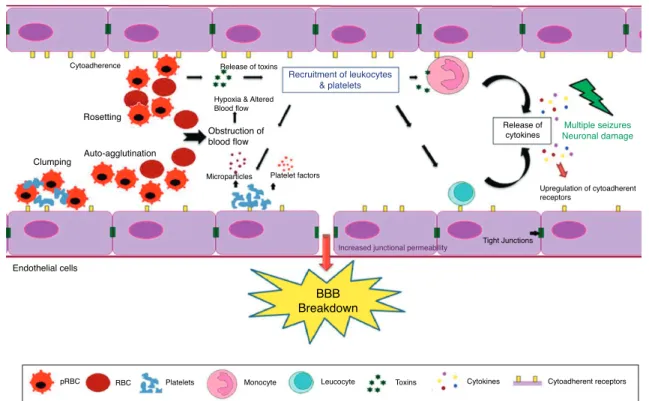
The pathogenesis of CM results from the adherence and sequestration of parasitized red blood cells (PRBCs), immune cells and platelets to vascular endothelial cells (ECs) lining the small blood vessels of the brain leading to their blockade.11
This is further accompanied by over production of type-1 pro-inflammatory cytokines followed by upregulation of endothelial adhesion molecules (Fig. 1).12It leads to
micro-and ring-hemorrhages micro-and necrosis of the surrounding tis-sues and brain edema resulting in a significant compression of cerebral arteries that could ultimately represent the cause of death.13The symptoms of CM range from confusion or stupor
to obtundation, convulsions and deep coma with long-term neurological deficits such as cortical blindness.14
Cytoadherence
Cytoadherence is defined as adherence of PRBCs to the vascu-lar endothelium, mediated byPlasmodiumderived proteins on the surface of PRBCs and modified erythrocyte cell wall pro-teins and ligands on the ECs.15 The best described adhesin
protein is parasite protein PfEMP-1 (P. falciparumerythrocyte membrane protein-1) which is a large transmembrane protein (200–350 kDa) comprising several external Duffy-binding-like (DBL) domains. It is encoded by a family of more than 150 highly variable (“var”) genes.16These are knob-like electron
dense structures protruding from the PRBCs surface. The role of the knobs in cytoadherence was confirmed when knob-less variant of P. falciparum were shown to lack adherence
to host cells in vitro as well as in vivo.17 Surface
poten-tial spectroscopy studies of knobs revealed that they were positively charged and hence better electrostatic interaction with negatively charged endothelial plasma membranes and receptors.18 These high molecular (200–350 kDa)
transmem-brane proteins are encoded PfEMP-1 mediated cytoadhesion begins at approximately 12 h of parasite development and is highly effective in the second half ofP. falciparumlife cycle. As a result, late stage of parasite (trophozoites and schizonts) is rarely seen in peripheral blood because of their seques-tration in various organs.19 The large number of variants
of PfEMP-1 results in potential of infected RBCs to bind to many different host receptors expressed on vascular ECs in peripheral microvascular beds.20There are several cell
adhe-sion receptors that have been characterized including ICAM-1 (intercellular adhesion molecule-1), VCAM-1 (vascular cell adhesion molecule), CD36, CD31, thrombospondin, E-selectin, hyaluronic acid and chondroitin sulfate A (CSA).8
Cytoadher-ence prevents malarial parasites from being recognized and cleared off the circulation by the spleen.21
CD36 seems to be constitutionally expressed on wide range of vascular beds but remarkably absent in brain vessels and thus its expression is not related qualitatively or quantitatively to disease severity.22This receptor is present in the
microvas-culature and its expression is not affected by inflammatory cytokines like tumour necrosis factor-␣(TNF-␣).23 However,
platelets expressing CD36 may serve as a stick bridge between PRBCs and endothelium which could be important in cerebral vessels lacking CD36 CSA is the main receptor in the placenta which in addition to CD36, are able to support firm adhesion under flow conditions.24The E-selectin mediates initial
tether-ing to the ECs and rolltether-ing of leukocytes. Plasma concentration of soluble E-selectin is raised inP. falciparummalaria.25
Main receptor for the cytoadherence is ICAM-1, which has been implicated in the pathogenesis of CM. It is a mem-brane glycoprotein expressed on lymphocytes, macrophages and vascular endothelium. Its primary role is to mediate cellular adhesion within the immune system via the leu-cocyte integrins LFA-1 and MAC-1.26 Expression of ICAM-1
Cytoadherence
Rosetting
Auto-agglutination
Endothelial cells
Obstruction of blood flow
Release of cytokines
BBB Breakdown
Hypoxia & Altered Blood flow
Release of toxins
Platelet factors
Toxins Leucocyte
Monocyte Platelets
pRBC RBC Cytokines
Tight Junctions
Increased junctional permeability
Cytoadherent receptors Upregulation of cytoadherent receptors
Multiple seizures Neuronal damage
Microparticles
Recruitment of leukocytes & platelets
Clumping
Fig. 1 – The blood–brain barrier (BBB) breakdown during cerebral malaria pathogenesis. The diagram shows the events and the possible mechanisms that play a vital role in CM. The mature forms (trophozoites and schizonts) of parasitized red blood cells (PRBCs), host leukocytes and platelet-fibrin thrombi adhere to the cerebral endothelial cells and sequester in large numbers in the brain. This cytoadherence, combined with other events such as rosetting, auto-agglutination, clumping and decreased RBCs and PRBCs deformability causes altered blood flow leading to impaired tissue perfusion and hypoxia. Further, sequestered parasites produce local toxins which lead to recruitment of leukocytes and platelets followed by the release of inflammatory cytokines (IL-1, IL-6, TNF, LT, and NO) and microparticles. These mediators lead to
endothelial cells activation and apoptosis, BBB breakdown, increased junctional permeability, followed by secondary neuropathological events that can lead to cerebral edema or coma. Sequestration of PRBCs within microvasculature increases cerebral volume, which together with increased cerebral blood flow from seizures, anemia and hyperthermia and altered BBB function lead to cerebral edema and raised intracranial pressure. This may result in death or neuronal damage with consequent neurocognitive sequelae in the survivors.
involve the endothelial nuclear factor kappa beta (NFB) sys-tem and may involve reactive oxygen species (ROS).28NFB is a
transcription factor located within cytoplasm and responsible for regulation of cytokine production. Normally NFB stays inactivated by an inhibitory protein IB. Once activated, NFB enters the nucleus and increases the transcription of different inflammatory mediators such as cytokines and chemokines (TNF-␣, IL, etc.) thereby causing neuroinflammation. Several molecules are capable of activating NFB including TNF-␣29
Monoclonal antibodies have been developed against ICAM-1 to improve microcirculatory flow inex vivomodels of malaria sequestration but have not been investigated in humans.30
Recently, it has been reported that cytoadherence, a potential pathogenic property ofP. falciparuminfected red blood cells, continues long after the parasite has been killed.31
Sequestration
Sequestration of PRBCs containing mature forms of the para-site (trophozoites and meronts) in the cerebral microvessels
has been found on post-mortem examination of the CM patients.32 The pathogenesis of fatal CM is associated with
sequestration of red blood cells. It has been confirmed that severe malaria pathology is related to sequestered parasite biomass.33 Sequestration occurs principally during the
sec-ond half of the intra-erythrocytic asexual growth phase of the parasite, following the cytoadherence. The accumulation of mature PRBCs is observed in almost all organs, but par-ticularly in the brain viz. cerebrum, cerebellum and medulla oblongata.11,32This in turn, reduces the vascular lumen to
cre-ate a mechanical obstruction for the erythrocytes transit. It is the sequestered parasites that cause considerable obstruction of blood flow, decreases tissue perfusion and removal of waste products like lactic acid and generates hypoxia.9,33
The relatively hypoxic venous beds provide optimal envi-ronment for the optimal growth and multiplication of parasite and prevents the PRBC from being destroyed by the spleen.34 Tissue hypoxia further causes hyperlactemia with
Red cell deformability, rosetting and
auto-agglutination
Parasitized erythrocytes and non-parasitized erythrocytes (NPRBCs) sequestration in the cerebral microvasculature reduces the microvascular flow. In addition, with progres-sive maturation of parasites inside erythrocytes decreases their ability to deform which may contribute to the eryth-rocyte destruction and impair the microcirculatory flow.15
Decreased deformability of PRBCs is mainly due to change in the cytoskeleton, increased stiffness of the membrane and rigidity of growing intracellular parasite. Such PRBCs have more difficulty in passing through in vitro micropore filters as demonstrated for Plasmodium knowlesi malaria in monkeys.36 Furthermore, it has been observed that NPRBCs
have to undergo considerable deformation as they squeeze through the sequestered microcirculation. It can be concluded that deformability of both PRBCs and NPRBCs is markedly reduced in severe malaria which is partly contributing to the pathogenesis of CM.37
The adherence of NPRBCs to PRBCs (rosetting) and PRBCs to PRBCs (agglutination) could further compromise microcircula-tory flow that leads to impaired tissue perfusion, hypoxia and creates a toxic local environment.15This phenomenon is more
prevalent in patients with CM than in patients of uncompli-cated malaria. Thevargenes seem to be responsible for roset-ting and are sensitive to pH and heparin.38 Rosettes can be
disrupted by antibodies againstP. falciparum, glycosaminogly-cans and sulfated glycoconjugates which is strain-specific.39,40
Rosette formationin vitroindicates the clogging of vascular lumen by interaction of NPRBCs and PRBCs with endothelium. However, PRBCs causes the cytoadherence but not all form rosettes.41Interestingly, epidemiological studies have shown
the association of rosette forming strains with the develop-ment of severe disease but most clinical studies have failed to show such an association.38,42Red blood cells in thalassemic
and sickle cell anemia patients have weak binding abilities forming small and weak rosettes. This decreased rosetting ability provides natural protection against development of CM in such patients.43 Thus, it can be concluded that a
com-promised microcirculation, with sequestration of parasitized erythrocytes due to reduced red cell deformability and the sticky forces related to rosetting and auto-agglutination, is central in the pathogenesis.
Inflammatory mediators
P. falciparuminfection causes an increase in blood concen-tration of both pro-inflammatory cytokines such as TNF-␣, IL-1, IL-6, IL-18 and anti-inflammatory Th2 cytokines IL-4 and IL-10. The balance between these inflammatory medi-ators is critical to parasite control. However, their role in pathogenesis of disease remains unclear.44 Studies have
shown that glycosylphosphatidylinositol (GPI), a malarial toxin, binds to the pattern recognition receptors of the innate immune system and stimulates pro-inflammatory cytokine production.45 GPI stimulates the production of TNF-␣ and
lymphotoxin which further cause upregulation of expression
of cytoadherence receptors notably ICAM-1 and VCAM-1 on ECs. Thus, they promote sequestration of PRBCs and leukocytes in brain leading to coma. The biological effects of TNF-␣during malaria are both protective and pathogenic.37,46
At low concentration, TNF-␣kills parasite by causing cytokine release from macrophages. Higher concentration of TNF-␣
is associated with hypoglycemia, hyperparasitemia, anemia, pulmonary edema and death.47
It has also been found that TNF activation leads to the production of microparticles which have pro-coagulant and pro-inflammatory properties and this is specifically seen in patients suffering from CM and does not occur in uncom-plicated malaria or severe anemia.48TNF-␣also induces the
release of nitric oxide (NO) via inducible NO synthase (iNOS) which has been proposed as cause of coma due to interference with synaptic transmission.49
The effect of NO in malaria could be either beneficial or harmful depending upon the amount and site of production. Beneficial effect of NO is involved in host defence by killing intracellular organisms, maintaining vascular status and neurotransmission.9Although, the direct killing effect of NO
on the parasite was not observed, but oxidation products of NO were found to be toxic to the malarial parasite.50Moreover,
it was also shown that human monocytes stimulated with IFN-␥via the secretion of reactive nitrogen intermediates had inhibitory action on parasite growthin vitro.51Measurement
of reactive nitrogen intermediates in various biological fluids such as plasma, cerebrospinal fluid and urine is very difficult since NO is short lived, unstable and reacts rapidly with several molecular targets. This observation correlates poorly within vivoproduction of NO at microvascular level. NO can easily diffuse across blood–brain barrier (BBB) and reduces the level of consciousness, impair neuronal signalling at higher levels and causes oxidative damage of ECs. Furthermore, NO may damage RBCs and increase anemia. To counteract the damage caused by the radical, NO scavengers might repre-sent an effective adjunct therapy. An alternative hypothesis states that NO production is limited and it is synthesized by scavenging molecules such as superoxides and plasma hemoglobin. Thus, enhancing NO bioavailability has been proposed as anti-malarial therapy.52 However, the role of
NO in CM pathogenesis has not been elucidated clearly and conflicting observations have been reported.8
The cause of impaired consciousness is still unclear but it is likely to result from the in homogenous obstruction of the cerebral microcirculation by sequestered parasitized erythro-cytes causing dysoxia and results in lactate production by the brain.53 Apart from NO mediated loss of consciousness
various host and parasite derived factors may be involved in pathogenesis of coma. Further, autopsy studies indicate the axonal dysfunction which may arise due to accumulation of
-amyloid precursor protein. This could in turn contribute to the reversible neurological dysfunction in CM.54However, the
cause of coma in CM and the role of dysoxia and axonal dys-function still remains unclear.
Reactive oxygen species
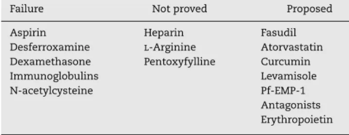
Table 3 – Adjunctive therapy in cerebral malaria.
Failure Not proved Proposed
Aspirin Heparin Fasudil
Desferroxamine l-Arginine Atorvastatin
Dexamethasone Pentoxyfylline Curcumin
Immunoglobulins Levamisole
N-acetylcysteine Pf-EMP-1
Antagonists Erythropoietin
responsible for the tissue damage. However, it was suggested that ROS plays a role in host defense mechanism against the parasite.55Thus, likewise NO, ROS too can have both beneficial
and pathological roles.
Oxidative stress (OS) plays a vital role in malaria parasite-infected erythrocytes and at the same time OS represents a most promising rationale for designing the anti-malarial chemotherapy. This has been indicated by the glucose-6-phosphate dehydrogenase deficiency which makes the malarial parasites highly susceptible to alterations in the redox equilibrium. This in turn contributes to disease mani-festation including sequestration, cerebral pathology, anemia and respiratory distress.56 However, the redox interactions
between the malarial parasite and that of their host are complex. Due to high metabolic rate of the rapidly growing and multiplying parasite, detoxification of ROS becomes a key challenge for infected erythrocytes. Central to the gen-eration of toxic redox-active-by-products is the degradation of host hemoglobin by the Plasmodium. Hemoglobin repre-sents the major source of aminoacids forPlasmodium, but its degradation in an acidic food vacuole results in the produc-tion of toxic free heme (ferri/ferroprotoporphyrin IX; FP) and ROS which in turn confers oxidative insult on the host cell. Hemozoin or malaria pigment is a crystalline form of free heme. Even if only a small amount (e.g. 0.5%) of the free heme escapes the neutralization processes, it could cause OS to host proteins and membranes, inhibit parasite enzymes and hemolysis.57 In addition, the production of ROS by the
host immune system also contributes to the overall oxida-tive burden of the infected erythrocyte. In order to maintain the redox status, malarial parasite possesses an efficient enzymatic antioxidant defense system including glutathione-and thioredoxin-dependent proteins as well as superoxide dismutase.56In this regard, targeting the parasite antioxidant
defense system might be useful for designing antiparasitic medication.
Indeed, a number of potential new drugs currently in clinical use, especially the artemisinins appear to act by alkylation of heme or protein to form a cytotoxic carbon-centred radical intermediate that further interfere with the redox metabolism of malarial parasite.58It seems likely that
the lethal effects of endoperoxide antimalarial are due to targeted damage of particular enzymes or membrane pro-teins by free radical species rather than non-specific damage caused by freely diffusing ROS.59Some antioxidants
adjunc-tive therapy have been evaluated in humans and mice (Table 3).
Increased permeability and evidence of
endothelial damage
The BBB is a highly specialized structural and functional inter-face between the intraerythrocytic stages ofP. falciparumand the human host.60 BBB maintains functional integrity with
the support of pericytes, microglial processes of neurons and specialized perivascular macrophages. Perivascular cells are involved in maintaining the normal neuronal function of the CNS and provide immunity.61There is growing evidence that
combination of parasite-induced sequestration of PRBCs and NPRBCs, cytokine response, host environment, activation of ECs and hemostasis changes BBB function and its perme-ability. This has been implicated in the onset of a clinically complex syndrome of coma with reversible encephalopathy, associated with high mortality rate and increasingly recog-nized as long-term neurological sequelae in survivors.8,62
Pathological changes to BBB dysfunction may be broadly divided into systemic effects of severe disease such as acidosis or hypoxia due to anemia and local factors include adher-ence of PRBCs, host leukocytes and platelets to the ECs, rosetting, sequestration, microvascular obstruction, release of cytokines and parasite toxins. These cascade of events cause direct cellular damage and may initiate signalling within ECs that could influence junctional protein expression, and also directly activate or damage peri- and paravascular cells such as microglia, astrocytes and neurons and parenchy-mal effector cells.14It is believed that cerebral microvascular
engorged by cytoadherent parasitized erythrocytes results in a cascade of intracellular signalling events that disrupts the cytoskeleton-cell junction structure and increases the junc-tional permeability of the BBB and suppresses activation of dendritic cell and macrophages.12 Thus, dysfunction of
BBB initiates the progression of secondary processes of neu-ropathological injury.
Even thoughP. falciparumdisturbs the BBB integrity, it never enters the brain parenchyma and yet it can cause severe neu-rological dysfunction and death.14,61Focal disruptions at sites
of PRBCs sequestration along the ECs of BBB could result in the exposure of sensitive perivascular neuronal cells to plasma proteins and further leads to localized hypoxia and hemor-rhaging of surrounding brain parenchyma.12,14 Apart from
systemic effects, increased concentration of cytokines and parasite toxins have also been shown to contribute to reduced consciousness and seizure activity. Thus causing direct dam-age to BBB.63
Studies have shown that PRBCs adhesion to ECs induces cell apoptosis via over expression of cytoadherent molecules, expression of iNOS and OS.64Further,in vivostudies in mice
model of CM have implicated cytotoxic effector CD8+T lym-phocytes in development of murine CM pathogenesis by direct cytotoxicity against ECs.65 Thus, apoptosis and cytotoxicity
petechial hemorrhage particularly of white matter and ring hemorrhages in white matter.9,15
The leakage of plasma into CSF and consequent elevation of its protein concentration due to breakdown of BBB has been demonstrated by parenteral administration of radioac-tive albumin and isothioacyanite in animal models.66
BBB disruption can contribute to severe increased intracra-nial hypertension which is associated with neurological sequelae or death. However, the most likely cause of raised intracranial hypertension includes increased cerebral blood volume due to sequestration and increased blood flow from seizures, hyperthermia and anemia. Mannitol is an effec-tive osmotic diuretic in reducing the burden of brain edema and intracranial pressure in children with mild degree of intracranial hypertension. It may decrease the mortality or neurological deficits in surviving patients of CM.67
Neurocognitive deficits
Though most of the patients suffering from CM make a full recovery, survivors have been found to be associated with neurological sequelae.9 CM sequelae seem to be of
multi-factorial origin, although their cause is largely unknown. These impairments are rarely observed in adults (<1%) but are more common in children (approx∼12%) particularly
in sub-Saharan Africa.68 The symptoms include hemiplegia
or quadriparesis, cortical blindness, deafness, ataxia, severe hypotension and aphasia.69 The sequelae reported include
protracted seizures, prolonged and deep coma, hypoglycemia and sometimes severe anemia.9 Some of these symptoms
resolve over a period of 1–6 months in 50% of the children. However, 25% of children with successfully treated CM are left with residual cognitive and neurological deficits such as memory disturbances, speech and language difficulties, disor-ders of attention, seizures, visuo-spatial and motor deficits.70
These deficits have been determined to persist after cessa-tion of therapy with follow-up periods extending to nine years after infection.71These cognitive deficits occur as a result of
complicated host inflammatory response in conjunction with vascular impairment, causing abnormalities in neuronal cells, which adversely affect cognitive function.
Prolonged febrile seizures not precipitated by malaria may lead to damage in the hippocampus associated with later episodic memory impairments which may be underreported.9,15,72 Further decreased in blood flow and
delivery of oxygen may lead to brain damage.15 The other
reasons include generation of excitotoxins, ROS, and toxins produced by the parasite.73
To date, there is no therapy targeting the cognitive and motor deficits seen after successful antimalarial therapy in experimental CM. Hence, the role of immunomodulator needs to be assessed as an adjunctive therapy in CM.
Severe
Plasmodium vivax
infections and
neurological complications
P. vivaxinfections considered as a benign disease for a long time is the major cause of morbidity in Asia, Central and South America and in the horn of Africa.74It is responsible
for more than 400 million infections each year thus repre-senting most widespread malarial species.75In 2010, 306,908
cases of malaria were recorded in Brazil, 85% of which were caused byP. vivax.76P. vivaxinfections are characterized by
relapse of malaria due to persistent latent forms in liver called as hypnozoites. In most of endemic areas,P. vivax infec-tions co-exist withP. falciparuminfections. However,P. vivax
infections are more difficult to control because of their ten-dency to relapse and is a major cause of malaria in young children.74SevereP. vivaxinfections are uncommon but have
been reported at regular intervals. The number of vivax infec-tions particularly those with serious clinical complicainfec-tions has been increasing. The most frequent complications are severe anemia, acute respiratory distress syndrome (ARDS), thrombocytopenia, renal and hepatic dysfunction, coma, CM and death.77 P. vivaxinfections have been rarely associated
with CM and to date less than 50 cases of central nervous systemP. vivaxinfections have been reported.78Severe
pul-monary involvement is frequent inP. falciparuminfection but rare in vivax malaria. Among the few cases reported involving vivax, the pulmonary complications appear from six to eight days after the initiation of antimalarial treatment. This is due to exacerbation of inflammatory response.79However, in
con-trast toP. falciparumassociated respiratory distress, the lungs are the sole organ system involved in vivax malaria. It is asso-ciated with acute lung edema and neutrophil accumulation in interalveolar space with or without sequestration ofP. vivax
infected RBCs.77,80
The mechanism of ARDS in vivax malaria is less well studied compared tofalciparuminfections. The level of pro-inflammatory mediators is elevated in vivax infections thus signifying the role of inflammatory dysregulation. There is a cytokine related increase in alveolar permeability and altered fluid clearance.81 In vivax infections, anemia persists even
after the parasite clearance. The severe anemia in vivax infec-tions does not result from destruction of infected RBCs alone. Studies have shown that for every infected RBC destroyed in vivax malaria, 32 uninfected RBCs are removed from circula-tion compared to eight RBCs in falciparum malaria.82
Immunopathogenesis of vivax malaria is poorly under-stood. However, it was found that Brazilian patients with severe vivax malaria had elevated levels of inflammatory cytokines TNF-␣and IFN-␥, increased IFN-␥/IL-10 ratio and superoxide dismutase (SOD-I).83The level of TNF-␣and other
inflammatory cytokines are higher duringP. vivaxinfections compared toP. falciparuminfections with same parasite load. Plasma concentration of microparticles was found to be ele-vated in individual withP. vivaxinfections.84 In contrast to P. falciparum, P. vivax has low parasite biomass and is con-sidered unable to cytoadhere or sequester. However, recently
P. vivax infected RBCs have been shown to cytoadhere to
ECs via chondroitin sulfate-A and ICAM-1 although at 10-fold lesser frequency thanP. falciparum.85This could be reason for
lower incidence of coma in vivax malaria and coma is perhaps of systemic metabolic origin.86The degree of endothelial
acti-vation is greater invivaxthanfalciparummalaria. Rosetting in
cases. In areas where both multidrug resistantP. falciparum
andP. vivaxare prevalent, infants and young children are at greater risk of acquiringP. vivaxinfection thanP. falciparum
infection.87Thus, an immunomodulator may have a potential
role in severe vivax infections.
Need of adjunctive therapy in cerebral malaria
Apart fromP. falciparuminduced cerebral injuries, CM is also associated with non-parasite events including OS and inflam-matory cascade which is responsible for mortality in spite of antimalarial treatment during the acute phase.88CM is
asso-ciated with elevated pro-inflammatory cytokines in serum and brain tissues, as well as leukocyte recruitment to sites of parasite sequestration in the cerebral microvasculature. The harmful, deregulated immune response leading to CM is mainly of the Th1 type, with overproduction of Interferon-␥
(IFN-␥) and TNF-␣.89These inflammatory processes promote
pathological changes in the brain that contribute to CM, such as endothelial damage and neuronal dysfunction. In spite of treatment with current antimalarials, CM is associated with mortality in 20–25% of cases and those surviving have been associated with neurological and cognitive deficits.71OS
has been found as a contributing factor to persistent cog-nitive deficits.90 ROS are generated in malaria as a result
of host immune response and may oxidize vital cellular components such as lipids, proteins, and DNA. Oxidative damage to membranes of parasitized and uninfected red blood cells may contribute to sequestration of red blood cells in microvascular cells.91 The current therapy utilizing
anti-malarials controls parasitic events alone and do not treat the inflammatory cascade. The control of these deleterious processes is probably one of the most challenging goals to reach for a full rate recovery suggesting that adjunctive ther-apies are urgently needed with the aim of providing a new therapeutic scheme for CM using a combination of a rapid antimalarial and immunomodulators. Also, the reversal of BBB leakage or its downstream effects presents a potential opportunity for neuroprotective therapy for patients with CM. Apart from killing the parasite effectively, there is a need to improve survival rate and overcome cognitive deficits in CM patients and therefore reduce the likelihood of perma-nent neurological disability or death. Thus, the objectives of the adjunctive therapy are mainly to decrease the mortal-ity, shorten the coma and reduce the rate and the severity of neurological sequelae. The first adjunctive intervention was using anti-TNF antibody in the murine model and since then more than 48 adjunctive interventions have been evalu-ated showing 92% success rate in ameliorating or preventing CM.92The adjunctive therapy may involve the identification
of immune modifying compounds such as curcumin, with ability to downregulate excessive pro-inflammatory response, reducing the expression of the adhesion molecules and sub-sequent sequestration of PRBCs in cerebral endothelium, thus improving the survival of CM patients. In contrast to many adjunctive therapies which have been investigated, curcumin has an added advantage of possessing antimalarial efficacy of its own. An important point to be considered for admin-istration of adjunctive therapy using an immunomodulator
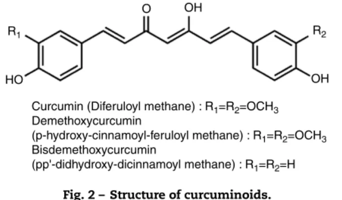
R1 R2
Curcumin (Diferuloyl methane) : R1=R2=OCH3 Demethoxycurcumin
(p-hydroxy-cinnamoyl-feruloyl methane) : R1=R2=OCH3 Bisdemethoxycurcumin
(pp'-didhydroxy-dicinnamoyl methane) : R1=R2=H HO
OH
OH O
Fig. 2 – Structure of curcuminoids.
is that it should be administered together with antimalarial drugs rather than alone in order to delay immunopathology and allow a longer time frame for antimalarial to work.4We are
working on intranasal delivery of curcumin loaded nanoemul-sions in combination with artemether for management of CM.93
Curcumin and cerebral malaria
Curcumin, a small molecular weight polyphenolic compound isolated from the rhizomes of Turmeric (Curcuma longaL.) and related species (familyZingiberaceae), has been used tradition-ally as a spice. The major curcuminoids present in turmeric are Curcumin (Curcumin I), Demethoxy curcumin (Curcumin II) and Bisdemethoxy curcumin (Curcumin III) (Fig. 2). Com-mercial curcumin contains curcumin I (77%), curcumin II (17%) and curcumin III (3%).94
Curcumin is from natural sources of long-term use, and as such, no resistance is known to curcumin that is present in a dietary supplement. It is tolerated at very high doses, and as much as 8 g/day has been given for three months to cancer patients on trial without toxic side effects.95A large number
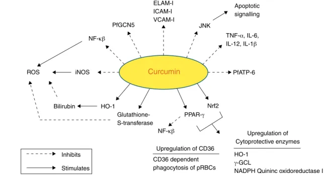
ofin vitro andin vivo studies in both animals and humans have indicated that curcumin exhibits promising pharma-cological activities including antioxidant, anti-inflammatory, angiogenic, spasmodic, microbial and anti-plasmodial activities. Accumulating evidence suggests that curcumin has a diverse range of molecular targets, suppor-ting the concept that it acts upon numerous biochemical and molecular cascades (Fig. 3).
In the context of malaria, curcumin has been shown to possess moderate antimalarial activity with IC50 value of
5–18M.96,97 Further, curcumin analogues were synthesized
having much more potent antimalarial activity with IC50value
of 400 nM. With the use of the CM model in mice, it has been shown that curcumin have potent activity against Plasmo-dium bergheiand it is able to prevent CM and delay death of animals by about 10 days.4,98 In addition to having a direct
killing effect as an antimalarial, curcumin is also able to prime the immune system againstP. berghei.98Curcumin
pos-sesses both anti-oxidant and pro-oxidant activity. Curcumin has shown to enhance ROS generation in concentration range of 20–100M.99The efficacy of curcumin has been tested in
Apoptotic signalling
TNF-α, IL-6, IL-12, IL-1β
PfATP-6
PfGCN5 JNK
HO-1 Bilirubin
ROS iNOS
Inhibits
Stimulates
Glutathione-S-transferase
PPAR-γ
NF-κβ NF-κβ
Nrf2 ELAM-I
ICAM-I VCAM-I
CD36 dependent phagocytosis of pRBCs
HO-1 γ-GCL
NADPH Quininc oxidoreductase I Upregulation of CD36
Upregulation of Cytoprotective enzymes
Curcumin
Fig. 3 – Molecular targets of curcumin in cerebral malaria.
Targets of curcumin
Curcumin has numerous molecular targets in the context of CM. It possesses itself antimalarial activity and acts as a neu-roprotective agent:
i. NF-B (nuclear factorbeta) plays a vital role in malaria as evidenced by PRBCs that have shown to induce
NF-B regulated inflammatory pathway in human cerebral endothelium. The deciphering of anti-malarial activity in curcumin against the blood phase of the life cycle may leads to its role in regulation of NF-B. Curcumin has profound anti-inflammatory activity and might exert its therapeutic effects by inflammatory cytokine pro-duction and expression of cytoadhesion molecules on ECs.100Curcumin has shown to reduce the production of
proinflammatory cytokines such as TNF, IL12 and IL6in
vitro.101It can be concluded that curcumin can effectively
control neuroinflammation and damage due to inflam-matory cascade as a result of host immune response and thereby prevent cognitive deficits in CM survivors. ii. Curcumin activates PPAR-␥(peroxisome proliferator
acti-vated receptor ␥). PPAR-␥is a transcription factor that regulates inflammatory responses. Its activation inhibits translocation of NF-B and inhibits microglia from pro-ducing proinflammatory cytokines.29 It also regulates
scavenger receptor CD36, which mediates non-opsonic phagocytosis of PRBCs. Pharmacological upregulation of CD36 in monocytes/macrophages by PPAR-␥ agonists has shown to increase CD36 dependent phagocytosis of PRBCs in vitro.102 CD36 is also activated by
acti-vation of redox-sensitive transcription factor nuclear related {erythroid-derived 2} factor (Nrf2). These two transcription factors have been found to be activated in macrophages/monocytes treated with curcumin.103
Activation of Nrf2 pathway has been demonstrated as potential target regarding brain inflammation.104
iii. It inhibits expression of various cell surface adhesion molecules such as ICAM-1, VCAM-1 and endothelial leukocyte adhesion molecule (ELAM-1) on ECs.94 These
data support the development of adjunctive thera-pies to reverse the pathophysiological consequences of cytoadherence.31 Curcumin also inhibits adhesion of
platelets to brain ECsin vitrowhich accumulate in brain blood vessels in CM patients.105
iv. Curcumin causes inhibition ofP. falciparumgeneral con-trol nondepressed 5 (PfGCN5). It is a histone acetyl transferase (HAT) that preferentially acetylates K9 and K14 of histone H3. Various post-translational modi-fications of histone tails lead to regulation of gene expression. Curcumin inhibits HAT and causes parasite chromatin modifications and have antiparasitic effects.99
v. Another target of curcumin is PfATP6 (P. falciparum
ATP6), the parasite analogue of SERCA (Sarcoplas-mic/endoplasmic reticulum Ca2+ ATPase). PfATP6 is the
only SERCA-type Ca2+ ATPase in P. falciparum which
is responsible for the maintenance of calcium ion concentrations for the generation of calcium-mediated signalling and correct folding and post-translational processing of the proteins.106,107
vi. Curcumin inhibits iNOS by suppression of IFN-␥and IL-12 production. iNOS has shown to mediate production of ROS.108
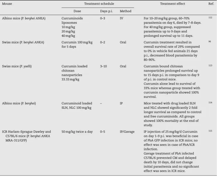
Table 4 – Curcumin examined for possible effects on animal models of malaria.
Mouse Treatment schedule Treatment effect Ref.
Dose Days p.i. Method
Albino mice (P. berghei ANKA) Curcuminoids
liposomes 10 mg/kg 20 mg/kg 40 mg/kg
0–3 IV For 10–20 mg/kg group, 60–70%
parasitemia on day 6, died by 7–8 days. For 40 mg/kg group, suppressed parasitemia up to 9 days and prolonged survival up to 11 days.
112
Swiss mice (P. berghei ANKA) Curcumin 100 mg/kg
for 5 days
0–2 Oral Curcumin treatment resulted in
overall survival rate of 29% compared to 0% in vehicle fed animals 21 days p.i., decreased blood parasitemia by 80–90%.
96
Swiss mice (P. yoelli) Curcumin loaded
chitosan nanoparticles 33.33 mg/kg
3–10 Oral Curcumin bound chitosan
nanoparticles prolonged survival up to 15 days p.i. in comparison to day 9 of p.i. in control mice.
Curcumin alone lead to survival of 33% mice whereas group treated with curcumin nanoparticle showed 100% survival.
113
Albino mice (P. berghei) Curcuminoid loaded
SLN, NLC 100 mg/kg
– IP Mice treated with drug loaded SLN
and NLC showed significantly 2 fold longer survival as compared to control and free curcuminoids. All groups showed 100% mortality at the end of study.
114
ICR Harlam-Sprague Dawley and C57BL/6 mice (P. berghei ANKA MRA-311/GFP)
50 mg/kg twice a day 0–5 IP/Gavage IP injection of 25 mg/kg/d Curcumin
on day 1–9 p.i. was beneficial in case of PbA GFP infection in ICR mice; no effect was seen in case of PbA/ICR infection.
Gavage treatment of PbA infected C57BL/6 prevented CM and delayed death by 10 days, did not change initial parasitemia and no significant effect was seen in ICR mice.
115
viii. C-Jun N terminal kinases (JNK) belongs to the family of mitogen activated kinases called MAP Kinases, which are activated in response to inflammatory cytokines and environmental stress conditions. Its activation induces the transcription-dependent apoptotic signalling path-way resulting in neuronal cell death during experimental CM.111Curcumin suppresses JNK activation and thus
pro-tects the neurons
ix. Curcumin enhances the activity of detoxifying enzymes like Glutathione-S-transferase.29
All these mechanisms suggest curcumin as an immunomodulator which may alleviate CM.
Conclusion
Paradoxically the rate of new drug delivery in case of malaria has been very low. The global response to this crisis has been inadequate. There are few new chemical entities in preclinical and clinical phases but these efforts could take considerable
time to fructify. So far the attempts have been limited to explore the use of immunomodulator in combination with conventional drugs for CM. Thus, this review highlights the potential of a plant based immunomodulator curcumin as an adjunctive therapy for better management and improvement of survival and overcoming the neurocognitive deficits in CM patients. It may be a useful strategy to complement the direct antiparasitic efficacy of currently used antimalarials and also limit the emergence of resistant parasites.
Conflict of interest
The authors declare no conflicts of interest.
Acknowledgements
gratitude to Department of Science and Technology (DST), New Delhi, India for award of INSPIRE Fellowship (IF10316).
r e f e r e n c e s
1. Joshi M, Pathak S, Sharma S, Patravale V. Design andin vivo
pharmacodynamic evaluation of nanostructured lipid carriers for parenteral delivery of Artemether: Nanoject. Int J Pharm. 2008;364:119–26.
2. World Malaria Report 2010. World Health Organization, Geneva.
3. Kiszewski A, Johns B, Schapira A, et al. Estimated global resources needed to attain international malaria control goals. Bull World Health Organ. 2007;85:623–30.
4. Waknine-Grinberg JH, Mcquillan JA, Hunt N, Ginsburg H, Golenser J. Modulation of cerebral malaria by fasudil and other immune-modifying compounds. Exp Parasitol. 2012;125:141–6.
5. WHO. Guidelines for the treatment of malaria. second edition; 2010. Available from: http://www.who.int/malaria/ publications/atoz/9789241547925/en/index.html
6. Dondorp AM. Pathophysiology, clinical presentation and treatment of cerebral malaria. Neurol Asia. 2005;10:67–77. 7. Idro R, Marsh K, John CC, Newton CRJ. Cerebral malaria:
mechanisms of brain injury and strategies for improved neuro-cognitive outcome. Pediatr Res. 2010;68:267–74. 8. Gay F, Zougbédé S, N’Dilimabaka N, Rebollo A, Mazier D,
Moreno A. Cerebral malaria: what is known and what is on research. Rev Neurol (Paris). 2012;168:239–56.
9. Idro R, Jenkins NE, Newton CR. Pathogenesis, clinical features, and neurological outcome of cerebral malaria. Lancet Neurol. 2005;4:827–40.
10. Beales PF, Brabin B, Dorman E, et al. Severe falciparum malaria. Trans R Soc Trop Med Hyg. 2000;94:S1–90. 11. Coltel N, Combes V, Hunt NH, Grau GE. Cerebral malaria – a
neurovascular pathology with many riddles still to be solved. Curr Neurovasc Res. 2004;1:91–110.
12. Schofield L, Grau GE. Immunological processes in malaria pathogenesis. Nat Rev Immunol. 2005;5:722–35.
13. Combes V, Coltel N, Faille D, Wassmer SC, Grau GE. Cerebral malaria: role of microparticles and platelets in alteration of the blood–brain barrier. Int J Parasitol. 2006;36:541–6. 14. Medana IM, Turner GDH. Human cerebral malaria and
blood–brain barrier. Int J Parasitol. 2006;36:555–68. 15. Newton RJC, Hien TT, White N. Cerebral malaria. J Neurol
Neurosurg Psychiatry. 2000;69:433–41.
16. Borst P, Bitter W, McCulloch R, Leeuwen F, Rudenko G. Antigenic variation in malaria: mini review. Cell. 1995;82:1–4. 17. David PH, Hommel M, Miller RH, Udeinya IJ, Oligino LD.
Parasite sequestration inPlasmodium falciparummalaria: spleen and antibody modulation of cytoadherence of infected erythrocytes. Proc Natl Acad Sci USA. 1983;80:5075–9.
18. Aikawa M, Kamanura K, Shiraishi S, et al. Membrane knobs of unfixedPlasmodium falciparuminfected erythrocytes: new findings as revealed by atomic force microscopy and surface potential spectroscopy. Exp Parasitol. 1996;84:339–43. 19. Silamut K, Phu NH, Whitty C, et al. A quantitative analysis of
the microvascular sequestration of malaria parasites in the human brain. Am J Pathol. 1999;155:395–410.
20. Kyes SA, Kraemer SM, Smith JD. Antigenic variation in
Plasmodium falciparum: gene organization and regulation of the var multigene family. Eukaryot Cell. 2007;6:1511–20. 21. Howard RJ, Barnwell JW. Roles of surface antigens on
malaria-infected red blood cells in evasion of immunity. Contemp Top Immunobiol. 1984;12:127–200.
22. Cooke B, Coppel R, Wahlgren M. Falciparum malaria: sticking up, standing out and out-standing. Parasitol Today. 2000;10:416–20.
23. Turner GDH, Morrison H, Jones M, et al. An
immunohistochemical study of the pathology of cerebral malaria. Evidence for widespread endothelial activation and a potential role for intercellular adhesion molecule-1 in cerebral sequestration. Am J Pathol. 1994;145:1057–69. 24. Wassmer SC, Lepolard C, Traore B, Pouvelle B, Gysin J, Grau
GE. Platelets reorientPlasmodium falciparum-infected erythrocyte cytoadhesion to activated endothelial cells. J Infect Dis. 2004;189:180–9.
25. Hviid L, Theander TG, Elhassan IM, Jensen JB. Increased plasma levels of soluble ICAM-1 and ELAM-1 (E-selectin) during acutePlasmodium falciparummalaria. Immunol Lett. 1993;36:51–8.
26. Diamond MS, Staunton DE, Fougerolles AR, et al. ICAM-1 (CD54): a counter receptor for Mac-1 (CD11b/CD18). J Cell Biol. 1990;111:3129–39.
27. Dustin ML, Rothlein R, Bhan AK, Dinarello CA, Springer TA. Induction of IL-l and interferon gamma: tissue distribution, biochemistry, and function of a natural adherence molecule (ICAM-1). J Immunol. 1986;137:245–54.
28. Collins T, Read MA, Neish AS, et al. Transcriptional
regulation of endothelial cell adhesion molecules: NF-kappa Band cytokine-inducible enhancers. FASEB J. 1995;9:899–909. 29. Ray B, Lahiri DK. Neuroinflammation in Alzheimer’s disease: different molecular targets and potential therapeutic agents including curcumin. Curr Opin Pharmacol. 2009;9:
434–44.
30. Kaul DK, Liu XD, Nagel RL, Shear HL. Microvascular hemodynamic and in vivo evidence for the role of intercellular adhesion molecule-1 in the sequestration of infected red blood cells in a mouse model of lethal malaria. Am J Trop Med Hyg. 1998;58:240–7.
31. Hughes KR, Biagini GA, Craig AG. Continued cytoadherence ofPlasmodium falciparuminfected red blood cells after antimalarial treatment. Mol Biochem Parasitol. 2010;169:71–8.
32. Taylor TE, Fu WJ, Carr RA, et al. Differentiating the pathologies of cerebral malaria by postmortem parasite counts. Nat Med. 2004;10:143–5.
33. Dondorp AM, Desakorn V, Pongtavornpinyo W, et al. Estimation of the total parasite biomass in acute falciparum malaria from plasma PfHRP2. PLoS Med. 2005;2:e204. 34. Marsh K, Marsh VM, Brown J, Whittle HC, Greenwood BM.
Plasmodium falciparum: the behavior of clinical isolates in an in vitro model of infected red blood cell sequestration. Exp Parasitol. 1988;65:202–8.
35. Dondorp AM, Ince C, Charunwatthana P, et al. Direct in vivo assessment of microcirculatory dysfunction in severe falciparum malaria. J Infect Dis. 2008;197:79–84. 36. Miller LH, Usami S, Chien S. Alteration in the rheologic
properties ofPlasmodium knowlesiinfected red cell. A possible mechanism for capillary obstruction. J Clin Invest. 1971;50:1451–5.
37. Dondorp AM, Angus BJ, Hardeman MR, et al. Prognostic significance of reduced red cell deformability in severe falciparum malaria. Am J Trop Med Hyg. 1997;57:507–11. 38. Carlson J, Helmby H, Wahlgren M, Hill AVS, Brewster D,
Greenwood BM. Human cerebral malaria: association with erythrocyte rosetting and lack of anti-rosetting antibodies. Lancet. 1990;336:1457–60.
39. Treutiger CJ, Hedlund I, Helmby H, et al. Rosette formation inPlasmodium falciparumisolates and anti-rosette activity of sera from Gambians with cerebral or uncomplicated malaria. Am J Trop Med Hyg. 1992;46:503–10. 40. Barragan A, Spillmann D, Kremsner PG, Wahlgren M,
strain-specific rosette disruption by glycosaminoglycans and sulfated glycoconjugates. Exp Parasitol. 1999;91:133–43. 41. David PH, Handunetti SM, Leech JH, Gamage P, Mendis KN.
Rosetting: a new cytoadherence property of malaria infected erythrocytes. Am J Trop Med Hyg. 1988;38:289–97.
42. Al-Yaman F, Genton B, Mokela D, et al. Human cerebral malaria: lack of significant association between erythrocyte rosetting and disease severity. Trans R Soc Trop Med Hyg. 1995;89:55–8.
43. Carlson J, Nash GB, Gabutti V, al-Yaman F, Wahlgren M. Natural protection against severePlasmodium falciparum
malaria due to impaired rosette formation. Blood. 1994;84:3909–14.
44. Combes V, El-Assaad F, Faille D, Jambou R, Hunt NH, Grau GER. Microvesiculation and cell interactions at the brain-endothelial interface in cerebral malaria pathogenesis. Prog Neurobiol. 2010;91:140–51. 45. Nebl T, De Veer MJ, Schofield L. Stimulation of innate
immune responses by malarial glycosylphosphatidylinositol via pattern recognition receptors. Parasitology.
2005;130:S45–62.
46. Jacobs P, Radzioch D, Stevenson MM. A Th1-associated increase in tumor necrosis factor alpha expression in the spleen correlates with resistance to blood-stage malaria in mice. Infect Immun. 1996;64:535–41.
47. Grau GE, Taylor TE, Molyneux ME, et al. Tumor necrosis factor and disease severity in children with falciparum malaria. N Engl J Med. 1989;320:1586–91.
48. Combes V, Simon AC, Grau GE, et al. In vitro generation of endothelial microparticles and possible prothrombotic activity in patients with lupus anticoagulant. J Clin Invest. 1999;104:93–102.
49. Clark IA, Rockett KA, Cowden WB. Possible central role of nitric oxide in conditions clinically similar to cerebral malaria. Lancet. 1992;340:894–6.
50. Rockett KA, Awburn MM, Cowden WB, Clark IA. Killing of
Plasmodium falciparum in vitroby nitric oxide derivatives. Infect Immun. 1991;59:3280–3.
51. Gyan B, Troye-Blomberg M, Perlmann P, Björkman A. Human monocytes cultured with and without interferon-gamma inhibitPlasmodium falciparumparasite growthin vitro via
secretion of reactive nitrogen intermediates. Parasite Immunol. 1994;16:371–5.
52. Sobolewski P, Gramaglia I, Frangos J, Intaglietta M, van der Heyde HC. Nitric oxide bioavailability in malaria. Trends Parasitol. 2005;21:415–22.
53. Chotivanich K, Sritabal J, Udomsangpetch R, et al. Platelet induced autoagglutination ofPlasmodium falciparum-infected red blood cells and disease severity in Thailand. J Infect Dis. 2004;189:1052–5.
54. Medana IM, Day NP, Hien TT, et al. Axonal injury in cerebral malaria. Am J Pathol. 2002;160:655–66.
55. Descamps-Latscha B, Lunel-Fabiani F, Kara-Binis A, Druilhe P. Generation of ROS in whole blood from patients with acute falciparum malaria. Parasite Immunol. 1987;9: 275–9.
56. Becker K, Tilley L, Vennerstrom JL, Roberts D, Rogersone S, Ginsburg H. Oxidative stress in malaria parasite-infected erythrocytes: host–parasite interactions. Int J Parasitol. 2004;34:163–89.
57. Tilley L, Loria P, Foley M. Chloroquine and other quinoline antimalarials. In: Totowa RPJ, editor. Antimalarial chemotherapy. Totowa: Humana Press; 2001.
58. Olliaro PL, Haynes RK, Meunier B, Yuthavong Y. Possible modes of action of the artemisinin-type compounds. Trends Parasitol. 2001;17:122–6.
59. Robert A, Dechy-Cabaret O, Cazelles J, Meunier B. From mechanistic studies on artemisinin derivatives to new modular antimalarial drugs. Acc Chem Res. 2002;35:167–74.
60. Hawkins BT, Davis TP. The blood–brain barrier/neurovascular unit in health and disease. Pharmacol Rev. 2005;57:173–85. 61. Adams S, Brown H, Turner G. Breaking down the blood–brain
barrier: signaling a path to cerebral malaria. Trends Parasitol. 2002;18:360–6.
62. Medana IM, Chaudhri G, Chan-Ling T, Hunt NH. Central nervous system in cerebral malaria: ‘Innocent bystander’ or active participant in the induction of immunopathology. Immunol Cell Biol. 2001;79:101–20.
63. Esamai F, Mining S, Forsberg P, Lewis DH. A comparison of brain, core and skin temperature in children with complicated and uncomplicated malaria. J Trop Pediatr. 2001;47:170–5.
64. Pino P, Vouldoukis I, Kolb JP, et al.Plasmodium
falciparum-infected erythrocyte adhesion induces caspase activation and apoptosis in human endothelial cells. J Infect Dis. 2003;187:1283–90.
65. Claser C, Malleret B, Gun SY, et al. CD8+ T cells and IFN-gamma mediate the time-dependent accumulation of infected red blood cells in deep organs during experimental cerebral malaria. PLoS ONE. 2011;6:e18720.
66. Maegraith B, Migasena A. The movement of fluorescent isothiocyanate (F.I.T.C.) labelled human albumin from blood into brain tissue examined by fluorescent technique in normal andPlasmodium knowlesiinfectedMacaca mulatta. Med J Malaya. 1968;22:251.
67. Newton CR, Crawley J, Sowumni A, et al. Intracranial hypertension in Africans with cerebral malaria. Arch Dis Child. 1997;76:219–26.
68. The South east Asian quinine artesunate malaria trial (seaquamat) group. Artesunate versus quinine for treatment of severe falciparum malaria: a randomised trial. Lancet. 2005;366:717–25.
69. Brewster DR, Kwiatkowski D, White NJ. Neurological sequelae of cerebral malaria in children. Lancet. 1990;336:1039–43.
70. Holding PA, Stevenson J, Peshu N, Marsh K. Cognitive sequelae of severe malaria with impaired consciousness. Trans R Soc Trop Med Hyg. 1999;93:529–34.
71. Dai M, Reznik SE, Spray DC, et al. Persistent motor and cognitive deficits after successful antimalarial treatment in murine cerebral malaria. Microbes Infect. 2011;12:1198–207. 72. Kihara M, Carter JA, Holding PA, et al. Impaired everyday
associated with encephalopathy of severe malaria: the role of seizures and hippocampal damage. Malar J. 2009;8:273. 73. Newton CR, Krishna S. Severe falciparum malaria in
children: current understanding of pathophysiology and supportive treatment. Pharmacol Ther. 1998;79:1–53. 74. White NJ. Determinants of relapse periodicity inPlasmodium
vivaxmalaria. Malar J. 2011;10:297.
75. Price RN, Tjitra E, Guerra CA, Yeung S, White NJ, Anstey NM. Vivax malaria: neglected and not benign. Am J Trop Med Hyg. 2007;77:79–87.
76. Pereira EA, Ishikawa EAY, Fontes CJF. Adherence to
Plasmodium vivaxmalaria treatment in the Brazilian Amazon region. Malar J. 2011;10:355.
77. Costa FTM, Lopes SCP, Albrecht L, et al. On the pathogenesis ofPlasmodium vivaxmalaria: perspectives from the Brazilian field. Int J Parasitol. 2012;42:1099–105.
78. Thapa R, Patra V, Kundu R.Plasmodium vivaxcerebral malaria. Indian Pediatr. 2007;44:433–4.
79. World Health Organization. Severe falciparum malaria. Trans R Soc Trop Med Hyg. 2000;94:1–90.
80. Anstey NM, Handojo T, Pain MC. Lung injury in vivax malaria: pathophysiological evidence for pulmonary vascular sequestration and post-treatment alveolar capillary inflammation. J Infect Dis. 2007;195:589–96. 81. Price L, Planche T, Rayner C, Krishna S. Acute respiratory
and review of the literature. Trans R Soc Trop Med Hyg. 2007;101:655–9.
82. Anstey NM, Russell B, Yeo TW, Price RN. The pathophysiology of vivax malaria. Trends Parasitol. 2009;25:220–7.
83. Andrade BB, Reis-Filho A, Souza-Neto SM, et al. Severe
Plasmodium vivaxmalaria exhibits marked inflammatory imbalance. Malar J. 2010;9:13.
84. Campose FM, Franklin BS, Teixeira-Carvalho A, et al. Augmented plasma microparticles during acutePlasmodium vivaxinfection. Malar J. 2010;9:327.
85. Carvalho BO, Lopes SC, Nogueira PA, et al. On the cytoadhesion ofPlasmodium vivaxinfected erythrocytes. J Infect Dis. 2010;202:638–47.
86. Rogerson SJ, Carter R. Severe vivax malaria: newly recognised or rediscovered? PLoS Med. 2008;5:e136. 87. Poespoprodjo JR, Fobia W, Kenangalem E, et al. Vivax
malaria: a major cause of morbidity in early infancy. Clin Infect Dis. 2009;48:1704–12.
88. Bienvenu AL, Ferrandiz J, Kaiser K, Latour C, Picot S. Artesunate-erythropoietin combination for murine cerebral malaria outcome. Acta Trop. 2008;106:104–8.
89. Hunt NH, Grau GE. Cytokines: accelerators and brakes in the pathogenesis of cerebral malaria. Trends Immunol.
2003;249:491–9.
90. Reis PA, Comim CM, Hermani F. Cognitive dysfunction is sustained after rescue therapy in experimental cerebral malaria, and is reduced by additive antioxidant therapy. PLoS Pathog. 2010;6:e1000963.
91. Dondorp AM, Omodeo-Sale F, Chotivanich K, Taramelli D, White NJ. Oxidative stress and rheology in severe malaria. Redox Rep. 2003;8:292–4.
92. White NJ, Turner GD, Medana IM, Dondorp AM, Day NP. The murine cerebral malaria phenomenon. Trends Parasitol. 2010;26:11–5.
93. Jain K, Gowthamarajan K, Sood S, Elango K, Suresh B. Olfactory drug delivery of artemether-curcumin
combination for management of cerebral malaria. Malaria J. 2012;11 Suppl. 1:P51.
94. Kunnumakkara AB, Anand P, Aggarwal BB. Curcumin inhibits proliferation, invasion, angiogenesis and metastasis of different cancers through interaction with multiple cell signalling proteins. Cancer Lett. 2008;269:199–225. 95. Cheng AL, Hsu CH, Lin JK, Hsu MM, Ho YF, Shen TS. Phase I
clinical trial of curcumin, a chemopreventive agent, in the patients with high-risk or pre-malignant lesions. Anticancer Res. 2001;21:2895–900.
96. Reddy RC, Vatsala PG, Keshamouni VG, Padmanaban G, Rangarajan PN. Curcumin for malaria therapy. Biochem Biophys Res Commun. 2005;326:472–4.
97. Nandakumar DN, Nagaraj VN, Vathsala PK, Rangarajan P, Padamanaban G. Curcumin-artemisinin combination therapy for malaria. Antimicrob Agents Chemother. 2006;50:1859–60.
98. Vathsala PG, Dende C, Nagaraj VA, et al. Curcumin-arteether combination therapy ofPlasmodium berghei-infected mice prevents recrudescence through immunomodulation. PLoS ONE. 2012;7:1–10.
99. Cui L, Miao J, Cui L. Cytotoxic effect of curcumin on malarial parasitePlasmodium falciparum: inhibition of histone acetylation and generation of reactive oxygen species. Antimicrob Agents Chemother. 2007;51:488–94.
100. Mimche PN, Taramelli D, Vivas L. The plant-based curcumin as a potential candidate for the development of an
adjunctive therapy for cerebral malaria. Malar J. 2011;10 Suppl. 1:S10.
101. Golenser J, McQuillan J, Hee L, Mitchell AJ, Hunt NH. Conventional and experimental treatment of cerebral malaria. Int J Parasitol. 2006;36:583–93.
102. Serghides L, Kain KC. Peroxisome proliferator-activated receptor gamma-retinoid X receptor agonists increase CD36-dependent phagocytosis ofPlasmodium
falciparum-parasitized erythrocytes and decrease malaria induced TNF-alpha secretion by monocytes/macrophages. J Immunol. 2001;166:6742–8.
103. Rushworth SA, Ogborne RM, Charalambos CA, O’Connell MA. Role of protein kinase C [delta] in curcumin-induced antioxidant response element-mediated gene expression in human monocytes. Biochem Biophys Res Commun. 2006;341:1007–16.
104. Innamorato NG, Rojo AI, Garcia-Yague AJ, Yamamoto M, de Ceballos ML, Cuadrado A. The transcription factor Nrf2 is a therapeutic target against brain inflammation. J Immunol. 2008;181:680–9.
105. Zhang L, Gu ZL, Qin ZH, Liang ZQ. Effect of curcumin on the adhesion of platelets to brain microvascular endothelial cells in vitro. Acta Pharmacol Sin. 2008;29:800–7.
106. Bilmen JG, Khan SZ, Javed MH, Michelangeli F. Inhibition of the SERCA Ca2+pumps by curcumin: curcumin putatively
stabilizes the interaction between the nucleotide binding and phosphorylation domains in the absence of ATP. Eur J Biochem. 2001;268:6318–27.
107. Eckstein-Ludwig U, Webb RJ, Van Goethem ID, et al. Artemisinins target the SERCA ofPlasmodium falciparum. Nature. 2003;424:957–61.
108. Pan MH, Lin-Shiau SY, Lin JK. Comparative studies on the suppression of nitric oxide synthase by curcumin and its hydrogenated metabolites through down-regulation of IkappaB kinase and NFkappaB activation in macrophages. Biochem Pharmacol. 2000;60:1665–76.
109. Morse D, Choi AM. Heme oxygenase-1: the emerging molecule has arrived. Am J Respir Cell Mol Biol. 2002;27:8–16. 110. Motterlini R, Foresti R, Bassi R, Green CJ. Curcumin, an
antioxidant and anti inflammatory agent, induces heme oxygenase-1 and protects endothelial cells against oxidative stress. Free Radic Biol Med. 2000;28:1303–12.
111. Anand SS, Babu PP. c-Jun N terminal kinases (JNK) are activated in the brain during the pathology of experimental cerebral malaria. Neurosci Lett. 2011;488:118–22.
112. Aditya NP, Chimote G, Gunalan K, Banerjee R, Patankar S, Madhusudhan B. Curcuminoids-loaded liposomes in combination with arteether protects againstPlasmodium bergheiinfection in mice. Exp Parasitol. 2012;131:292–9. 113. Akhtar F, Rizwi MMA, Kar SK. Oral delivery of curcumin
bound to chitosan nanoparticles curedPlasmodium yoelii
infected mice. Biotechnol Adv. 2011;30:310–20.
114. Nayak AP, Tiyaboonchai W, Patankar S, Madhusudhan B, Souto EB. Curcuminoids-loaded lipid nanoparticles: novel approach towards malaria treatment. Colloids Surf B. 2010;81:263–73.