Effe cts o f subm axim al e xe rcise with
wate r inge stio n o n intrao cular pre ssure
in he althy hum an m ale s
1Faculdade de Medicina and 2Laboratório de Fisiologia do Exercício,
Universidade Federal de Minas Gerais, Belo Horizonte, MG, Brasil M.A. Moura1,
L.O .C. Rodrigues2,
Y. Waisberg1,
H.G. de Almeida1
and E. Silami-Garcia2
Abstract
The effects of exercise and water replacement on intraocular pressure (IOP) have not been well established. Furthermore, it is not known whether the temperature of the fluid ingested influences the IOP response. In the present study we determined the effect of water ingestion at three temperatures (10, 24 and 38ºC; 600 ml 15 min before and 240 ml 15, 30 and 45 min after the beginning of each experimental session) on the IOP of six healthy male volunteers (age = 24.0 ± 3.5 years, weight = 67.0 ± 4.8 kg, peak oxygen uptake (VO2peak) = 47.8 ±
9.1 ml kg-1 min-1). The subjects exercised until exhaustion on a cycle
ergometer at a 60% VO2peak in a thermoneutral environment. IOP was
measured before and after exercise and during recovery (15, 30 and 45 min) using the applanation tonometry method. Skin and rectal tem-peratures, heart rate and oxygen uptake were measured continuously. IOP was similar for the right eye and the left eye and increased post-water ingestion under both exercising and resting conditions (P<0.05) but did not differ between resting and exercising situations, or be-tween the three water temperatures. Time to exhaustion was not affected by the different water temperatures. Rectal temperature, hydration status, heart rate, oxygen uptake, carbon dioxide extraction and lactate concentration were increased by exercise but were not affected by water temperature. We conclude that IOP was not affected by exercise and that water ingestion increased IOP as expected, regardless of water temperature.
Co rre spo nde nce
L.O .C. Rodrigues
Av. Presidente Carlos Luz, 4664 31310-250 Belo Horizonte, MG Brasil
Fax: + 55-31-3499-2325 E-mail: lor@ eef.ufmg.br
Research supported by CNPq and
CAPES.
Received July 25, 2001 Accepted O ctober 2, 2001
Ke y wo rds
·Exercise
·Intraocular pressure ·Thermal regulation ·Water ingestion
Physical effort is an integral part of all sports, recreational and professional activi-ties and programs for the prevention and treatment of diseases. Exercise is recom-mended for patients with cardiac disease, osteoporosis and several other health prob-lems (1). Many of these patients are older and more prone to suffer from ocular
hyper-tension or glaucoma. Water replacement dur-ing exercise is recommended in many situa-tions (1). The effects of exercise, coupled with the ingestion of water upon intraocular pressure (IOP), are yet to be clearly estab-lished.
be-ginning of the 20th century (2) and there have been reports suggesting that IOP de-creases during exercise (2-9). However, these studies often failed to control for variables such as the type, intensity and duration of exercise, or subject age and level of fitness and possible tonometric errors (10). Thus, definite conclusions concerning IOP changes due to exercise cannot be reached.
The metabolic muscular heat produced during exercise increases body temperature and activates thermoregulatory mechanisms to dissipate excess heat. In hot environments, sweat is the main mechanism for heat loss during exercise, leading to weight loss. Sweat contains small amounts of minerals such as sodium and potassium (1). Profuse sweating may lead to changes in plasma osmolarity, which could affect IOP.
Under certain conditions, hyperthermia induced by physical exercise may be hazard-ous to health and physical performance. To counteract the effects of dehydration, sweat losses during exercise must be replaced with adequate amounts of water (1).
In experimental studies on rabbits, se-vere hypothermia caused a decrease in IOP, while hyperthermia caused an increase in IOP (10) and a decrease in IOP was reported after exposing the cornea to cold air.
The literature shows that water ingestion affects IOP in humans. It was observed that 78% of healthy volunteers showed an in-crease of 2.7 mmHg in IOP 10 min after drinking 1000 ml of waterand progressive increases in IOP during a period of 30 min, and a return to basal levels within the next 30 min (11).
The purpose of the present study was to investigate the IOP responses of a group of healthy male volunteers who ingested water at three different temperatures during pro-longed submaximal exercise.
Six healthy male volunteers participated in this study (age: 24.0 ± 3.5 years; weight: 67.0 ± 4.8 kg; peak oxygen uptake (VO2peak):
47.8 ± 9.1 ml kg-1 min-1, and body fat: 9.5 ±
2.0%). A routine ophthalmologic examina-tion consisting of refracexamina-tion, biomicroscopy, indirect binocular ophthalmoscopy, keratom-etry and applanation tonomkeratom-etry was per-formed prior to participation in the experi-ment.
The criteria for participant inclusion in the study were: 1) informed written consent to participate in the study; 2) having a nor-mal eye and clinical examination; 3) aged 20 to 30 years; 4) a non-smoker; 5) having VO2peak equal to or greater than 35 ml kg-1
min-1.
The research conformed to the Interna-tional Ethical Guidelines for Biomedical Research Involving Human Subjects (WHO) and was approved by the Human Subjects Committee of the School of Physical Educa-tion of the Federal University of Minas Gerais. The participants were instructed to maintain a standard diet and to be alcohol and caffeine free while they participated in the study. They were also asked to refrain from any intense physical exercise during the 48 h preceding each experiment. All experimental procedures were performed in the exercise physiology laboratory.
All volunteers completed a questionnaire about their daily life for the three days pre-ceding the experiment. After they reported to the laboratory, they rested for 20 min before measurement of basal IOP, heart rate and rectal and skin temperatures. After the basal measurements, the participants began each experimental session.
The subjects were submitted to six exper-imental conditions: exercising with an in-take of 1320 ml of water at 38ºC, resting for 45 min with an intake of 1320 ml of water at 38ºC, exercising with an intake of 1320 ml of water at 24ºC, resting with an intake of 1320 ml of water at 24ºC, exercising with an intake of 1320 ml of water at 10ºC, and resting with an intake of 1320 ml of water at 10ºC.
The exercise was discontinued when the volunteers could no longer maintain the ini-tial power output. During the resting condi-tion, the subjects remained seated for 45 min.
Water ingestion was 600 ml 15 min be-fore each experimental session and 240 ml 15, 30 and 45 min after the beginning of each experimental session (exercise or rest).
The water temperatures were selected to produce physical cooling (10ºC), to simulate body temperature (38ºC) and to reach an intermediate point (24ºC).
IOP was measured by the applanation tonometry method using a Haag-Streit Gold-mann tonometer, model R 900, prior to the intake of 600 ml of water, 10 min after the intake of 600 ml of water, just after the end of each experimental session (exercising or resting), and 15, 30 and 45 min afterthe end of each session.
There was a 3-day interval between the different experimental conditions and each volunteer performed the sequence of experi-ments at the same time of day.
Skin and rectal temperatures were moni-tored continuously with Yellow Springs ther-mistors, a probe and a telethermometer. Heart rate was monitored continuously and re-corded at 3-min intervals using a Polar
Vantageâ XLTM monitor. Oxygen uptake
was measured using a gas analyzer (Erich-Jaeger, Cologne, Germany). The tempera-ture and air relative humidity during the experiments ranged from 22.4 to 22.8ºC and 62.6 to 65%, respectively.
The experimental design followed a Latin square model in which six volunteers were submitted to six experimental conditions. In each experimental condition, six measure-ments of IOP were made to provide 216 samples. Data were analyzed by ANOVA and by the Tukey test for post hoc compari-sons. The significance level adopted was P<0.05.
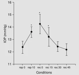
The results were similar when the right eye and the left eye were compared. A
statis-IO
P
(
m
m
H
g
)
16
15
14
13
12
11
rep 0 rep 10 rec 0 rec 15 rec 30 rec 45
Conditions *
*
*
Figure 1. Pre- and post-exer-cise intraocular pressure (IOP) and w ater intake for both the right and left eye. Data are re-port ed as m eans ± SD of mmHg. rep 0 = first measure-ment of IOP at rest; rep 10 = IOP 10 min after 600 ml of w a-ter ingestion; rec 0 to rec 45 = IOP at 15-min intervals after the end of exercise on a bicycle or after the equivalent period of rest. Least significant difference betw een data = 1.20; * P<0.05 compared to rep 0 (Tukey test).
Table 1. Physiological variables.
H2O Condition ETmax DTrec % H HR VO2 VCO2 La Tenv
10ºC Resting - -0.7 ± 0.3 0.9 ± 0.5 60.3 ± 10.4 0.26 ± 0.07 0.25 ± 0.03 1.02 ± 0.09 22.8 ± 0.75
24ºC Resting - -0.5 ± 0.4 1.0 ± 0.9 63.0 ± 6.5 0.23 ± 0.04 0.20 ± 0.05 0.96 ± 0.13 22.8 ± 0.26
38ºC Resting - -0.3 ± 0.4 0.7 ± 0.6 59.8 ± 11.6 0.21 ± 0.05 0.19 ± 0.03 0.76 ± 0.20 22.5 ± 0.55
10ºC Exercise 91.5 ± 23.7 0.6 ± 0.3 -0.7 ± 0.6 155.8 ± 5.9 2.01 ± 0.23 1.93 ± 0.29 1.89 ± 1.50 22.5 ± 0.97
24ºC Exercise 93.9 ± 24.5 0.8 ± 0.2 -0.6 ± 0.9 157.8 ± 8.7 1.97 ± 0.17 2.06 ± 0.32 1.73 ± 0.32 22.5 ± 0.49
38ºC Exercise 93.8 ± 27.0 0.7 ± 0.3 -0.5 ± 0.6 149.8 ± 8.1 2.04 ± 0.34 1.88 ± 0.27 1.47 ± 0.25 22.4 ± 0.49
Rest x exercise - * * * * * * NS
H2O temp NS NS NS NS NS NS NS NS
Data are reported as means ± SEM for 6 subjects. H2O temp = temperature of ingested w ater; ETmax = maximal exercise time (min); DTrec = difference betw een final and initial rectal temperature (oC); % H = final hydration status (% of corporal w eight); HR = final heart rate (min); VO2 = final oxygen consumption (l/min); VCO2 = final carbonic gas extraction (l/min); La = final plasma lactate (mmol/l); Tenv = environmental temperature (oC).
tically significant increase in IOP was ob-served between the first and the second, the first and the third, and the first and the fourth measurements of IOP, in both exercising and resting sessions. There were no significant differences in IOP between the resting and exercising conditions (Figure 1).
Time to exhaustion was not affected by the different water temperatures. Rectal tem-perature, hydration status, heart rate, oxygen consumption, carbonic gas extraction, and lactate concentration were increased by ex-ercise but were not affected by the different water temperatures. The subjects had an av-erage loss of 0.3 kg body weight during exercise and an average increase of 0.5 kg during the resting period. All experiments were performed under similar environmen-tal conditions (Table 1).
The present study addressed some of the possible variables related to IOP responses to physical exercise, i.e., heart rate, blood lactate, blood gases, hydration status, envi-ronmental conditions and body temperature, and their relations to the temperature of the ingested water.
Most papers reviewed report a decrease in IOP after different types of exercises (2-9,12-18). Some studies showed increases of IOP after exercise (1,19), while others found either no effect on IOP or variable results (20).
The lack of standardization in the exper-imental procedures appears to be the main reason for the lack of agreement in the litera-ture concerning the effects of exercise on IOP. Factors like food or water ingestion and postural variations were not well controlled previously or even considered. These meth-odological errors and dissimilarities between published data make it difficult to reach definitive conclusions about the effect of exercise on IOP.
Certain inverse relationships have been found between osmolarity and IOP (5.8) and these changes in IOP have been attributed to serum lactate or blood pH by some authors
(5,6,8).
The loss of water and electrolytes that occurs during exercise may affect plasma osmolarity and volume, which could cause changes in IOP. In the present study, heart rate, blood lactate, mean skin temperature, oxygen consumption, carbonic gas extrac-tion, and hydration status were consistently different between rest and exercise, but no significant differences in IOP were found between exercising and resting conditions. Furthermore, the temperature of the water ingested by the volunteers during the differ-ent experimdiffer-ental treatmdiffer-ents did not influ-ence the IOP results.
The variations in IOP - an initial increase followed by a return to basal values (Figure 1) - were similar during exercise and rest and were probably due to the ingestion of water, a result previously reported in the literature (11).
The volunteers had an average loss of 0.3 kg body weight during exercise, caused by ingestion of 1320 ml of water. During the resting period there was an average increase of 0.5 kg in body weight. On the other hand, IOP showed the same pattern and magnitude of variation in both situations, i.e., exercise and rest. This fact suggests that the effect of drinking water on IOP was dependent on the initial ingestion of water, as reported previ-ously. The return of IOP toward basal values occurred both after exercise and rest regard-less of the relative level of hydration, as shown by the differences in mean body weight. It seems, therefore, that the rapid intake of water caused the variations ob-served in IOP and that exercise or rest did not influence IOP. The temperature of the water also had no influence on these results.
In the present study, a difference in rectal temperature was observed between rest and exercise. The behavior of IOP was the same in both situations, indicating that the varia-tions in body temperature within these physi-ological limits did not influence IOP.
induced by exercise under conditions of water ingestion or the temperature of the ingested water had no significant effects on IOP. In
fact, only a transient increase in IOP was observed, related to the ingestion of water.
Re fe re nce s
1. American College of Sports M edicine (1987). Position stand - The prevention of thermal injuries during distance running.
M edicine and Science in Sports and Exer-cise, 19: 529-533.
2. Lem pert P, Cooper KH, Culver JF & Tredict TJ (1967). The effect of exercise on intraocular pressure. American Journal of Ophthalmology, 63: 1673-1676. 3. Leighton DA & Phillips CI (1970). Effect of
moderate exercise on the ocular tension.
British Journal of Ophthalmology, 54: 599-605.
4. M arcus DF, Krupin T, Podos SM & Becker B (1970). The effect of exercise on in-traocular pressure. I. Human beings. In-vestigative Ophthalmology and Visual Sci-ence, 9: 749-752.
5. M arcus DF, Edelhauser HF, M aksud M G & Wiley RL (1974). Effects of a sustained muscular contraction on human intraocu-lar pressure. Clinical Science and M olecu-lar M edicine, 47: 249-257.
6. Qureshi IA (1995). Effects of mild, moder-ate and severe exercise on intraocular pressure of sedentary subjects. Annals of Human Biology, 22: 545-553.
7. Stew art RH, Leblanc R & Becker B (1970). Effects of exercise on aqueous dynamics.
American Journal of Ophthalmology, 69: 245-248.
8. M artin B, Harris A, Hammel T & M
ali-novsky V (1999). M echanism of exercise-induced ocular hypotension. Investigative Ophthalmology and Visual Science, 40: 1011-1015.
9. Krupin T, Bass J, Oestrich C, Podos SM & Becker B (1977). The effect of hyperther-mia on aqueous humor dynamics in rab-bits. American Journal of Ophthalmology, 83: 561-564.
10. Whitacre M M & Stein R (1993). Sources of error w it h use of Goldm ann-t ype tonometers. Survey of Ophthalmology, 38: 1-30.
11. Drance SM (1963). Studies w ith applana-tion w ater tests. Archives of Ophthalmol-ogy, 69: 39-43.
12. Shapiro A, Shoenfeld Y & Shapiro Y (1978). The effect of standardized sub-maximal w orkload on intraocular pres-sure. British Journal of Ophthalmology, 62: 679-681.
13. Passo M S, Goldberg L & Elliot DL (1987). Exercise conditioning and intraocular pres-sure. American Journal of Ophthalmology, 103: 754-757.
14. Passo M S, Goldberg L, Elliot DL & Buskirk EM (1991). Exercise training reduces in-tra-ocular pressure among subjects sus-pected of having glaucoma. Archives of Ophthalmology, 109: 1096-1098. 15. Harris A, M alinovsky VE, Cantor LB,
Hen-derson PA & M artin BJ (1992). Isocapnia
blocks exercise-induced reductions in ocular tension. Investigative Ophthalmol-ogy and Visual Science, 33: 2229-2232. 16. Kiuchi Y, M ishim a HK, Hot eham a Y,
Furumoto A, Hirota A & Onari K (1994). Exercise intensity determines the magni-tude of IOP decrease after running. Japa-nese Journal of Ophthalmology, 38: 191-195.
17. Orgul S & Flammer J (1994). M oderate exertion lasting only seconds reduces in-traocular pressure. Graefe’s Archive for Clinical and Experimental Ophthalmology, 232: 262-264.
18. Qureshi IA (1996). Effects of exercise on intraocular pressure in physically fit sub-jects. Clinical and Experimental Pharma-cology and Physiology, 23: 648-652. 19. Dickerman RD, Smith GH, Langham-Roof
L, M cConathy WJ, East JW & Smith AB (1999). Intra-ocular pressure changes dur-ing maximal isometric contraction: does this reflect intra-cranial pressure or retinal venous pressure? Neurological Research, 21: 243-246.