Acta bot. bras. 23(3): 780-785. 2009
Stigmatic surface in the Vochysiaceae: reproductive and
taxonomic implications
Renata Carmo-Oliveira1,3 and Berta Lange de Morretes2
Received: June 11, 2008. Accepted: November 26, 2008
RESUMO – (Superfície estigmática das Vochysiaceae: implicações reprodutivas e taxonômicas). Espécies de Vochysiaceae são árvores e arbustos, principalmente do neotrópico, e constituem elementos característicos dos cerrados do Brasil Central. A família foi subdividida nas tribos Erismeae, com três gêneros e Vochysieae, com cinco gêneros. A superfície estigmática de seis espécies de Vochysiaceae foi caracterizada, representando quatro gêneros de Vochysieae (Vochysia, Salvertia, Callisthene e Qualea). Foram coletados botões em diferentes fases de desenvolvimento e flores abertas. Características gerais foram observadas no material fresco e a receptividade estigmática foi definida pela ação esterásica. Pistilos, fixados e incluídos em “paraplast”, foram secionados e corados para análises histológicas. Estigmas de flores abertas foram observados em microscopia eletrônica de varredura. Os estigmas de todas as espécies eram úmidos e apresentavam ação esterásica em pré-antese e na antese. Apresentavam, ainda, continuidade com o tecido transmissor de natureza secretora. Em Vochysia e Salvertia, a superfície estigmática era formada por tricomas multicelulares uniseriados, enquanto nas espécies dos demais gêneros, a superfície era formada por papilas unicelulares. A secreção que recobre os estigmas de todas as espécies fluía sem que houvesse ruptura da superfície estigmática e os tubos polínicos cresceram entre as papilas ou tricomas. As diferenças na superfície estigmática observadas corroboram a reconstrução filogenética separando dois clados e indicando que a tribo Vochysieae não é monofilética. Entretanto, não foi possível associar as características estigmáticas com os sistemas de polinização presentes na família.
Palavras-chave: Callisthene, Qualea, receptividade do estigma, Salvertia, Vochysia
ABSTRACT – (Stigmatic surface, reproductive biology and taxonomy of the Vochysiaceae). The Vochysiaceae are Neotropical trees and shrubs, common in the savanna areas in Central Brazil (Cerrados). The family has been traditionally divided into two tribes: Erismeae, with three genera, and Vochysieae, with five genera. We investigated the stigmatic surface of six Vochysiaceae species, belonging to four genera of Vochysieae: Vochysia, Salvertia, Callisthene and Qualea. Flowers and buds at different developmental stages were collected. Morphological features were observed on fresh material and stigmatic receptivity was inferred based on esterasic activity. Pistils were fixed and embedded in paraplast and sectioned on a rotary microtome; the sections were stained before histological analysis. Stigmas of open flowers were also observed by scanning electron microscopy. Stigmas of all species were wet and showed esterasic activity at pre-anthesis and anthesis stages. Stigmatic surface was continuous with transmitting tissue of glandular nature. Vochysia and Salvertia stigmatic surfaces were formed by multicelular uniseriate hairs, and species of the remaining genera showed papillate surface. The exudate over mature stigmas in all species flowed without rupture of stigmatic surface and pollen tubes grew down between hairs or papillae. Differences on the stigmatic surface agreed with a phylogenetic reconstruction that separated two clades and indicated that Vochysieae is not monophyletic. Stigmatic features could not be associated with pollination and breeding systems.
Kew words: Callisthene, Qualea, Salvertia, stigma receptivity, Vochysia
Introduction
Vochysiaceae includes eight genera and ca. 200 tropical species of trees and shrubs (Heywood 1985; Litt & Cheek 2002). Six genera occur in America and are well represented in the Brazilian flora. The dispersal centre is in the Guyana-Amazonian region and on the Central Brazilian Plateau, from where they seem to have spread to other Neotropical and Paleotropical areas (Barroso et al. 1984; Kawasaki 1998; Litt 1999). The family has been traditionally divided in two tribes: Vochysieae with five genera, Callisthene, Qualea, Ruizteranea, Salvertia and Vochysia (Stafleu 1948; 1952; Marcano-Berti 1969) and Erismeae with two genera Erisma
and Erismadelphus, the latter occurring in tropical Africa (Stafleu 1953; Kawasaki 1998). A new monospecific genus,
Korupodendron was recently described for western Africa (Litt & Cheek 2002). All genera are well defined and clearly separated by floral features. Flowers are mostly zygomorphic and with a single fertile stamen, but they differ in the number and other petal features. Vochysiaceae was traditionally included in the order Polygalales (Takhtajan 1969; Cronquist
1981; Dahlgren 1983) based on their zygomorphic flowers and/or the presence of hairs on the seed integument (Boesewinkel & Venturelli 1987), but these features could as well be the result of homoplasies. Conti et al. (1996; 1997) argued against the inclusion of the Vochysiaceae in the Polygalales and, based on DNA analysis, suggested it would fit better inside the Myrtales. This relationship is corroborated by the fact that the Vochysiaceae are one of the few families presenting a combination of bicollateral vascular bundles in the primary stem and vestured pits in the secondary xylem, very rare features outside the Myrtales. This phylogenetic association has been supported by further studies by Litt (1999), who revised morphological data and increased the molecular database for the family. Based on these studies, Litt (loc. cit.) proposed to divide the family in three clades, two of which have been previously included in the tribe Vochysieae. Moreover, the Vochysieae is characterized by semi-inferior or superior ovaries, which contrast with the inferior ovaries in the tribe Erismeae. Superior ovaries in the tribe Vochysieae were viewed as of secondary origin, since in the Myrtales flowers are mostly
epigynous (Litt & Stevenson 2003a).
Reproductive biology and floral specialization have been discussed for many Cerrado species of Vochysiaceae (Silberbauer-Gottsberger & Gottsberger 1975; Oliveira & Gibbs 1994; Oliveira 1996; Santos et al. 1997). These studies have shown varied pollination and breeding systems in the family: Salvertia convallariodora is hawkmoth pollinated and self-compatible (Oliveira 1996); Vochysia species were visited and pollinated mainly by large bees (sensu Frankie
et al. 1983) and secondarily by hawkmoths and hummingbirds. Most of them are self-incompatible and present the classic gametophytic self-incompatibility, with selfing pollen tube arresting along the style (Oliveira & Gibbs 1994; Santos et al. 1997); The genus Qualea has more varied flower structure, with species pollinated by large bees (Fischer & Gordo 1993; Oliveira 1998) and hawkmoths (Silberbauer-Gottsberger & Gottsberger 1975; Oliveira et al.
2004). The corolla is zygomorphic with a single petal, and stamen and stigma position alternate from flower to flower and characterize enantiostyly (Oliveira 1998; Litt & Stevenson 2003b); Finally, Callistene major is pollinated by small (less than 12 mm long), mostly social bees, while
C. fasciculate, with slightly larger flowers, is pollinated by medium to large bees (Santos 1997; Oliveira 1998). Both species are self-incompatible and present enantiostyly. These reproductive studies have provided basic data to understand the evolution and taxonomy of the family (Oliveira 1998).
Reproductive features and self-incompatibility mechanisms have been related to functional aspects of pollen/stigma interaction (Gibbs 1988; 1990; Sage et al. 2006). Investigation of angiosperm stigma structure revealed variable characters, some of them with clear taxonomic and phylogenetic meaning, particularly concerning the receptivity mechanisms of the stigmatic surface (Heslop-Harrison 1981). The receptivity and general stigmatic surface structure are known only partially for the Vochysiaceae (Oliveira 1998). Wet and papillate stigmas are common among the species studied, but the area of the stigmatic surface and its position in relation to the flower axis may vary between taxa. Here we studied the morphology, receptivity and anatomical structure of the stigma of some species of Vochysiaceae, and relate the results to reproductive features (summarized in Oliveira 1998) and to the phylogenetic reconstruction proposed recently for the family (Litt 1999; Litt & Stevenson 2003a; b).
Materials and methods
We studied species of Vochysiaceae in the Triângulo Mineiro region in Central Brazil. The botanical material was collected according to blooming period of each species inside or in areas around the Reserva Particular do Clube Caça e Pesca Itororó (CCPIU), the Panga Ecological Station (PES) and also in the Parque do Sabiá, all in Uberlândia, Minas Gerais state (19o09’20”-19o11’10”S and 48o23’20”-48o24’35”W,
average altitude 800 m). Plant vouchers were deposited in the Herbarium Uberlandensis (HUFU), with duplicate sheets placed at SPF. Voucher details are Callisthene major Mart., P.E. Oliveira 3073; Qualea
grandiflora Mart., R.C. Oliveira 03, 06 and 07; Q. multiflora Mart., R.C. Oliveira 05 and 10; Q. parviflora Mart., R.C. Oliveira 04 and 09; Salvertia convallariodora St. Hil., R.C. Oliveira 01; Vochysia cinnamomea Pohl., P.E. Oliveira 3095 and R.C. Oliveira 02 and 08. The study was carried out from May 1992 to August 2001, involving discontinuous sessions of fieldwork and laboratory analysis. Flowering phenology and floral features of the species are already published or summarized elsewhere (Oliveira 1998; Oliveira & Gibbs 2000; Oliveira et al. 2004). Most flowers last one day; they open at dawn and by the end of the day have released most pollen. Hawkmoth pollinated Salvertia convallariodora and Qualea grandiflora have basically one-night flowers, although the stigma may remain receptive the following night. Based on field experience, herbarium data and literature on phenology, we scheduled observations and collections during the flowering period of the species, whenever possible, along the study period.
Flower buds and flowers were collected, measured and separated into size classes. Previous experience allowed us to estimate time before anthesis based on flower bud size (Carmo-Oliveira, pers. obs.). The material was dipped in distilled water and placed in a vacuum chamber at 20 psi for 10 min before fixation. The samples were then fixed in Allen Bouin (Sass 1951), FAA 50% and FPA 50% (Johansen 1940), buffered neutral formalin (Lillie 1948 apud Clark 1981), and mainly in modified FAA with glutaraldehyde (Lersten & Curtis 1988). After fixation, the samples were kept in 70% ethanol or butanol. Some developmental stages were selected for sectioning and slide preparation. Buds and flowers selected were dehydrated in a butilic series (Johansen 1940; Sass 1951) and embedded in Paraplast (Sass 1951). In some cases we previously dissected the pistils to improve embedding and sectioning. The material was sectioned in a rotary microtome at 4 to 10 mm. Serial longitudinal sections were double stained with fuchsin and astra blue (Roeser 1972; Luque et al. 1996). The sections were observed in bright field light microscope, on some occasions with differential interference contrast (DIC), and photographed.
Stigmatic receptivity was inferred by using the alpha-naftol test, which detects esterasic activity (Dafni 1992). Esterasic activity on the stigma is commonly used to indicate receptivity and also to delimit specific stigmatic area (Kearns & Inouye 1993). For the test, styles with intact stigmas were carefully excised from pre-anthesis floral buds (a day before anthesis), recently open flowers and smaller floral buds at different developmental stages. These samples were submitted to a -naftol acetate solution in phosphate buffer, with acetone and Fast Blue B for about 5 minutes. Afterwards, stigmas were washed in distilled water and observed under stereomicroscope. Esterasic activity and receptivity is indicated by dark blue staining of the stigmatic surface and/or papillae (Dafni 1992). Blanks were tested with a solution without α-naftol. In order to characterize the stigmatic surface as wet or dry, besides careful observations, a quick test was carried out in the field using fresh material. The stigmatic surface was pressed lightly or swept across a clean glass slide so that wet stigma secretion would leave a mark on the slide (Heslop-Harrison & Shivanna 1977)
For observations using Scanning Electronic Microscopy (SEM), we selected pistils from pre-anthesis and open flowers, previously fixed. The pistils were carefully dissected and dehydrated to 100% ethanol. The samples were further dehydrated at critical point in liquid CO2 in a Balzers CPD 030. Dehydrated materials were mounted
in stubs and sputter-coated with gold in a Balzers CPD 050 (Sputter Coater). Observations and SEM micrographs were done at the Instituto de Biociências - USP, São Paulo, using a Zeiss Digital Scanning Microscope (DSM 940) and a Sinar 67 camera. Images were both digitalized and copied to photo film. Graphical scales were obtained under the same optical conditions.
Results
Carmo-Oliveira & Morretes: Stigmatic surface, reproductive biology and taxonomy of the Vochysiaceae
782
species. A description of the stigmatic surface of each species is presented bellow.
Vochysia cinnamomea – The stigmatic surface covers a sub-apical portion of the style and it was positioned sideways and opposite to the dehiscence of the anther (Fig. 1). The stigma was formed by pluricellular, uniseriate hairs (Fig. 2). The hairs were loosely organized and mostly free at anthesis. It was possible to notice, even with the naked eye, that the stigma was slightly groovy at the central region. The uniseriate hairs were formed by cells with prominent nuclei at the central portion flanked by vacuoles (Fig. 2). The stigmatic region continued through the transmitting tissue down to the ovary, were it forms a placentary obturator. The receptivity test with a-naftol showed that stigmas in pre-anthesis buds seemed to be already receptive.
Salvertia convallariodora – This species presented a relatively large and evident stigma at the apex of the style. Its bright yellow color contrasted with the whitish style and white petals (Fig. 3). As in Vochysia cinnamomea, the stigmatic surface was lateral and opposite the anther. It was also formed by pluricellular, uniseriate hairs which were loosely organized. In pre-anthesis and open flowers, the stigmatic surface was clearly wet, covered by abundant secretion. Uniseriated hairs also presented prominent nuclei and vacuoles (Fig. 4). In pre-anthesis and open flowers, the hairs appeared in SEM images covered by a precipitate that may be the secretion itself or some fixation artifact, which impaired more detailed observations. The α-naftol test
showed the stigmatic surface was already receptive in fairly young floral buds, about 19.0 mm long and estimated 12 days before anthesis.
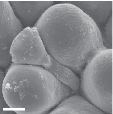
Callisthene major – The stigmatic surface was papillate and covered the apex of the style (Fig. 5). In some points it was possible to see that some papillae were formed down the style, below the continuous stigmatic region. The papillae (Fig. 6) were loosely disposed and egg shaped, with a central nucleus flanked by apical and basal vacuoles. The papillae showed clear continuity with the transmitting tissue that was also continuous with the placentary obturator, which is prominent in this species. The receptivity test showed that floral buds 8.0-8.75 mm long (ca. two days before anthesis) were already receptive. At pre-anthesis (one-day before anthesis) esterasic activity was apparent all over the stigma and putative receptivity was even clearer. On the stigma of open flowers, different from that observed in Vochysia and Salvertia, the secretion was not so copious and it was not evident to the naked eye. As in the other species, pollen tubes penetrated and grew into the papillae (Fig. 5, 7) without rupture of the papilla or the pellicle that involves these structures.
Qualea species – Stigma structure of the studied Qualea
species (Fig. 8 to 13) was similar to that observed in
Callisthene and different from that observed in Vochysia
and Salvertia. In Q. multiflora and Q. parviflora, the apex of the style was completely covered by the stigmatic surface
(Fig. 10, 12), as observed in Callisthene. In Q. grandiflora, however, the stigmatic surface occupied only one side of the style apex (Fig. 8). The stigmatic surface was constituted by short ovoid papillae in Q. parviflora (Fig. 12) and by longer papilla with enlarged cell base in Q. multiflora and
Q. grandiflora (Fig. 9, 11). As in Callisthene, the cells presented prominent nucleus flanked by vacuoles. In mature stigmas, the enlarged papilla bases were united while it was possible to observe in both light microscopy and SEM, as its distal portion was free (Fig. 8 to 11). In Q. parviflora, we observed esterasic activity in some parts of the stigma in about 6.3 mm buds (two-days before anthesis). Pre-anthesis buds and open flowers presented esterasic activity and possible receptivity all over the stigmatic surface. In flowers 24h after opening, the stigmatic surface presented
degenerative cells. In Q. multiflora and Q. grandiflora, that test revealed receptivity only in stigmas of pre-anthesis buds and open flowers. In freshly open flowers of species of Qualea the stigma was clearly wet but secretion was not as copious as observed in the stigmas of Vochysia and
Salvertia species.
Discussion
The stigmatic surface of angiosperms is the portion of the gynoecium responsible for pollen reception and for providing the conditions for its germination and initial growth (Heslop-Harrison & Shivanna 1977). The nature of the stigma and its role in the reproductive process have been studied since the 19th century and typification of the morphological diversity of these structures has been proposed by various authors since Capus (1878 apud
Heslop-Harrison & Shivanna 1977). The typifications paid attention mainly to the differences between wet and dry stigmas, presence or absence of stigmatic papillae, number of cells forming these papillae, and to the relationship between the stigma and the transmitting tissue (Heslop-Harrison & Shivanna 1977). The different types of stigma were related to angiosperm systematics (Heslop-Harrison 1981; Owens & Stirton 1989) and to its reproductive features (Heslop-Harrison 1975; Heslop-Harrison & Shivanna 1977; Pacini 1981). The stigma of the species of Vochysiaceae studied here can be classified as wet and papillate, and included in group III, as defined in Heslop-Harrison & Shivanna (1977). Esau (1977) and Fahn (1990) defined papillae as unicellular and hairs as bi- or pluricellular structures. In this sense, among the studied species, only
Callisthene major and the species of Qualea presented
true papillae. In Vochysia and Salvertia the stigmatic surface would be more properly defined as covered with multicellular hairs. However, Heslop-Harrison & Shivanna (1977) used the types of stigma by their general function and did not distinguish between papillae and hairs.
Wet stigmas, either hairy or papillate, are common in many Fabaceae, Rosaceae and other families in the Rosids (Heslop-Harrison & Shivanna 1977; Owens & Stirton 1989). Shivanna & Sastri (1981) observed that the exudate produced by these wet stigmas may or may not present esterasic activity. This activity seems to be associated with the role of stigmatic secretion in promoting pollen grain germination. In the species studied here, the secretion and esterasic activity seems to be clearly associated with receptivity, appearing mainly in buds one-day before anthesis and in open flowers.
Figure 7. SEM micrography of germinating pollen grain and pollen tube growing between intact stigmatic papillae in Callisthene major. Scale = 20 µm.
Carmo-Oliveira & Morretes: Stigmatic surface, reproductive biology and taxonomy of the Vochysiaceae
784
Only in S. convallariodora were secretion and esterasic activity present in floral buds several days before anthesis. On the stigmatic surface of the species studied here, we observed no evidence of pellicle, as present in wet stigmas of many Fabaceae (Owens 1989). In the Vochysiaceae, the exudate flows freely on the stigmatic surface without apparent damage or rupture of the cuticle or pellicle vesicles on the papillae or hairs. It was possible to observe pollen grains germinating on stigmas with entire, undamaged papillae and hairs. Unicellular papillae or multicellular hairs observed in the species studied did not seem to result in differential functioning of the stigma. Heslop-Harrison & Shivanna (1977) observed a great diversity of papilla structure in the species they studied; however, despite potentially important in taxonomic terms, they did not subdivide group III based on the occurrence of papillae or hairs.
Species with papillate, wet stigma may present either a solid style (O’Brien 1996) or a channel down to the carpellary chamber (Tangmitcharoen & Owens 1997). Different kinds of style may be present in the same group, as in the Caesalpinioideae (Owens 1989) and they may not be that different in their basically secretory nature. As for the species studied, the style is solid, filled by a secretory transmitting tissue, which shows a continuity between the stigma and the placentary obturator, similar to that described for Ornithogalum caudatum (Liliaceae) (Tilton& Horner 1980).
The different types of stigma can be related to incompatibility mechanisms (Heslop-Harrison 1975). Dry stigmas seem to occur mainly in groups with sporophytic self-incompatibility systems (SSI) and with incompatibility reaction on the stigma, while wet stigmas in group III would be related to gametophytic self-incompatibility systems (GSI) and incompatibility reaction occurring along the style (Heslop-Harrison & Shivanna 1977). These associations, although frequent, should be taken carefully, since there are many exceptions (Gibbs 1988; 1990). For the Vochysiaceae, two different kinds of self-incompatibility mechanisms have been observed based on pollen tube growth (Oliveira 1998). In Vochysia, pollen tubes from self-pollination are arrested along the style (Oliveira & Gibbs 1994; Santos et al. 1997), as observed in classic GSI, but in
Qualea and Callisthene, self-pollen tubes grow down to the ovary and were observed sometimes penetrating the micropile of the ovules, which would characterize a late-acting self-incompatibility mechanism (Oliveira 1998). Hence, the association between wet stigma and GSI can be applied to Vochysia cinnamomea which presents self-pollen tube arresting along the style (Santos et al. 1997), but any generalization is made difficult due to the late-acting self-incompatibility found in Callisthene major and Qualea parviflora (Oliveira 1998). It is possible that in Qualea and
Callisthene, the incompatibility reaction occurs in the placentary obturator. Since this structure maintains continuity with the transmitting tissue, incompatibility
mechanisms acting there could be functionally similar to the mechanisms observed in Vochysia. There was a discussion on the nature and functioning of late-acting incompatibility systems and suggestions that these systems would not be fundamentally different from classic GSI (Sage
et al. 1994; Lipow & Wyatt 2000). Further detailed studies in other Vochysiaceae may provide new insights on the evolution of self-incompatibility mechanisms and the role of the placentary obturator in these processes.
It is interesting that differences in the structure of the stigmatic surface, i.e. multicellulary hairs vs. papillae, and self-incompatibility mechanisms support very well the separation between the groups previously included in the tribe Vochysieae. Recent analyses, with both molecular and morphological data (Litt 1999), indicated that the tribe is not monophyletic and includes two distinct clades: one joins together Qualea, Ruizteranea and Callisthene (named QRC) and a second includes Vochysia and Salvertia. Although we have no data for Ruizteranea, stigma morphology presented here and incompatibility mechanisms seem to support this phylogenetic hypothesis. On the other hand, despite morphological differences, the stigmatic surface of species studied here is wet and functionally similar. There were no marked structural or functional differences, which could be associated with the different pollination systems described for the family (Oliveira 1998). Salvertia covallariodora presented stigma structurally different from
Q. grandiflora, although both species are moth pollinated (Oliveira et al. 2004). The stigma of Qualea grandiflora is, nevertheless, structurally similar to those of other species of Qualea that are bee pollinated (Oliveira 1998).
Acknowledgements
We thank the CNPq and FAPEMIG for, respectively, the doctoral and post-doctoral grants to RCO; Paulo E. Oliveira for critical reading of earlier manuscripts and for English translations; two anonymous referees and Acta Botanica Brasilica subject-editor, for the stimulus to translate the original paper into English in order to broaden its interest.
References
Barroso, G.M.; Peixoto, A.L.; Ichaso, C.L.F.; Costa, C.G.; Guimarães, E.F. & Lima, H.C. 1984. Sistemática de Angiospermas do Brasil. v.2. Viçosa, Imprensa Universitária.
Boesewinkel, F.D. & Venturelli, M. 1987. Ovule and seed structure in Vochysiaceae. Botanicher Jahrbrücher 108: 547-566. Clark, G. 1981. Staining Procedures. Baltimore, Willians and
Wilkins.
Conti, E.; Litt, A. & Sytsma, K.J. 1996. Circumscription of Myrtales and their relationships to other Rosids: evidence from rbcL sequence data. American Journal of Botany 83: 221-233. Conti, E.; Litt, A.; Wilson, P.G.; Graham, S.A.; Briggs, B.G.; Johnson,
L.A.S. & Sytsma, K.J. 1997. Interfamilial relationships in Myrtales: molecular phylogeny and patterns of morphological evolution. Systematic Botany 22: 629-647.
Cronquist, A. 1981. An integrated system of classification of flowering plants. New York, Columbia University Press. Dafni, A. 1992. Pollination Ecology: a Practical Approach.
Oxford. IRL Press at Oxford University Press.
Esau, K. 1977. Anatomy of seed plants.New York, John Wiley & Sons.
Fahn, A. 1990. Plant anatomy. Oxford, Pergamon Press. Fischer, E.A. & Gordo, M. 1993. Qualea cordata, pollination by the
territorial bee Centris tarsata in the “Campos Rupestres”. Ciência e Cultura 45: 144-147.
Frankie, G.W.; Haber, W.A.; Opler, P.A. & Bawa, K.S. 1983. Characteristics and organization of the large bee pollination system in the Costa Rican dry forest. Pp. 411-447. In: C.E. Jones & R.J. Little (eds.). Handbook of experimental pollination biology. New York, Van Nostrand Reinhold.
Gibbs, P.E. 1988. Self-incompatibility mechanisms in flowering plants: some complications and clarifications. Lagascalia 15: 17-28.
Gibbs, P.E. 1990. Self-incompatibility in flowering plants: a Neotropical perspective. Revista Brasileira de Botânica 13: 125-136.
Heslop-Harrison, J. 1975. Incompatibility and the pollen-stigma interaction. Annual Review of Plant Physiology 26: 403-425.
Heslop-Harrison, Y. 1981. Stigma characteristics and angiosperms taxonomy. Nordic Journal of Botany 1: 401-420.
Heslop-Harrison, Y. & Shivanna, K.R. 1977. The receptive surface of the Angiosperm stigma. Annals of Botany 41: 1 2 3 3 - 1 2 5 8 .
Heywood, V. 1985. Flowering Plants of the World. London, Croom Helm.
Johansen, D. 1940. Plant microtechnique. New York, McGraw-Hill.
Kawasaki, M.L. 1998. Systematics of Erisma (Vochysiaceae).
Memoirs of the New York Botanical Garden 81: 1-40. Kearns, C.A. & Inouye, D.W. 1993. Techniques for pollination
biologists. Niwot, University Press of Colorado.
Lersten, N.R. & Curtis, J.D. 1988. Secretory reservois (ducts) of two kinds in giant ragweed (Ambrosia trifida; Asteraceae). American Journal of Botany 75: 1313-1323.
Lipow, S.R. & Wyatt, R. 2000. Single gene control of post-zygotic self-incompatibility in Poke Milkweed, Asclepias exaltata L.
Genetics 154: 893-907.
Litt, A. 1999. Floral morphology and phylogeny of Vochysiaceae. PhD Thesis. New York, City University of New York.
Litt, A. & Cheek, M. 2002. Korupodendron songweanum, a new genus and species of Vochysiaceae from West-Central Africa.
Brittonia 54: 13-17.
Litt, A. & Stevenson, D. 2003a. Floral development and morphology of Vochysiaceae. I. The structure of the gynoecium. American Journal of Botany 90: 1533-1547.
Litt, A. & Stevenson, D. 2003b. Floral development and morphology of Vochysiaceae. II. The position of the single fertile stamen.
American Journal of Botany 90: 1548-1559.
Luque, R.; Souza, H.C. & Kraus, J.E. 1996. Método de coloração de Roeser (1972) - modificado - Kropp (1972) visando a substituição do azul de astra por azul de alcião 8GS ou 8GX. Acta Botanica Brasilica 10: 199-212.
Marcano-Berti, L. 1969. Ruizterania. Pittieria 2: 6-27.
O’Brien, S.P. 1996. Timetable of stigmatic receptivity and development and pollen tube growth in Chamelaucium uncinatum (Myrtaceae). Australian Journal of Botany 44: 49-59. Oliveira, P.E. 1996. Biologia floral de Salvertia convallariodora
(Vochysiaceae): uma espécie de cerrado polinizada por mariposas.
Revista Brasileira de Botânica 19: 49-53.
Oliveira, P.E. 1998. Reproductive Biology, Evolution and Taxonomy of the Vochysiaceae in Central Brazil. Pp. 381-393. In: S. Owens & P. Rudall (eds.). Reproductive biology: in systematics, conservation and economic botany. Richmond, Royal Botanical Gardens at Kew.
Oliveira, P.E. & Gibbs, P.E. 1994. Pollination and breeding systems of six Vochysia species (Vochysiaceae) in Central Brazil. Journal of Tropical Ecology 10: 509-522.
Oliveira, P.E.; Gibbs, P.E. & Barbosa, A.A. 2004. Moth pollination of woody species in the Cerrados of Central Brazil: a case of so much owed to so few? Plant Systematics and Evolution 245: 41-54. Owens, S. 1989. Stigma, style, pollen and the pollen-stigma interaction in Caesalpinioideae. Monographs on Systematic Botany of the Missouri Botanic Garden 29: 113-126.
Owens, S.J. & Stirton, C.H. 1989. Pollen, stigma, and style interactions in the Leguminosae. Monographs on Systematic Botany of the Missouri Botanic Garden 29: 105-112.
Pacini, E. 1981. Pollen-stigma interactions in plants with gametophytically controlled self-incompatibility.
Phytomorphology 31: 175-180.
Roeser, K.R. 1972. Die Nadel der Schwarzkiefer-Massenprodukt und Kunstwert der Natur. Mikrocosmos 61: 33-36.
Sage, T.L.; Bertin, R.I. & Willians, E.G. 1994. Ovarian and other late-acting self-incompatibility systems. Pp. 116-140. In: E.G. Williams, A.E. Clarke & R.B. Knox (eds.). Genetic control of self-incompatible and reproductive development in flowering plants. Amsterdam, Kluver Academic Publishers. Sage, T.L.; Price, M.V. & Waser, N.M. 2006. Self-sterility in Ipomopsis
aggregata (Polemoniaceae) is due to prezygotic ovule degeneration. American Journal of Botany 93: 254-262. Santos, M.L. 1997. Biologia de polinização e reprodução de
Callisthene fasciculata e Qualea parviflora em cerrado sobre afloramento basáltico, Araguari-MG. Dissertação de Mestrado. Brasília, Universidade de Brasília.
Santos, M.L.; Afonso, A.P. & Oliveira, P.E. 1997. Biologia floral de Vochysia cinnamomea Pohl. (Vochysiaceae) em cerrados do Triângulo Mineiro, MG. Revista Brasileira de Botânica 20: 127-132.
Sass, J.E. 1951. Botanical microtechnique. Iowa, The Iowa State College Press.
Shivanna, K.R. & Sastri, D.C. 1981. Stigma-surface esterase activity and stigma receptivity in some taxa characterized by wet stigmas.
Annals of Botany 47: 53-64.
Silberbauer-Gottsberger, I.& Gottsberger, G. 1975. Uber sphingophile Angiospermen Brasiliens. Plant Systematics and Evolution 123: 157-184.
Stafleu, F.A. 1948. A monography of the Vochysiaceae. I. Salvertia and Vochysia. Mededelingen van het Botanich Museum en Herbarium van de Rijksuniversiteit te Utrecht 95: 397-540. Stafleu, F.A. 1952. A monography of the Vochysiaceae. II. Callisthene.
Acta Botanica Neerlandica 1: 222-242.
Stafleu, F.A. 1953. A monography of the Vochysiaceae. III. Qualea.
Acta Botanica Neerlandica 2: 144-217.
Takhtajan, A. 1969. Flowering plants. Edinburgh, Oliver and Boyd. Tamgmitcharoen, S. & Owens, S.J. 1997. Floral biology, pollination, pistil receptivity, and pollen tube growth of teak (Tectona grandis Linn. F.). Annals of Botany 79: 227-241.
Tilton, V.R. & Horner, H.T. 1980. Stigma, style, and obturator of Ornithogalum caudatum (Liliaceae) and their function in reproductive process. American Journal of Botany 67: 1113-1131.