Human Lung Cancer
Sarah C. Mutka1*, Lucia H. Green1, Evie L. Verderber1, Jane P. Richards1, Doug L. Looker1, Elizabeth A. Chlipala2, Gary J. Rosenthal1
1N30 Pharmaceuticals, Inc., Boulder, Colorado, United States of America,2Premier Laboratory, LLC, Longmont, Colorado, United States of America
Abstract
Endogenous S-nitrosothiols, including S-nitrosoglutathione (GSNO), mediate nitric oxide (NO)-based signaling, inflammatory responses, and smooth muscle function. Reduced GSNO levels have been implicated in several respiratory diseases, and inhibition of GSNO reductase, (GSNOR) the primary enzyme that metabolizes GSNO, represents a novel approach to treating inflammatory lung diseases. Recently, an association between decreased GSNOR expression and human lung cancer risk was proposed in part based on immunohistochemical staining using a polyclonal GSNOR antibody. GSNOR is an isozyme of the alcohol dehydrogenase (ADH) family, and we demonstrate that the antibody used in those studies cross reacts substantially with other ADH proteins and may not be an appropriate reagent. We evaluated human lung cancer tissue arrays using monoclonal antibodies highly specific for human GSNOR with minimal cross reactivity to other ADH proteins. We verified the presence of GSNOR in$85% of specimens examined, and extensive analysis of these samples demonstrated no difference in GSNOR protein expression between cancerous and normal lung tissues. Additionally, GSNOR and other ADH mRNA levels were evaluated quantitatively in lung cancer cDNA arrays by qPCR. Consistent with our immunohistochemical findings, GSNOR mRNA levels were not changed in lung cancer tissues, however the expression levels of other ADH genes were decreased. ADH IB mRNA levels were reduced (.10-fold) in 65% of the lung cancer cDNA specimens. We conclude that the previously reported results showed an incorrect association of GSNOR and human lung cancer risk, and a decrease in ADH IB, rather than GSNOR, correlates with human lung cancer.
Citation:Mutka SC, Green LH, Verderber EL, Richards JP, Looker DL, et al. (2012) ADH IB Expression, but Not ADH III, Is Decreased in Human Lung Cancer. PLoS ONE 7(12): e52995. doi:10.1371/journal.pone.0052995
Editor:Klaus Roemer, University of Saarland Medical School, Germany
ReceivedOctober 23, 2012;AcceptedNovember 27, 2012;PublishedDecember 28, 2012
Copyright:ß2012 Mutka et al. This is an open-access article distributed under the terms of the Creative Commons Attribution License, which permits unrestricted use, distribution, and reproduction in any medium, provided the original author and source are credited.
Funding:N30 Pharmaceuticals, Inc is a privately held company. The funders had no role in study design, data collection and analysis, decision to publish, or preparation of the manuscript.
Competing Interests:All authors except Elizabeth Chlipala are or were employees of N30 Pharmaceuticals, the funder of this study. Elizabeth Chlipala (Premier Laboratory) was paid to perform the immunohistochemistry work reported in this manuscript. N30 Pharma is developing a novel class of disease modifying therapies that preserve intracellular GSNO (Snitrosoglutathione), a key regulator of organ repair, regeneration, and healing. There are no further patents, products in development or marketed products relevant to this paper to declare. This does not alter the authors’ adherence to all the PLOS ONE policies on sharing data and materials, as detailed online in the guide for authors.
* E-mail: [email protected]
Introduction
S-nitrosoglutathione (GSNO) is an endogenous nitric oxide donor that serves as a depot for nitric oxide (NO) in the body and plays an integral role in communicating NO mediated signaling functions. Decreased levels of GSNO have been correlated to a variety of diseases and its restoration has been proposed as a therapeutic approach to cystic fibrosis [1] and asthma [2,3]. The oxidoreductase, S-nitrosoglutathione reductase (GSNOR), is the primary enzyme involved in the catabolism of intracellular GSNO, and its pharmacologic inhibition provides a therapeutic mechanism for preserving intracellular GSNO levels. High potency GSNOR inhibitors are currently under clinical develop-ment [4–7].
GSNOR, also known as the alcohol dehydrogenase class III enzyme (ADH III) and formaldehyde dehydrogenase, is evolu-tionarily the oldest member of the ADH protein family, and all other ADHs are thought to derive from GSNOR by gene duplication [8,9]. In humans, the ADH isozymes are homologous and show up to 60% amino acid sequence identity. They are also highly conserved between species. This high amino acid sequence
Materials and Methods
Antibodies and purified proteins
ADH IA protein was purchased from Abnova (Taipei City, Taiwan,#H00000124-Q01), but some degradation was noted in this preparation. Other proteins used in this study were prepared for us by Emerald BioStructures (Bainbridge Island, WA) as described previously [14]. Briefly, GSNOR, ADH IB, ADH II, and ADH IV were expressed with an N-terminal 66-histidine affinity tag and a Smt-fusion sequence which was removed by
Ulp-1 cleavage after Ni affinity chromatography to produce the full-length recombinant proteins. The approximate molecular weights for the ADH proteins before and after removal of the His-Smt tag are: ADH IB (51 kDa, 40 kDa), ADH II (51 kDa, 40 kDa), and ADH IV (53 kDa, 41 kDa). The His-Smt-GSNOR fusion protein is approximately 50 kDa. The GSNOR protein preparations used for antibody generation were confirmed by Emerald BioStructures to be full length by mass spectrometry [4] with a molecular weight of 39.6 kDa. We and others [15] have shown that GSNOR, whether purified or from human cell lysates, consistently migrates slightly faster than its predicted molecular weight on SDS-PAGE. A polyclonal antibody was generated from serum of rats immunized with purified, recombinant, full length human GSNOR protein at Biomodels (Watertown, MA) for N30 Pharmaceuticals. Three distinct monoclonal antibodies to human GSNOR (N30-C3 rat GSNOR, N30-F6 mouse anti-GSNOR, and N30-G11 mouse anti-GSNOR) were generated by immunization of mice or rats with purified, recombinant, full length human GSNOR protein at ProMab Biotechnologies (Richmond, CA) for N30 Pharmaceuticals. Serum titer was evaluated by ELISA prior to selection for hybridization. Hybrid-oma fusion with myelHybrid-oma cells (ProMab Biotechnologies; Richmond, CA) was performed with the splenocytes from the mouse or rat with the best titer. Supernatants from the hybridoma wells were screened by ELISA for positive reactivity for GSNOR. Supernatants were also screened by ELISA for negative reactivity for ADH IB, ADH II and ADH IV. Supernatant from 10 clones was then screened at N30 Pharmaceuticals by western blot for specificity for the GSNOR isozyme and minimal non-specific protein binding. Two of the 10 clones for both the mouse and the rat were selected and then subcloned at ProMab by limiting dilution. For the mouse monoclonal antibodies, a supply of antibody was prepared by ascites production in 10 mice, and the antibody was harvested and purified by IgG chromatography. For the rat monoclonal antibody, antibody was produced in vitro by cell culture and IgG purified. Rabbit polyclonal anti-GSNOR was purchased from Proteintech (Chicago, IL;#11051-1-AP).
GSNOR immunoblotting
Purified ADH proteins (22.5 ng/well) were analyzed by SDS-PAGE and immunoblotting using the polyclonal antibody (Proteintech; 1:50 dilution to final concentration of 1.4mg/mL) or monoclonal antibodies (rat anti-GSNOR N30-C3, 2.4mg/mL;
Figure 1. Polyclonal GSNOR antibodies react with other ADHs.
22.5 ng purified recombinant proteins were separated by SDS-PAGE, and immunoblots were performed to determine antibody reactivity to GSNOR, ADH IA, ADH IB, ADH II, and ADH IV. With the exception of ADH IA, the purified recombinant proteins were generated with a fusion protein tag during cloning, and the tag was cleaved off during purification as described in the Materials and Methods. The molecular weights of the fusion proteins are approximately 50–51 kDa, and the final, purified proteins are 39–41 kDa. Human GSNOR consistently migrates faster than the calculated molecular weight. As seen by for the presence of both 50 and 40 kDa bands for ADH II, the majority of the protein in this preparation still contains the fusion protein tag. However, the purified GSNOR protein used for immunization was confirmed to be full length, and free of additional tag sequence as described in the Materials and Methods. A) Commercially available rabbit polyclonal GSNOR antibody (Proteintech #11051-1-AP). B) In-house polyclonal antibody generated by immunization of rats with purified, recombinant, full length human GSNOR protein at Biomodels (Watertown, MA) for N30 Pharmaceuticals.
doi:10.1371/journal.pone.0052995.g001
Figure 2. Monoclonal GSNOR antibodies are specific for GSNOR. Purified recombinant proteins were separated by SDS-PAGE, and immunoblots were performed to determine antibody reactivity to GSNOR, ADH IB, ADH II, and ADH IV. Three independent monoclonal antibodies were tested: A) N30-F6 mouse anti-GSNOR; B) N30-G11 mouse anti-GSNOR; C) N30-C3 rat anti-GSNOR.
mouse anti-GSNOR N30-F6 and N30-G11 10 ng/mL). HRP conjugated secondary antibodies (Santa Cruz Biotechnology, Santa Cruz, CA) were used for detection.
GSNOR immunohistochemistry
Immunohistochemical analysis for GSNOR was performed using formalin-fixed, paraffin-embedded human lung tissue microarrays (Folio Bioscience, Columbus, OH;#ARY-HH0178,
#ARY-HH0176). Slides were deparaffinized in xylene and rehydrated to water through a series of alcohol gradients. The slides were pretreated in a 95uC citrate pH 6.1 antigenic retrieval solution (Dako, Carpinteria, CA). After allowing the slides to cool to room temperature, a 3.0% hydrogen peroxide solution (EMD Chemicals, Gibbstown, NJ) was added to quench endogenous peroxidase activity. Slides were preincubated with Serum Free Protein Block (Dako), followed by incubation with a GSNOR antibody or the appropriate non-specific immunoglobulin control: rat IgG control (AbD Serotec, Raleigh, NC), mouse IgG2B (Dako), or rabbit Ig fraction (Dako). Slides labeled with the N30-C3 antibody were then incubated with rabbit anti rat immuno-globulin (Dako), and all slides were then labeled with a goat anti rabbit or anti mouse polymer (Dako). DAB+(Dako) was used for detection, and slides were counter stained with hematoxylin (Dako).
Analysis of mRNA levels in lung tissue
cDNA arrays made from 23 matched pairs of normal and cancerous lung tissue were purchased from Origene (Rockville, MD; #HLRT504, lot #0411). The pairs included lung cancer tissue covering Stage IA (n = 4), IB (n = 4), IIA (n = 2), IIB (n = 8), IIIA (n = 3), and IIIB (n = 2) each paired with adjacent normal tissue. One Stage IV tissue sample was not paired with a normal sample, and was not included in the analysis. Gene expression of GSNOR (ADH III; gene name ADH5), ADH IB (gene ADHIB),
ADH II (geneADH4), ADH IV (geneADH7), and beta-actin was
quantified by qPCR using TaqMan primer probe sets (Applied Biosystems, Carlsbad, CA): Human B-Actin (Hs00357333_g1),
Human GSNOR (Hs00605185_m1), Human ADH IB
(Hs00605175_m1), Human ADH II (Hs00923466_m1), and Human ADH IV (Hs00609447_m1). Quantitative PCR (qPCR) was performed with an ABI 7300 RT PCR instrument (Applied Biosystems) using Perfecta qPCR fastmix (Quanta Biosciences, Gaithersburg, MD) according to the manufacturer’s instructions. Samples were subjected to 1 cycle at 95uC for 3 minutes, followed by 40 cycles at 95uC for 15 sec and 60uC for 1 minute. For data analysis, comparative threshold cycle values (Ct) for beta-actin were used to normalize loading variations and calculate DCt values.DDCtvalues were calculated by subtracting theDCtvalue for the control (normal) tissue from theDCtvalue for the matched tumor tissue.DDCtvalues were converted to fold differences using the formula 22DDCt.
Results
Commercial polyclonal GSNOR antibody cross-reacts with other ADH proteins
Recent work examining GSNOR in human lung cancer [12] employed immunohistochemical staining of human lung cancer samples with a commercially available polyclonal GSNOR antibody (see materials and methods). Because the protein sequence of GSNOR is highly conserved with other ADH isozymes, we determined its specificity for GSNOR compared with its specificity toward other ADHs. Cross reactivity was examined by immunoblotting using purified human recombinant
GSNOR (ADH III), ADH IA, ADH IB, ADH II, and/or ADH IV. As shown in Figure 1A, a commonly used commercial polyclonal antibody strongly cross-reacts with other ADH proteins, particularly ADH IB, ADH II, and ADH IV. We tested other commercially available polyclonal antibodies and an in-house prepared rat polyclonal GSNOR antibody and found all polyclonal antibodies tested also strongly cross-reacted with ADH proteins (data not shown and Figure 1B). To address this issue, monoclonal antibodies specific for GSNOR were developed by immunization of mice and rats with purified, full length, recombinant human GSNOR as described in the methods. This effort also included a counter-screening step to minimize cross reactivity to other ADH proteins. As shown in Figure 2, three of the new monoclonal antibodies against GSNOR have either greatly reduced or no cross reactivity to other ADH proteins. To our knowledge, this is the first paper to describe antibodies with this level of specificity toward GSNOR.
Monoclonal GSNOR antibodies detect expression in a wide variety of human lung cancers
We examined GSNOR protein levels in various lung cancer tissues by immunohistochemistry using the above described three GSNOR-specific monoclonal antibodies and compared the tissue staining pattern to the commercially available polyclonal antibody used in a previous publication [12]. We also compared the antibody signal in normal lung tissue. Using the four antibodies, we stained four identical human lung cancer tissue microarrays each containing human tissue cores from over 100 cases of lung cancer at various stages of progression. GSNOR was strongly detected in normal human lung tissue including in bronchial epithelial cells, alveolar macrophages, and type 2 pneumocytes in
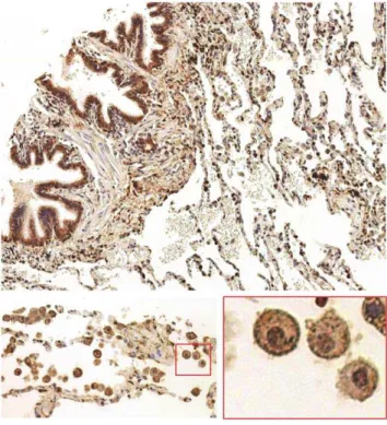
Figure 3. GSNOR is present in normal lung tissue.Normal human lung tissue microarrays were stained with N30-C3 monoclonal GSNOR antibody (1mg/mL) followed by DAB detection. Bronchial epithelial
cells, alveolar macrophages, and type 2 pneumocytes in alveoli are strongly stained.
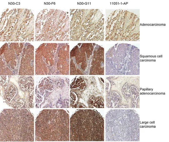
alveoli (Figure 3). GSNOR was also detected in a wide variety of lung cancer tissues including adenocarcinoma, squamous cell carcinoma, papillary adenocarcinoma, and large cell carcinoma when tissues were stained with the monoclonal GSNOR antibodies (Figure 4, N30-C3, N30-F6, N30-G11). Staining in lung cancer tissues was fainter when the polyclonal antibody was used (Figure 4, 11051-1-AP). Tissue sections were scored by three reviewers for GSNOR staining for each antibody (Table 1). Using the three specific monoclonal antibodies, GSNOR protein levels were not different in lung cancer specimens compared to normal tissue; whereas the polyclonal antibody with demonstrated ADH
cross reactivity suggested that GSNOR expression was higher in normal lung tissue.
GSNOR mRNA levels are not decreased in human lung cancer tissue versus adjacent normal tissue
Due to the potential for subjectivity in grading tissue staining, GSNOR and ADH mRNA levels were evaluated quantitatively in human lung cancer cDNA arrays. Commercially available cDNA arrays (see materials and methods) were used that contained 23 matched pairs of normal and cancerous lung tissue. The pairs included Stage IA (n = 4), IB (n = 4), IIA (n = 2), IIB (n = 8), IIIA
Figure 4. GSNOR is found in human lung cancer tissues stained with monoclonal GSNOR antibodies.Normal human lung cancer tissue microarrays were stained with monoclonal GSNOR antibodies (N30-C3, N30-F6, N30-G11) or a commercially available polyclonal GSNOR antibody (11051-1-AP) followed by DAB detection. Various human lung cancer tissues are strongly stained by all three GSNOR monoclonal antibodies, but staining is less prominent when the non-specific polyclonal GSNOR antibody is used.
doi:10.1371/journal.pone.0052995.g004
Table 1.Quantitation of GSNOR positive lung cancer tissue sections.
Antibody % tumors positive for GSNOR % normal lung positive for GSNOR
N30-C3 85 83
N30-F6 95 83
N30-G11 99 92
11051-1-AP 28 83
Four identical sets of 120 lung tissue cores (12 normal, 108 cancer) were stained with the GSNOR antibodies indicated below. The stained tissue cores were graded as positive or negative for GSNOR expression by three reviewers blinded to antibody used (SCM, GJR, JPR). The percent of tumor and normal lung cores graded as positive for GSNOR expression are shown below.
(n = 3), and IIIB (n = 2) lung cancer tissues each paired with adjacent normal tissue. GSNOR mRNA levels were quantitated using qPCR, and relative quantities of GSNOR in tumor versus adjacent normal tissue were calculated as described in the supplementary methods. No correlation was observed between GSNOR mRNA expression and lung cancer. In this analysis, 11 of 23 tumor samples had relative tumor/normal expression ratios ,1 and 12 of 23 samples had expression ratios.1. Furthermore, evidence of dramatically decreased GSNOR expression was rare in the tested lung cancer samples; only 3 of the tumor samples were found with GSNOR expression decreased by more than 3-fold (ratio,0.33; Figure 5). Conversely, when expression of ADH IB, ADH II, and ADH IV was analyzed, more of the samples demonstrated larger expression decreases in the tumor samples compared to GSNOR. Moreover, we observed substantial decreases in ADH IB expression in lung cancer tissues, with 12 of 23 pairs having$10-fold decrease in ADH IB expression in the lung cancer samples (relative tumor/normal expression ratio #0.1). Only 3 of 23 pairs had increased expression of ADH IB in the lung cancer samples compared to the adjacent normal tissue (Figure 5).
Discussion
In the lung, endogenous S-nitrosothiols including GSNO mediate NO-based signaling, inflammatory responses, and smooth muscle function. Inhibition of GSNOR represents a novel approach to treating respiratory disease via preserving endogenous GSNO, and a novel GSNOR inhibitor is currently in clinical development for the treatment of asthma and cystic fibrosis [4].
In this study, we demonstrate no consistent differences in the expression of GSNOR when comparing lung cancer tissue to normal lung tissue. These findings contradict previously published
work by Marozkina and colleagues [12] who reported that GSNOR expression was decreased in lung cancer tissues. In their analysis, the authors observed that 69% of human lung cancer biopsies demonstrated significantly reduced GSNOR levels. In their assessment, GSNOR levels were significantly lower in late stage tumors compared to early stage tumors, and they concluded that reduced GSNOR activity is correlated with later stages of tumor maintenance. It is likely that a primary discrepancy between our report and previous reports stems from the lack of specificity of the polyclonal antibodies used in their analysis. We demonstrated that the polyclonal antibody strongly cross reacts with other, closely related proteins, including ADHIB, ADH II, and ADH IV. In fact, we have found that every polyclonal antibody we have tested suffers from this same lack of specificity, and conclusions about GSNOR expression and localization drawn from experiments using polyclonal antibodies have great potential to be misinterpreted. The lack of specificity in polyclonal antibodies is not surprising given the high sequence identity between GSNOR and other ADH isozymes. When developing a monoclonal antibody to GSNOR, we found it necessary to counter-screen the clones against several other purified ADH proteins to obtain the required specificity. Using GSNOR specific monoclonal antibodies, we found no difference in the staining of cancerous versus normal lung tissue.
Additionally, we explored the expression of not only GSNOR but also other ADHs in lung cancer tissue using more definitive qPCR methods. No change in GSNOR mRNA expression between normal and cancerous lung tissue was observed. Conversely, when expression of ADH IB, ADH II, and ADH IV were analyzed, we saw more and larger expression decreases in the tumor samples. Moreover, we observed substantial decreases in ADH IB expression in lung cancer tissues with 12 of 23 pairs
Figure 5. GSNOR and ADH gene expression in lung cancer cDNA arrays.GSNOR (gene nameADH5), ADH IB (ADHIB), ADH II (ADH4), and ADH
IV (ADH7) mRNA levels were evaluated by qPCR in human lung cancer cDNA arrays (Origene,#HLRT504, lot#0411, Rockville, MD) and normalized to b-actin levels. Arrays contained 23 matched pairs of normal and cancerous lung tissue. Tumor expression relative to the matched normal sample was calculated using theDDCt method. Relative quantities,1 represent decreased expression in the tumor sample. Lung cancer specimens included Stage IA (n = 4), IB (n = 4), IIA (n = 2), IIB (n = 8), IIIA (n = 3), and IIIB (n = 2) with each sample paired with adjacent normal tissue. No correlation between GSNOR mRNA levels and lung cancer was observed, while expression of ADH IB was strongly reduced in lung cancer samples.
having $10-fold decrease in ADH IB expression in the lung cancer samples (relative tumor/normal expression ratio#0.1).
That ADH IB expression shows some relationship to lung cancer is not surprising. ADH IB is integral in a number of pathways with known influence in lung cancer including fatty acid, retinol and tyrosine metabolism, as well as in glycolysis and gluconeogenesis [16]. In a meta-analysis of published studies looking at the relationship of ADH IB in lung cancer, ADH IB expression was decreased in lung cancer in an overwhelming number of samples and analysis [16] and clearly polymorphisms of ADH IB have shown correlations to other types of cancer including esophageal [17]. Unlike ADH IB where there has been substantial investigation in its relation to cancer, GSNOR expression levels have recently been suggested to correlate with hepatocellular carcinoma [13]. These studies in part relied on GSNOR knock-out mice that have substantial phenotypic differences when compared to wild type controls, and recent studies from our lab have shown this any purported relationship between genetic deletion of GSNOR and hepatocellular carcino-ma is not seen in a haplosufficient model (GRJ and D. Colagiovanni, manuscript in preparation, N30 Pharmaceuticals).
In summary, it appears that a decrease in ADH IB, rather than GSNOR, correlates with human lung cancer, and the previously reported results showed an incorrect association of GSNOR and human lung cancer risk. In further support of this conclusion, published studies of mice with homozygous genetic deletion of GSNOR have found no increase in murine lung cancer in both untreated GSNOR2/2mice nor GSNOR2/2 mice treated with the carcinogen diethylnitrosamine [13]. Thus GSNOR expression and inhibition do not appear to be associated with risk of human lung cancer.
Acknowledgments
The authors thank Ross Benik for technical assistance with tissue staining.
Author Contributions
Conceived and designed the experiments: SCM JPR DLL GJR. Performed the experiments: SCM LHG ELV EAC. Analyzed the data: SCM LHG ELV JPR EAC GJR. Contributed reagents/materials/analysis tools: EAC. Wrote the paper: SCM GJR.
References
1. Snyder AH, McPherson ME, Hunt JF, Johnson M, Stamler JS, et al. (2002) Acute effects of aerosolized S-nitrosoglutathione in cystic fibrosis. American journal of respiratory and critical care medicine 165: 922–926.
2. Wu H, Romieu I, Sienra-Monge JJ, Estela DR-N, Anderson DM, et al. (2007) Genetic variation in S-nitrosoglutathione reductase (GSNOR) and childhood asthma. J Allergy Clin Immunol 120: 322–328.
3. Que LG, Liu L, Yan Y, Whitehead GS, Gavett SH, et al. (2005) Protection from experimental asthma by an endogenous bronchodilator. Science 308: 1618– 1621.
4. Sun X, Wasley J, Qiu J, Stout A, Green L, et al. (2011) Discovery of S-Nitrosoglutathione Reductase Inhibitors: Potential Agents for the Treatment of Asthma and other Inflammatory Diseases. ACS Med Chem Letters 21: 5849– 5853.
5. Sun X, Qiu J, Strong SA, Green LS, Wasley JW, et al. (2011) Structure-activity relationships of pyrrole based S-nitrosoglutathione reductase inhibitors: Pyrrole regioisomers and propionic acid replacement. Bioorg Med Chem Lett 21: 3671– 3675.
6. Sun X, Qiu J, Strong SA, Green LS, Wasley JW, et al. (2011) Discovery of potent and novel S-nitrosoglutathione reductase inhibitors devoid of cytochrome P450 activities. Bioorg Med Chem Lett 21: 5849–5853.
7. Sun X, Qiu J, Strong SA, Green LS, Wasley JW, et al. (2012) Structure-activity relationship of pyrrole based S-nitrosoglutathione reductase inhibitors: carbox-amide modification. Bioorg Med Chem Lett 22: 2338–2342.
8. Danielsson O, Jornvall H (1992) ‘‘Enzymogenesis’’: classical liver alcohol dehydrogenase origin from the glutathione-dependent formaldehyde dehydro-genase line. Proc Natl Acad Sci U S A 89: 9247–9251.
9. Hoog JO, Hedberg JJ, Stromberg P, Svensson S (2001) Mammalian alcohol dehydrogenase - functional and structural implications. J Biomed Sci 8: 71–76. 10. Straub AC, Billaud M, Johnstone SR, Best AK, Yemen S, et al. (2011) Compartmentalized connexin 43 s-nitrosylation/denitrosylation regulates het-erocellular communication in the vessel wall. Arterioscler Thromb Vasc Biol 31: 399–407.
11. Wu X, Du L, Xu X, Tan L, Li R (2010) Increased nitrosoglutathione reductase activity in hypoxic pulmonary hypertension in mice. J Pharmacol Sci 113: 32– 40.
12. Marozkina NV, Wei C, Yemen S, Wallrabe H, Nagji AS, et al. (2012) S-nitrosoglutathione reductase in human lung cancer. Am J Respir Cell Mol Biol 46: 63–70.
13. Wei W, Yang Z, Tang CH, Liu L (2011) Targeted deletion of GSNOR in hepatocytes of mice causes nitrosative inactivation of O6-alkylguanine-DNA alkyltransferase and increased sensitivity to genotoxic diethylnitrosamine. Carcinogenesis 32: 973–977.
14. Green LS, Chun LE, Patton AK, Sun X, Rosenthal GJ, et al. (2012) Mechanism of inhibition for N6022, a first-in-class drug targeting S-nitrosoglutathione reductase. Biochemistry 51: 2157–2168.
15. Lee SP, Chiang CP, Lee SL, Hsia YJ, Chuang TL, et al. (2006) Immunochemical features in the classification of human alcohol dehydrogenase family. Alcohol 39: 13–20.
16. Wang L, Xiong Y, Sun Y, Fang Z, Li L, et al. (2010) HLungDB: an integrated database of human lung cancer research. Nucleic Acids Res 38: D665–D669. 17. Yang SJ, Yokoyama A, Yokoyama T, Huang YC, Wu SY, et al. (2010)