Rev. bras. farmacogn. vol.24 número5
Texto
Imagem

Documentos relacionados
no domínio da inclusão social, o Governo dispôs-se, no quadro da Resolução do.. Nesta quarta fase conta-se com a implementação de 130 novos projectos, com a
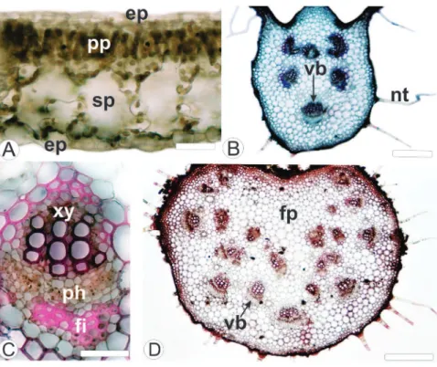
Leaf blade - The uniseriate epidermis - Ep - (Figu- re 1A) of all seven species features cells with sinuous an- ticlinal walls in paradermic section (Figure 1B - arrow) and thin
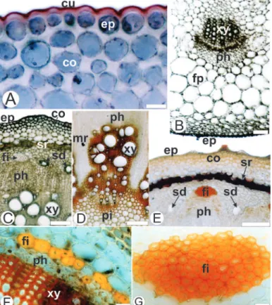
The tepals (Figs. 2A, C) presented uniseriate epidermis with cuticle flange, and periclinal thick- walled cells in the abaxial face and thin-walled cells in the
Glandular capitate-stalked and capitate- sessile, uniseriate multicellular non-glandular trichome with tapered apical cell, conical non-glandular trichome, isobilateral
S: Spherical capitate trichomes, with circular apical cells, have unicellular stalks; E: Ellipsoid capitate trichomes have oblong shaped heads, and unicellular stalks; B:
Due to their great diversity of forms and functions, this study aimed to inventory the glandular trichomes present in the aerial vegetative axis of Amphilophium
Hesperozygis ringens presents remarkable characteristics such as the presence of glandular and non-glandular trichomes in leaf and stem, two morphs of diallelocytic stomata
Lipids are detected in the epidermis, non-glandular trichomes, and parenchymatic cells of outer and inner compartments (figure 14-15), and nutritive tissue.. Lignins
