ISSN 0102-695X http://dx.doi.org/10.1590/S0102-695X2012005000118 Received 19 Mar 2012 Accepted 6 Jul 2012 Available online 9 Oct 2012
bark roots of Guettarda platypoda
Evelyn M. L. Pina,
1,2Fernando W. C. Araújo,
1Ivone A. Souza,
1Isla V. G. A. Bastos,
1Teresinha G. Silva,
1Silene. C. Nascimento,
1Gardenia C. G. Militão,
1Luiz A. L. Soares,
2Haroudo S. Xavier,
2Sebastião J. Melo
*,1¹Departamento de Antibióticos, Universidade Federal de Pernambuco, Brazil, ²Departamento de Farmácia, Universidade Federal de Pernambuco, Brazil.
Abstract: Due to its folk use, scientii c reports and phytochemical screening, the
purpose of this work was to study the phytochemical and the biological properties of
the methanol extract and to evaluate the anti-inl ammatory activity as well as determine
the acute toxicity, antitumor and cytotoxic activity of the root barks of Guettarda platypoda DC., Rubiaceae. In this analysis the presence of l avonoids and therpenoids were identii ed. These data and the ones in the literature indicated it as a potential
antioxidant and motivated the cytotoxic analysis related with three tumoral cell strains as well as to evaluate its antitumoral activity (sarcoma 180 and Ehrlich carcinoma) in female mice. Due to the presence of esteroids and the previous study of the ethanolic
extract, its anti-inl ammatory activity and toxicity were also evaluated. Absence or low toxicity in 2000 mg/kg doses was verii ed and the attention to their phytochemical and
pharmacological properties is constantly increasing.
Keywords:
acute toxicity anti-inflammatory activity antitumor activity cytotoxicity Guettarda platypoda
Introduction
The chemical and pharmacological potential of higher plants due to the identification of their secondary metabolites has long been known and the attention to their phytochemical and pharmacological properties is constantly increasing. Stems, flowers, leaves, roots, tubercle, seeds and fruits are targets of interdisciplinary studies in order to broaden the knowledge of their properties.
It is reported that from nearly 400000 plant species existing on our planet (IGEduca, 2010), Brazil has about 55000 of these in its territory. Thus, Brazil has one of the highest biodiversity in the world, featuring a variety of biomes that reflects the richness of flora and fauna. For this reason, it is one of the most important regions among the so-called mega-diverse and, many species found in Brazilian biomass are unique (endemic) (Portal Brasil, 2011). Due to this, it is easy to deduce the potential of bioactive products from the plants, and a large number of plant species in Brazil has still not been studied from the chemical and/ or pharmacological point of view (Cunha et al., 2003; David et al., 2006).
Among the angiosperms, the most representative is the Rubiaceae family, being the fourth largest in Dicotyledons, comprising about 13000 species in 650 genera and 44 tribes (Delprete, 2004). Its species are found
in tropical and subtropical regions, and can also reach the temperate and cold regions of Europe and northern Canada (Pessoa, 2009). In the Americas, the family Rubiaceae is represented by 217 genera and 5000 species and in Brazil, about 120 genera and 2000 species, representing one of the leading families of our l ora. This family is present in almost all natural formations: forest, Cerrado, Caatinga, shoal, rainforest, forest and mountain altitude, trays, dunes and open i elds (Delprete, 2004; Lima et al., 2010; Pereira, 2002). According to Barbosa et al. (2011), the Rubiaceae family also stands out among the most diverse in the Brazilian Northeast, with 66 genera and some 310 species.
One of the representatives of this family is the genus Guettarda, composed of about 180 species known widely distributed in the tropical and neotropical zone, with the occurrence of 24 species distributed throughout the Brazilian territory (Mól, 2010). They are used in folk medicine for various purposes, mainly in the treatment of wounds and inflammation (Agra et al., 2007, 2008; Bertucci et al., 2008; Capasso et al., 1998).
Guettarda platypoda DC., Rubiaceae is found in the coastal plain of northeast of Brazil and is known as “angelica” or “angelica-do-mato” (Andersson, 1992; Pereira & Barbosa, 2004). G. platypoda is a shrub and its root is used in folk medicine as a febrifuge (Aquino et al., 1988a,b; Bhattacharyya & Almeida, 1985; Ferrari
et al., 1986), as well as during the puerperal period (Bhattacharyya & Almeida, 1985; Matos, 1997) and to correct menstrual irregularities (Bahia, 1979).
The objective of this work was to perform the phytochemical analysis and to evaluate the anti-inflammatory activity as well as to determine the acute toxicity, antitumor and cytotoxic activity. As the population of the northeast of Brazil uses this plant for medicinal purposes, and very few data were available, it was decided to evaluate its pharmacological potential, toxicity and safety levels of employment.
Materials and Methods
Plant material
The barks of Guettarda platypoda DC., Rubiaceae, were collected in August 2009 in the Island Itamaracá (Pernambuco, Brazil). The plant was identified by Rita de Cássia Araújo Pereira and a voucher specimen was deposited at Dárdano de Andrade Lima Herbarium, Agronomic Institute of Pernambuco (IPA), under the number 86565.
Preparation of methanolic extract
After G. platypoda roots were collected the barks were immediately removed from them and left to dry at room temperature (25±2 ºC) for seven days. They were weighed and triturated in an electric mill to obtain a ine granulated powder. The preparation of the methanolic extract was obtained by an infusion of 700 g of bark roots in 2000 mL of methanol, followed by 72 h maceration with sporadic agitation. The extract was iltered and the plant residue was re-extracted two more times. The iltered was concentrated in a rotary evaporator at 50 ºC, to obtain the Crude Dry Extract (CDE).
Phytochemical analysis
Using silica gel plates (Alugram® SIL G/UV254, Ref: 818133), 10 μL aliquots were subjected to thin layer chromatography (TLC) to analyze the presence of the main secondary metabolites: lavonoids, cinnamic derivatives, phenylpropanoid glycoside (Neu, 1956; Wagner & Bladt, 1996), condensed proanthocyanidins, leuco-anthocyanins (Roberts et al., 1957), monoterpenes, sesquiterpenes, diterpenes (Wagner & Bladt, 1996), triterpenes, steroids (Harbone, 1998; Sharma & Dawra, 1991), cardiac glycosides, alkaloids (Wagner & Bladt, 1996) and reducing sugars (Russel, 1982), employing the respective standards and speciic revelators. The saponin presence was tested by afrogenicity, according to the methodology by Costa (2001).
Animals
Adult female mice (Mus musculus) weighing 25-35 g were obtained from the Antibiotics Department of the Federal University of Pernambuco. The animals were kept in polypropilene cages with water and ration (Labina Purina Brazil) ad libitum, under controlled lightness, light and dark cycles of 12 h and of temperature (23±2 ºC).
The experimental procedure was performed according to the ethical principles of CEEA (Ethics Committee of Animal Experiments) of Federal University of Pernambuco under the number 23076.013788/2009-82.
Acute toxicity evaluation
The mice were submitted to acute toxicity essay according the OECD 423 protocol (2001). The CDE was given orally. After 8 h of fasting, the animals received a 2000 mg/kg and 3000 mg/kg doses and were observed during the first 24 h and daily for fourteen days period. At the end of fourteen days, they were sacrificed in a CO2 chamber and taken to autopsy for macroscopic observation of the organs (liver, kidney and spleen).
Anti-inflammatory activity evaluation
The carrageenan-induced peritonitis experiment was conducted in female mice (n=6) as describe by Uchôa et al. (2009). One hour before the experiment, the animals received the G. platypoda CDE orally in doses of 30, 50, 100, 150 and 200 mg/kg. The standard group received the vehicle (10% propylene glycol in saline, orally). Mice were injected intraperitoneally with 0.25 mL of carrageenan (1% in saline solution). After 4 h, the animals were sacriiced in CO2 chamber and immediately submitted to surgery to have their chest opened, which was lushed with 3% of saline solution containing 3 µM of EDTA. The collected exudates were centrifuged and re-suspended in saline solution and the count of polymorph nuclear leucocytes (PMNL) was performed in cell automatic counter (Micros 60) (Vital et al., 2009).
Antitumor evaluation (Sarcoma 180 and Ehrlich carcinoma)
animals were then divided in three groups (six animals per group). During the experiment (seven days), the animals were weighed and the control group received 0.5 mL of physiological solution orally, the positive group was treated with methotrexate (2.5 mg/kg oral way) and to the test group G. platypoda CDE in doses of 100 and 200 mg/kg was administered. The experimental chemotherapy was started 48 h after the transplant. At the end of the treatment (8th day) all the animals were weighed and sacrificed, and the organs (liver, kidneys, heart, lung and spleen) were removed and weighed, as well as the tumors were also excised, dissected and weighed. The tumor inhibition was calculated as: Inhibition ratio (%) was calculated as: inhibition ratio (%)=[(A-B)⁄A]x100, where A is the average tumor weitht of the negative control, and B is that of the treated group.
Cytotoxicity evaluation
The cytotoxic potential of Crude Dry Extract was evaluated by the MTT assay (Mossman, 1983), comparated with three human tumor cell lines: HT29 (human colon carcinoma), MCF-7 (breast human carcinoma) and NCI H-292 (lung human cancer) obtained from Rio de Janeiro´s Cell Bank Brazil. All cell lines were maintained in DMEN medium supplemented with 10% fetal bovine serum, 100 U/mL penicillin and 100 mg/mL streptomycin, at 37 ºC with 5% CO2. The cells were plated in 96-well plates at 1x 10⁵ cells/mL. The G. platypoda CDE was diluted in DMSO 0.1% and tested in quadruplicate in 50 µg/mL concentration. After 72 h of incubation, 25 μL of MTT (5 mg/mL) was added. Three hours later the MTT formazan product was dissolved in 100 mL DMSO and absorbance was measured at 595 nm. The experiments were carried out in duplicate and they were analyzed according to their averages and standard deviations employing the Graph Pad Prism program.
Statistical analysis
All results were expressed as mean values±SD for each experimental group. Statistical analysis between groups was performed by one-way analysis of variance (ANOVA), followed by Tukey’s test. For a confidence interval of 95%, only p values less than 0.05 (p<0.05), as determined by BioEstat (version 5.0), were considered to be indicative of statistical significance.
Results
Phytochemical analysis
The phytochemical analysis enables the
identification of the secondary metabolites, which may be related to biological activities. The phytochemical profile from the methanol extract of the root bark of Guettarda platypoda DC., Rubiaceae, detected the presence of flavonoids, cinnamic derivatives, phenylpropanoid glycoside, monoterpenes, sesquiterpenes, diterpenes, triterpenes, steroids, saponins and reducing sugars. The other metabolites (condensed proanthocyanidins, leuco-anthocyanins, cardiac glycosides and alkaloids) were not detected in the tests. A strong violet coloration was observed in the chromatographic test for triterpenes/steroids identification, which may indicate the presence of triterpene acids (oleanolic acid or ursolic acid). The presence of steroid beta-sitosterol was found in the extract, afrogenicity was found in the test, and the presence of saponins was indicated by the existence of an intense foam which lasted for more than 24 h.
Acute toxicity evaluation
Some signs and unusual symptoms during the first 60 min of the acute toxicity protocol were observed (Table 1): as exciting behavioral (agitation, touch response, tachycardia and tremor), inhibitory (sleepiness) and others characteristics (piloerection and tail quivering).
However, in this study, no death was observed fourteen days after acute treatment with high doses (2000 and 3000 mg/kg) of G. platypoda CDE, so it was not possible to calculate the LD50, considered here as greater than 3000 mg/kg. The treated animals did not show any significant alteration in water or food consumption, or in body weight (Table 2) during the experimental period. In addition, macroscopic examination revealed that the organs taken presented aspect of color, size and texture, showing no difference from the control group, with the remaining aspects preserved.
Anti-inflammatory activity evaluation
The results obtained in the carrageenan-induced peritonitis test are shown in Table 3. The data revealed that the doses tested with the Groups 2, 3, 4 and 5 (50, 100, 150 and 200 mg/kg, respectively) caused a significant reduction in the migration of polymorphonuclear leukocytes in comparison to the control group, presenting a promising anti-inflammatory activity. But, between these groups no statistically significant difference was observed.
Antitumor activity evaluation
transplanted with the tumor sarcoma 180 and Ehrlich carcinoma are shown in Figure 1 and 2, respectively. Compared with the mean tumor weight control group treated with saline solution, the groups treated with 100 mg⁄kg (74,26%) and 200 mg⁄kg (84,77%) doses, had the men weight of sarcoma 180 tumors reduced (Figure 1). The positive control group, metotrexato (2.5 mg/ kg, oral way) had 89% tumor inhibition. In the animals transplanted with Ehrlich carcinoma, of the treatment with G. platypoda CDE presented a slight increase of the average weight of the tumor, however this decrease was not statistically significant. The positive control group (metrotexate) showed 92% inhibition in the growth of Ehrlich carcinoma.
Table 1. Clinical signs of acute toxicity observed in adult female mice (Mus musculus), treated with different doses CDE of Guettarda platypoda by oral doses.
Parameters Groups
2,000 mg/kg 3,000 mg/kg Group control
Stimulants
Agitation +++ +++ ++
Convulsions ─ ─ ─
Vocal fremitus + ─ ─
Irritatacion ─ ─ ─
Stereotyped movements
─ ++ ─
Touch Response ++ ++ ─
Salivation ─ ─ ─
Tachycardia ++ +++ ─
Tremors ++ ++ ─
Inhibitory
Abdominal writhing ─ + ─
Stretch ─ ─ ─
Ptosis ─ ─ ─
Sleepiness ++ +++ +
Hindquarter ─ + ─
Other
Cyanosis ─ ─ ─
Defecation ++ ++ +
Diarrhea ─ ─ ─
Tearing ─ ─ ─
Urination + + ─
Piloerection + + ─
Nº of deaths 0 0 0
─: no effect; +: light effect; ++: moderate effect; +++: major effect.
During the experiments it was observed that all the animals presented no significant weight changes Mice with Sarcoma 180: 90% of the ST1 or ST2 experimental groups presented hyperemic, friable, fibrous-looking lungs, one of the ST1 group presented
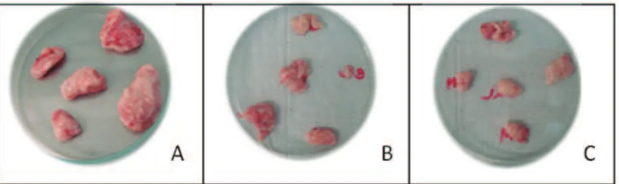
kidneys with a cystic appearance Mice with Ehrlich carcinoma; it was macroscopically verified that two animals of CT1 group presented lung hemorrhage. With these exceptions, the mice of both strains, and independent of treatment, showed no macroscopic changes in their organs, with no fibrous consistency, thrombus, lesions and atrophy on the edge of the organs, preserving aspects in relation to structure, texture and shape. The tumors were well-defined and had a solid consistency (Figure 3).
Figure 1. Antitumor activity of Guettarda platypoda CDE in Mus musculus female mice after implantation of Sarcoma
180. Saline (0.5 mL) was orally administered in the Control Group (SCG) and experimental treated groups (ST1=100 mg/ kg; ST2=200 mg/kg) for seven days. *p<0.05.
Figure 2. Antitumor activity of Guettarda platypoda CDE in Mus musculus mice after implantation of Ehrlich carcinoma.
Saline (0.5 mL) was orally administered in the Control Group (SCG) and experimental treated groups (CT1=100 mg/kg; CT2=200 mg/kg) for seven days. *p<0.05.
Cytotoxicity evaluation
to 50%) with moderate activity (cell growth inhibition ranging from 50 to 70%) and with large activity (cell growth inhibition ranging from 70 to 100%). The average cell growth inhibition (IC%) of cell treated with 50 μg/mL CDE is shown below (Table 4).
Table 3. Effect of Guettarda platypoda CDE, in different doses, on cell migration into peritoneal cavity on carrageenan-induced peritonitis in mice. Data are the mean±SD of at least six animals.
Treated Dose (mg/kg) Nº of PMNL/mL
(x106) Inhibition (%)
Group 1 30 9.48±2.0 6.7
Group 2 50 3.35±0.7 * 67.0
Group 3 100 4.72±1.4 * 53.5
Group 4 150 3.40±0.7 * 66.5
Group 5 200 6.54±1.7 * 35.6
Indomethacin 10 4.90±0.5 * 51.8
Control - 10.16±3.1
-*p<0.01. Significance was determined with ANOVA one-way
followed by Tukey’s Test when compared with control group.
Discussion
The phytochemical analysis indicated the presence of significant secondary metabolites in the Guettarda platypoda DC., Rubiaceae, CDE. Some authors have reported the presence of several triterpene saponins, such as quinovic acid derivatives (Aquino et al., 1988a, 1989; Almeida, 1982; Bhattacharyya & Almeida, 1985; Ferrari et al., 1986, Melo et al., 2008). In this study alkaloids were not identified but
their presence was described in the ethanol extract by Almeida (1982) and detected the presence of 5α-carboxistrictosidina was detected by Ferrari et al. (1986) and Melo et al. (2008).
Results indicated that G. platypoda CDE has low oral acute toxicity. According to the criteria for acute toxicity by the GHS (Globally Harmonized Classification System) the fact that no serious injury or death occurred in any animal tested at the highest recommended dose (2000 mg/kg), indicate that the compounds were fit in class 5 (very low or no toxicity) (GHS, 2005). The absence of atypical behavior is also another indication of lack of toxicity of the compounds (Vital et al., 2009).
In order to evalute the effect of G. platypoda CDE on cell migration to the peritoneal cavity, we tested its effects on the induction of peritonitis by carrageenan. The statistical analysis revealed also that G. platypoda CDE evaluated, exhibited an inhibitory profile similar to indomethacin in the doses 50, 100, 150 and 200 mg/kg. The potential anti-inflammatory activity by CDE may be due to synergistic action of quinovic acid glycosides already reported in the literature (Bhattacharyya & Almeida, 1985; Aquino et al., 1988a, 1989, 1991; Ferrari et al., 1986) and the presence of beta-sitosterol (Bhattacharyya & Almeida, 1985), flavonoids and the triterpnes identified in this study, since anti-inflammatory action of the metabolites mentioned above have been already described.
The action of β-sitosterol is similar to the hydrocortisone (Gupta, 1980) and the action of flavonoids was explained in part by its inhibitory effect on arachidonic acid metabolism (Silva et al., 2002). Table 2. Weight gain and physiological habits (consumption of water/diet) in adult female mice (Mus musculus), treated with different doses of G. platypoda CDE in acute toxicity by oral doses.
Groups Body weight Consumption of water (mL) Consumption of diet (g)
Initial (g) Final (g)
Control 22.6±0.85 27.7±1.11 31.07±6.59 11.28±4.69
2000 mg/kg 24.6±0.52 30.6±1.39 30.71±9.83 12.77±2.21
3000 mg/kg 23.9±1.41 29.7±1.38 31.78±8.64 11.66±7.96
Some flavonoids can block the biosynthetic processes of eicosanoids, which lead to an anti-inflammatory action, because it causes inhibition of COX-2 and nitric oxide synthase (Machado et al., 2008). It is also reported that triterpenes have anti-inflammatory activity, due to inhibition of nitric oxide and prostaglandin E2 synthesis (Moraes et al., 2012).
However, other disorders can lead to a decrease in PMNL migration. This probably happened with doses above 150 mg/kg, observing an activity decrease anti-inflammatory, probably because exudate presented hemolysate due to the abundant presence of saponins in the roots of G. platypoda as described in this paper and by several other authors (Aquino et al., 1988a, 1989; Almeida, 1982; Bhattacharyya & Almeida, 1985; Ferrari et al., 1986, Melo et al., 2008).
In the antitumor activity evaluation, the G. platypoda CDE is quite promising for the treatment of Sarcoma 180, but did not provide satisfactory results against the Ehrlich carcinoma. Therefore, it is important to emphasize the positive results of this study, which may improve our understanding in the use of this plant as an alternative anti-cancer therapy. For Sarcoma 180, as this strain is the most difficult to treat, according to Almeida et al. (2005), who emphasizes that sarcomatose tumors are more difficult to treat because they affect of bone and muscle regions. The identification of active principles and their mechanisms of action still need to be studied in order to enhance the promising effects in treatment of this strain. Probably flavonoids are responsible for the antitumor action due to its antioxidant capacity, and cell growth inhibitory activity in several tumor cell lines and induce apoptosis (Machado et al., 2008).
It was veriied in this study that of G. platypoda CDE did not demonstrate cytotoxic activity. Analysis of cytotoxicity by MTT method has been used in the screening program of the National Cancer Institute of the USA (NCI), which tests more than 10000 samples each year (Skehan et al., 1990). It is a rapid, sensitive and inexpensive test. It was irst described by Mosman (1983) as having the potential to analyze the viability and metabolic state of the cell. This is a colorimetric analysis based on conversion of 3-(4,5-dimethyl-2-thiazole)-2,5-diphenyl-2-H-tetrazolium bromide salt (MTT) into blue formazan due
to mitochondrial enzymes present only in metabolically active cells. The study carried out employing cytotoxic the MTT method allows to deine the cytotoxicity easily, but not the action mechanism (Berridge et al., 1996).
Conclusions
The methanolic extract of Guettarda platypoda DC., Rubiaceae has low toxicity and promising anti-inflammatory activity at doses of 50, 100 and 150 mg/ kg and antitumor activity to Sarcoma 180 at doses of 100 mg/kg and 200 mg/kg. It’s necessary to continue the studies with fractions to determine which fraction may will be more active. The cytotoxic activity did not indicate satisfactory results. These tests don’t necessarily discard the potential of CDE against this activity and it is necessary additional tests using other cell strains and experimental models.
Acknowledgment
To CNPq for its financial support.
References
Agra MF, França PF, Barbosa-Filho JM 2007. Synopsis of the plants known as medicinal and poisonous in Northeast of Brazil. Rev Bras Farmacogn 17: 114-140.
Agra MF, Silva KN, Basílio IJLD, França PF, Barbosa-Filho
JM 2008. Survey of medicinal plants used in the region Northeast of Brazil. Rev Bras Farmacogn 18: 472-508.
Almeida MZ 1982. Estudo itoquímico e triagem farmacológica
da casca da raiz da Guettarda platypoda DC. João Pessoa, p.133. Dissertação de Mestrado, Programa de Pós-graduação em Produtos Naturais, Universidade Federal da Paraíba.
Almeida VL, Leitão A, Reina LCB, Montanari CA, Donnici CL, Lopes MTP 2005. Câncer e agentes
antineoplásicos celular específicos e ciclo-celular não específicos que interagem com o DNA: uma introdução. Quim Nova 28: 118-129.
Andersson L 1992. A provisional checklist of neotropical
Rubiaceae. Scripta Botan Belg 1: 1-199.
Aquino R, Feo V, Simone F, Pizza C, Cirino G 1991. Plant Table 4. Percent inhibition of cell growth (IC%) of the three tumor cell lines tested in a single dose (50 µg/mL) of Guettarda platypoda CDE.
Tumor lineage CDE (50 µg/mL)Mean (IC%) Standard Deviation (SD) CDE Doxorrubicina (15 μg/mL)Mean (IC%)
HT29 15.2 0.6 0.4
MCF-7 0 0 N.d.
NCI-H-292 1.7 0.5 0.2
Metabolites. New Compounds and Anti-Inflammatory Activity of Uncaria tomentosa. J Nat Prod 54: 453-459.
Aquino R, Simone F, Pizza C, Cerri R, Mello JF 1988a. Quinovic acid glycosides from Guettarda platypoda. Phytochemistry 27: 2927-2930.
Aquino R, Simone F, Pizza C, Mello JF 1989. Further quinovic acid glycosides from Guettarda platypoda. Phytochemistry 28: 199-201.
Aquino R, Simone F, Senatore F, Pizza C 1988b. Iridoids and secoiridoids from Guettarda platypoda. Pharmacol Res Commun 20: 105-108.
Bahia 1979. Governo do Estado. Seplantec - Subsecretaria
de Ciência e Tecnologia. Inventário de plantas medicinais do Estado da Bahia. Salvador.
Barbosa MRV, Souza EB, Jardim JG 2011. Rubiaceae. In:
Checklist das Plantas do Nordeste (Versão 1.5). http://
www.cnip.org.br/bdpn/checklistNE.pdf, accessed Jan 2011.
Berridge MV, Tan AS, Mccoy KD, Wang R 1996. The
Biochemical and Cellular Basis of Cell Proliferation
Assays that Use Tetrazolium Salts. Biochem J 4: 14-19.
Bertucci A, Haretche F, Olivaro C, Vázquez A 2008. Prospección
química del bosque de galería del río Uruguay. Rev Bras Farmacogn 18: 21-25.
Bhattacharyya J, Almeida MZ 1985. Isolation of the constituents of the root-bark of Guettarda platypoda. J Nat Prod 48: 148-149.
Capasso A, Balderrama L, Silvila SC, Tommasi N, Sorretino L 1998. Phytochemical and pharmacological studies
of Guettarda acreana. Planta Med 64: 348-352. Costa AF 2001. Farmacognosia. 3rd Ed., vol.3, Lisboa:
Calouste Gulbenkian.
Cunha AP, Silva AP, Roque OR 2003. Plantas e produtos vegetais em fitoterapia. Lisboa: Calouste Gulbenkian,
p. 21-61.
David JPL, David JM, Soares MBP, Costa JFO, Santos RR
2006. Introdução. In Lucchese AM (ed.). Instituto do Milênio do Semi-árido. Plantas da Caatinga: perfil
botânico, fitoquímica e atividade biológica. vol.4. Recife: Associação de Plantas do Nordeste, p. 15-17. Delprete PG 2004. Rubiaceae (Coffea or Quinine family). In
Smith N, Mori SA, Henderson A, Stevenson DWm.,
Head SV (ed.) Flowering plants of the neotropics.
Princeton: Princeton University Press, p. 328-333. Ferrari F, Mesana I, Botta B, Mello JF 1986 Constituents of
Guettarda platypoda. J Nat Prod 49: 1150-1151.
GHS 2005. A Guide to The Globally Harmonized System of Classification and Labeling of Chemicals (GHS).
http://www.osha.gov/dsg/hazcom/ghsguideoct05.pdf, accessed Feb 2011.
Gupta MB, Nath R, Srivastava N, Shanker K, Kishor K, Bhargava KP 1980. Anti-inflammatory and antipyretic
activities of β-sitosterol. Planta Med 39: 157-163.
Harbone JB 1998. Phytochemical Methods. 3rd ed., London:
Chapman & Hall.
IGEduca 2010. Biodiversidade. http://www.igeduca.com.br/ artigos/acontece/biodiversidade.html, accessed Feb 2011.
Lima LF, Lima PB, Almeida-Jr EB, Zickel CS 2010. Morfologia de frutos, sementes e plântulas de Guettarda platypoda
DC. (Rubiaceae). Biota Neotrop. 10: 155-160.
Machado H, Nagem TJ, Peters VM, Fonseca CS, Oliveira TT
2008. Flavonóides e seu potencial terapêutico. Bol Centro Biol Reprod 27: 33-39.
Matos FJA 1997. O formulário fitoterápico do professor Dias da Rocha. Fortaleza: UFC.
Melo SJ, Braz-Filho R, Lemos TLG, Santos HS, Fonseca
AM, Xavier HS 2008. Derivados terpênico e indólico glicosilados das raízes de Guettarda platypoda D.C. (Rubiaceae). 31ª Reunião Anual da Sociedade
Brasileira de Química. Águas de Lindóia, São Paulo.
Mól DFF 2010. Rubiaceae em um remanescente de floresta atlântica no Rio Grande do Norte, Brasil. Natal, 69 p. Dissertação de Mestrado, Programa de Pós-graduação em Ciências Biológicas, Universidade Federal do Rio Grande do Norte.
Moraes WF, Galdino PM, Nascimento MVM, Vanderlinde FA, Bara MTF, Costa EA, Paula JR 2012. Triterpenes
involved in the anti-inflammatory effect of ethanolic extract of Pterodon emarginatus Vogel stem bark. J Nat Med 66: 202-207.
Mossman T 1983. Rapid colorimetric assay for cellular
growth and survival:application to proliferation and cytotoxicity assays. J Immunol Methods 65: 55-63. Neu R 1956. A New reagent for differentiating and
determining flavones of paper chromatograms.
Naturwissenschaften 43: 82.
OECD (Organization for economicco-operation and development) 2001. Guideline for Testing of Chemicals: Acute Oral Toxicity-Acute Toxic Class Method.
Guideline: 423. http://iccvam.niehs.nih.gov/SuppDocs/
FedDocs/OECD/OECD_GL423.pdf, acessed Jan 2012.
Pereira MS 2002. A família Rubiaceae na reserva biológica
Guaribas, Paraíba, Brasil. Recife, 117 f. Dissertação de Mestrado, Programa de Pós-graduação em Biologia
Vegetal, Universidade Federal de Pernambuco. Pereira MS, Barbosa MRV 2004. A família Rubiaceae
na Reserva Biológica Guaribas, Paraíba, Brasil. Subfamílias Antirheoideae, Cinchonoideae e Ixoroideae. Acta Bot Bras 18: 305-18.
Pessoa MCR 2009. Diversidade e riqueza da família Rubiaceae
Juss. no Cariri paraibano. Recife, 83 f. Dissertação de Mestrado, Programa de Pós-graduação em Biologia
Vegetal em Biologia Vegetal, Universidade Federal de
Pernambuco.
Portal Brasil 2010. Por dentro do Brasil-Meio Ambiente.
download/por-dentro-do-brasil-2013-meio-ambiente-2013-setembro-2010, accessed Jan 2011.
Roberts EAH, Cartwright RA, Oldschool M 1957. Phenolic substances of manufactured tea. I. Fractionation and paper chromatography of water-soluble substances. J Sci Food Agr 8: 72-80.
Russel JE 1982. Contribuition a l’estude botanique et chimiotaxonomique du genre Verbascum en Languedoc-Cevennes. França. Thèse de Doctorat,
Université de Montpellier I.
Sharma OP, Dawra RK 1991. Thin-layer chromagraphye
separations of lantadens, the pentacyclic triterpenoids from (Lantana camara) plant. J. Chromatogr 587: 351-354.
Skehan P, Storeng R, Scudiero D, Monks A, Mcmahon J, Vistica D, Warren JT, Bodesch H, Kenney S, Boyd MR 1990.
New colorimetric cytotoxicity assay for anticancer-drug screening. J Natl Cancer I 82: 1107-1112.
Silva FRMB, Szpoganicz B, Pizzolatti MG, Willrich MAV,
Sousa E 2002. Acute effect of Bauhinia forficata on serum glucose levels in normal and alloxan-induced diabetic rats. J Ethnopharmacol 83: 33-37.
Sugiura K, Stock CC, Sugiura MM 1955. The effect of
phosphoramides on the growth of a variety of mouse
and rats tumors. Cancer Res 2: 38-50.
Uchôa FT, Silva TG, Lima MC, Galdino SL, Pitta Ida R, Dalla Costa T 2009. Preclinical pharmacokinetic and
pharmacodynamic evaluation of thiazolidinone PG15: an anti-inflammatory candidate. J Pharm Pharmacol 61: 339-345.
Vital FAC, Melo SJ, Silva TG, Carneiro S, Araújo JM,
Mendonça-Jr FJB 2009. Avaliação da toxidade aguda e das atividades citotóxica, antimicrobiana e
antiinflamatoria de 7-aril-2,3-diidrotiazolo[3,2-α]
pirimidin-5-ona-6-carbonitrila. Lat Am J Pharm 28: 507-512.
Wagner H, Bladt S 1996. Plant drugs analysis. A thin layer chromatography atlas. 2nd ed., Munich: Springer.
*Correspondence
Sebastião J. Melo
Departamento de Antibióticos, Universidade Federal de Pernambuco
Av. Artur Sá s/n, Cidade Universitária, 50670-901 Recife-PE, Brasil