The Structure of the T190M Mutant of
Murine
α
-Dystroglycan at High Resolution:
Insight into the Molecular Basis of a Primary
Dystroglycanopathy
Manuela Bozzi1☯, Alberto Cassetta2☯*, Sonia Covaceuszach2, Maria Giulia Bigotti3, Saskia Bannister4, Wolfgang Hübner4, Francesca Sciandra5, Doriano Lamba2, Andrea Brancaccio3,5*
1Istituto di Biochimica e Biochimica Clinica, UniversitàCattolica del Sacro Cuore, Roma 00168, Italy, 2Istituto di Cristallografia—CNR, Trieste Outstation, Trieste, 34149, Italy,3School of Biochemistry, University of Bristol, Bristol, BS8 1TD, United Kingdom,4Department of Physics, University of Bielefeld, 33615, Bielefeld, Germany,5Istituto di Chimica del Riconoscimento Molecolare—CNR, c/o Università Cattolica del Sacro Cuore, Roma, 00168, Italy
☯These authors contributed equally to this work.
*[email protected](AB);[email protected](AC)
Abstract
The severe dystroglycanopathy known as a form of limb-girdle muscular dystrophy (LGMD2P) is an autosomal recessive disease caused by the point mutation T192M inα -dystroglycan. Functional expression analysisin vitroandin vivoindicated that the mutation
was responsible for a decrease in posttranslational glycosylation of dystroglycan, eventual-ly interfering with its extracellular-matrix receptor function and laminin binding in skeletal muscle and brain. The X-ray crystal structure of the missense variant T190M of the murine N-terminal domain ofα-dystroglycan (50-313) has been determined, and showed an overall topology (Ig-like domain followed by a basket-shaped domain reminiscent of the small sub-unit ribosomal protein S6) very similar to that of the wild-type structure. The crystallographic analysis revealed a change of the conformation assumed by the highly flexible loop encom-passing residues 159–180. Moreover, a solvent shell reorganization around Met190 affects the interaction between the B1–B5 anti-parallel strands forming part of the floor of the bas-ket-shaped domain, with likely repercussions on the folding stability of the protein domain (s) and on the overall molecular flexibility. Chemical denaturation and limited proteolysis experiments point to a decreased stability of the T190M variant with respect to its wild-type counterpart. This mutation may render the entire L-shaped protein architecture less flexible. The overall reduced flexibility and stability may affect the functional properties of α-dystroglycanvianegatively influencing its binding behavior to factors needed for
dystroglycan maturation, and may lay the molecular basis of the T190M-driven primary dystroglycanopathy.
OPEN ACCESS
Citation:Bozzi M, Cassetta A, Covaceuszach S, Bigotti MG, Bannister S, Hübner W, et al. (2015) The Structure of the T190M Mutant of Murineα
-Dystroglycan at High Resolution: Insight into the Molecular Basis of a Primary Dystroglycanopathy. PLoS ONE 10(5): e0124277. doi:10.1371/journal. pone.0124277
Academic Editor:Annalisa Pastore, National Institute for Medical Research, Medical Research Council, London, UNITED KINGDOM
Received:December 18, 2014
Accepted:March 12, 2015
Published:May 1, 2015
Copyright:© 2015 Bozzi et al. This is an open access article distributed under the terms of the
Creative Commons Attribution License, which permits unrestricted use, distribution, and reproduction in any medium, provided the original author and source are credited.
Data Availability Statement:The structure solved has been deposited to the Protein Data Bank (www. pdb.org) and assigned the following accession number: 4WIQ.
Funding:This work was supported by a Wellcome Trust Career Re-entry fellowship to MGB.
Introduction
Dystroglycan (DG) is composed of two subunits,αandβ, that are formed from a unique pre-cursor which undergoes a proteolytic event within the endoplasmic reticulum during the first steps of its maturation pathway [1,2]. DG represents a widely expressed adhesion complex that plays a crucial role in offering stability to tissues, being at the crossroad between cytoskele-ton, plasma membrane and the surrounding extracellular matrix [3,4]. The DG main role is to bind with high-affinity laminins that are central organizers of the molecular network behind specialized basement membranes surrounding skeletal muscle. Besides, DG may form a pletho-ra of additional intepletho-ractions with other binding partners sharing laminin globular (LG) do-mains within epithelia, endothelia and Schwann cells. Indeed, its binding affinity may largely vary based on the degree of glycosylation of itsαsubunit [5].
DG has been particularly studied within skeletal muscle, a tissue where itsα-subunit is found to be highly glycosylated, since a number of neuromuscular diseases involving glycosyl-transferases active in muscle have been identified during the last years [6]. The commonly ac-cepted molecular scenario implies that hypoglycosylated DG cannot bind the skeletal muscle laminin-2 [7] with the usual high affinity, which would lead to the reduction of sarcolemma stability at the basis of a wide number of severe, or later onset, muscular dystrophies, defined as secondary dystroglycanopathies [6].
Theα-subunit plays multiple important roles in DG, in that i) it is crucially involved in the
maturation and posttranslational modification of the precursor; ii) it is highly modified with sugar chains especially in its central region (i.e. the mucin-like portion) from where some es-sential sugar moieties protrude to bind laminin. The first is based on the noncovalent binding activity ofα-DG towardβ-DG [8] while for the latter, it seems that the N-terminal domain
may be central in recognizing and directing the activity of important modifying enzymes, such as the like-acetylglucosaminyltransferase LARGE, that adds the repeating disaccharide block [-α3-glucuronic acid (GlcA)-β3-xylose (Xyl)-) required for laminin-binding [9] to a phosphor-ylated mannose ofα-DG [10].
We have pioneered studies onα-DG at the molecular level [11], having extensively charac-terized the structure of its N-terminal domain, which is shaped in two subdomains [12]. The first is a typical Ig-like domain, while the second one is similar to the S6 protein present in the small ribosomal subunit ofT.termophilus[13].
Although a plethora of interesting studies are currently unraveling the structure of the sugar blocks modifying the mucin-like domain ofα-DG as well as the exact residues ofα-DG
in-volved in these modifications [14–17], no ultimate structural information is currently available on the dystroglycan/laminin complex at the molecular level. The challenges in obtaining crys-tals of the complex mainly arise from the highly heterogeneous nature ofα-DG glycosylation, confirmed by the fact that the protein can be typically visualized in overlay assays or Western blots as a band with a broad smear appearance indicating a distribution of different molecular masses [1,3,4].
Nonetheless, we believe that the structural analysis of the T190M mutant ofα-DG is a
fun-damental step towards the understanding at the molecular level of the mechanism leading to muscular dystrophy. Following this basic idea, we inserted the murine mutant in our recombi-nant system for expression inE.coli, and purified it for crystallization studies aimed at possibly solving its structure and comparing it with that of the wild-type protein previously reported [13].
Materials and Methods
Mutagenesis and primers
Traditionally, we have worked on murine dystroglycan since it displays a very high degree of identity (93%) with humanα-DG. We introduced an additional mutation, R166H, within the N-terminal domain ofα-DG in order to make it more resistant to proteolysis [13]; both the
wild-type and the mutated variants herein analyzed harbor this mutation. The murineα-DG (50–313)R166H (hereinafter WT) DNA was cloned into a bacterial vector, pHis-Trx, for the expression of the protein as thioredoxin fusion product, also containing an N-terminal 6xHis tag and a thrombin cleavage site, as previously described [13]. The point mutation T190M was introduced into the WT DNA construct using the QuikChange site-directed mutagenesis kit (Stratagene) and appropriate primers. Briefly, the mutation was inserted within the coding re-gion of the WT N-terminal domain cloned in the vector pHis-Trx [13] for prokaryotic expres-sion aimed at crystallographic analysis, and within the entire cDNA sequence of the murine DG cloned in the pEGFP-N1 plasmid for expression in eukaryotic cells [19], respectively, using the following primers:
Forward:5’-CCAGTGACTGTCCTTATGGTGATTCTGGATGCT-3’ Reverse:5’-AGCATCCAGAATCACCATAAGGACAGTCACTGG-3’
The construct DGT190M-pEGFP-N1 allows to express DG with a Green Fluorescent Protein (GFP) fused at the C-terminus ofβ-DG. Moreover, a Myc tag is present, inserted after Lys498,
within the C-terminal domain ofα-DG [19]. All constructs were verified by automated sequencing.
Fusion protein expression and purification
The recombinantα-DG(50–313)R166H T190M (hereinafter T190M) fusion protein was ex-pressed inEscherichia coliBL21(DE3) Codon Plus RIL strain and purified using nickel affinity chromatography. The fragment of interest was obtained upon thrombin cleavage. Further puri-fication steps were carried out using anion exchange and gel filtration chromatography. Name-ly, after thrombin cleavage, the flow through of a HiTrap Chelating column (GE Healthcare) was applied onto a Hi-Trap Q HP column (GE Healthcare) pre-equilibrated with buffer A (25 mM Tris–HCl pH 7.5). T190M was eluted with a linear gradient of 0–0.5 M NaCl in buffer A. The fractions containing T190M were pooled, concentrated with Amicon Ultra 15 (Milli-pore) and loaded on Superdex 200 10/300 GL (GE Healthcare) pre-equilibrated with 25 mM Tris–HCl pH 7.5, 0.15M NaCl, at a flow rate of 0.4 mL/min: the core fractions of the peak were supplemented with 2.5% Glycerol and concentrated by Microcon GM10 (Millipore). The puri-ty of the protein was confirmed by Tricine/SDS-PAGE [20].
Crystallization, data collection, structure solution, and refinement
Crystals of T190M were grown by using the vapor diffusion hanging drop method, following the protocol used for the crystallization of WT [13]. Drops were prepared by mixing 1μL of
the protein solution (5.25 mg/mL in 25 mM Tris, 150 mM NaCl and 2.5% glycerol; pH 7.5) with 1μL of the precipitant solution (0.8 M citrate buffer; pH 7.0) and equilibrated against the
reservoir (1 mL) at 4°C.
Data collection was carried out at the European Synchrotron Radiation Facility Grenoble (France), ID23-1 beamline, using a detector Pilatus-6M (Dectris) and the wavelength of 1.00 Å. Data collection was carried out at -173°C. Before being exposed to the X-ray beam, crystals were quickly dipped in cryoprotectant solution (25% ethylene glycol, 1.0 M citrate buffer, pH 7.0) and flash frozen in liquid nitrogen.
Indexing, integration and data reduction of the diffraction data were carried out by using a combination of XDS [21] and CCP4 [22] programs. Data reduction statistics are reported in Table 1.
The structure solution of T190M was obtained by Patterson search methods with PHASER [23] using the crystal structure of WT as template (PDB id: 1U2C). Restrained refinement of the crystal structure was carried out by usingphenix.refine[24]. The improvement of the initial model underwent a protocol that included a rigid body fitting stage followed by simulated an-nealing, coordinates and individual B-factors refinement. The subsequent stages integrated a Translation-Libration-Screw (TLS) model parameterization before the individual B-factors re-finement. Automatic refinement cycles were alternated with manual rebuilding sessions per-formed with COOT [25]. Solvent molecules were identified by the automatic water-picking algorithm implemented inphenix.refine[24]. The overall quality of these automatically picked solvent molecules were manually checked. Multiple conformations were introduced for select-ed side chains during the last cycles of refinement.
Protein stereochemistry was monitored throughout the refinement process and during manual rebuilding with MolProbity [26]. Statistics of the crystallographic refinement are re-ported inTable 1.
Protein structures superposition was carried out using ProFit (Martin, A.C.R.,http://www. bioinf.org.uk/software/profit/). The STRIDE web-server was used to assign the protein second-ary structure [27]. Interfaces between domains, solvent accessible areas and solvation energies were analyzed by using PISA as part of the CCP4 package [28]. Waters-protein interactions were analyzed using the WAP web-server [29].
Coordinates and structure factors have been deposited in the PDB, with accession code: 4WIQ. Figures were prepared using PyMol [30].
Fluorescence analysis
Equilibrium fluorescence titrations were performed in 50 mM Tris HCl, pH 7.4, with spectra collected at each guanidine hydrochloride (GdnHCl) concentration (point by point, as to avoid photo-bleaching phenomena) at 25°C in a 1-cm quartz cuvette using a fluorescence spectro-photometer Cary Eclipse (Agilent Technologies). The final concentration of protein (both WT and T190M) was 0.1μM, and for each point spectra were taken after 20 minutes of
Limited proteolysis
WT and T190M at 30μM in 50 mM Tris (pH 8) buffer were subjected to limited proteolysis at
37°C by the addition of trypsin (Sigma) to a final concentration of 2μg/mL. The reaction was
stopped after 1, 5, 10, 20, 40 and 60 min by adding SDS sample buffer to aliquots of the reaction mixture. The samples were analyzed by performing 15% SDS-PAGE and Coomassie staining. As a control the same time course was performed in the absence of the enzyme.
Table 1. X-ray diffraction: data collection and model refinement statistics.
Data Collection
Space group H 3
Unit-cell parameters (Å) a = 71.879, c = 144.296
Molecules per asymmetric unit 1
Wavelength (Å) 1.00
Resolution (Å) 48.1–1.59 (1.65–1.59)a
Total observations 119373
Unique reflections 35504
Rmerge(%)b 4.3 (44.2)
<I/σ(I)> 15.28 (1.74)
Completeness (%) 95.0 (71.8)
Redundancy 3.4 (2.3)
Refinement
Number of reflections (work-set/test-set) 33720/ 1784
Rworkc/ Rfreed(%) 14.61 / 16.32
Number of non-H atoms
Protein 1741
Waters 194
Organic (ethylene glycol) 12
Ions (Cl-) 1
Average isotropic B factors (Å2) 32.9
Protein (main chain) 28.9
Protein (side chain) 35.8
Solvents 39.1
r.m.s. deviation
Bond length (Å) 0.010
Angle (deg) 1.245
Ramachandran plot
favored regions (%) 100
allowed regions (%) 0
disallowed regions (%) 0
PDB code 4WIQ
aValues in parenthesis are given for the highest resolution shell bR
merge=∑hkl∑j│Ihkl, j-<Ihkl>│/∑hkl∑jIhkl,j cR
work=∑work-set│Fobs-Fcal│/∑work-setFobs dR
free=∑test-set|Fobs-Fcal|/∑test-setFobs
Super Resolution Microscopy with 3D SIM
Human osteosarcoma U2OS cells cultured in DMEM, 10% FBS, were seeded on No. 1.5H pre-cision cover glass (Marienfeld-Superior). The cells were transfected with the corresponding pEGFP-N1 vectors expressing full-length DG, WT and T190M, with polyethylenimine (PEI) precipitates based on a 3:1 ratio of PEI (μg) to total DNA (μg). 48h post transfection the cells
were fixed in 4% PFA.
Lipid staining was achieved after 30 minutes incubation in a 1:2000 dilution of Cellmask Plasma Membrane Orange dye (Invitrogen). For the immunostaining of the Myc-tag fused to DG, cells were permeabilized for 90 seconds in 0.5% Triton X100 and washed in PBS. Cells were blocked in 5% BSA in PBS for 1h followed by an incubation with 1:1000 dilution of pri-mary rabbit polyclonal anti-c-Myc antibody (C3956 from Sigma-Aldrich) in PBS 0.1% Tween-20 for 1h at room temperature. Secondary goat anti-rabbit antibody conjugated to Alexa-Fluor647 (A-21244, Life Technologies) was applied at a 1:400 dilution for 1h after several washes in PBS 0.1% Tween-20. The cells were finally washed three times in PBS. The cover glass was mounted in Vectashield containing DAPI (Vector Laboratories) for nuclear counter stain and sealed with nail polish.
U2OS cells expressing WT-EGFP and T190M-EGFP were imaged on a 3D Structured Illumi-nation Microscope (3D-SIM) OMX v.4 (GE Healthcare). The refraction index of the immersion oil was chosen to avoid spherical aberrations in the green emission channel. DAPI was excited with a 405 nm, EGFP with 488 nm, Cellmask Plasma Membrane Orange with 568 nm and goat anti-rabbit antibodies bound to AlexaFluor647 with 647nm laser, while sCMOS cameras re-corded the corresponding emission in the following bandwidth of 436/31 nm, 528/48 nm, 609/ 37 nm and 683/40nm. The 3D structured illumination images were reconstructed in the Soft-worx v6.1.1 software (GE Healthcare) at a Wiener filter setting of 0.004 and using one week old recorded OTFs optimized for the 528 nm emission channel. For presentation here, the final im-ages were adjusted linearly in intensities with the software package Fiji [32] to minimize any visi-ble spherical aberrations in the blue and red emission channels. Raw data are availavisi-ble
upon request.
Results
Crystallization and crystal structure determination
Well shaped crystals of T190M grew in 7–10 days as regular slabs that diffracted X-rays. The crystal structure of T190M has been determined and refined up to a resolution of 1.6 Å. The final model includes residues (58–163) and (179–303), that could be traced in the 2Fo-Fc
mutation is also present in T190M, although this position falls into the region of the flexible loop that could not be traced in WT as well as in T190M (see below).
The crystallographic analysis confirmed the substitution of Thr190 with Met, as showed in Fig 2and, according to the refined model, the T190M mutation does not introduce significant
Fig 1. Cartoon depiction of the T190M crystal structure.The two domains are colored in violet (Ig-like domain) and green (small ribosomal S6 subunit domain), respectively; the B1 and B5β-strands are labeled for clarity. Spheres representation is employed for highlighting Met190. The loop regions connecting the two domains are shown for both WT (colored in cyan) and T190M (colored in red). Residues Ala58 and Thr303 (N- and C-term residues) together with Gln163 and Ala179 (N- and C-term of the modeled loop in the T190M crystallographic structure) are highlighted as stick-and-ball representation.
conformational changes in the structure. These findings altogether indicate that this mutation does not significantly alter the overall fold of T190M with respect to that of WT.
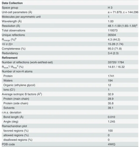
Despite showing very similar crystal structures, WT and T190M differ in the conformation of the flexible loop linking the two domains (residues 159–179). Indeed the conformation of the residues (158–163) in T190M, that has been best modeled based on the Fourier maps (Figs 3and4) and by monitoring the Rfree, differs with respect to the one being observed for residues Fig 2. CalculatedσA-weighted 2Fo-Fcmap (contoured at 1.0σand colored in light-blue) and Fo-Fcmap
(contoured at±3.0σand colored in green for positive values and in orange for negative values).The
maps have been calculated following the Patterson search stage using the WT as a model (PDB id: 1U2C). The final model of T190M (colored in blue) is superposed to the initial Patterson search solution (colored in pink-salmon). Both models are represented as ribbon except residues at position 190 (Thr for the template structure and Met for the mutated final model), which are represented as sticks.
(158–160) in WT (PDB id: 1U2C). The refined structure thus suggests that the main-chain loop conformation has changed in T190M with respect to WT. Moreover, in T190M the loop could be traced for three additional residues, up to Gln163, with respect to the WT structure, with residues (158–161) assuming aβ-Turn structure (Figs3and5). Being the WT and the
T190M crystallization conditions almost identical and the crystal structures exhibiting the same packing features, it is reasonable to correlate the observed differences in the loop confor-mation to the ThrvsMet mutation, which would result in a less flexible loop and in an in-creased proximity of the loop to the side chain of the mutated residue.
Residue 190 is located at the center of the B1β-strand, at the bottom of the basket-like do-main and is solvent exposed [13]. The point mutation Thr to Met does not introduce drastic
Fig 3. SA-omitσ
A-weighted 2Fo-Fcmap (contoured at 1.0σand colored in grey) and Fo-Fcmap (contoured at 3.0σand colored in green) overlaid
with the loop region encompassing residues (156–163) as the T190M final model (PDB id 4WIQ).Maps have been calculated using the T190M model,
where residues (156–163) have been omitted from the calculation. The loop of the T190M final model nicely fits the 2Fo-Fc maps, which is indicative of the soundness of the built model.
local changes in the structure. The Met residue nicely superposes on the position occupied by Thr in the WT (Fig 2), with the Cβ-Cγbond rotated by20° with respect to the corresponding
bond (Cβ-Cγ2) in Thr, while the Cγ-Sδ-Cεgroup is pointing towards the solvent region. Also the nearby residues geometries are not greatly affected by the point mutation.
Despite the limited effect of the Thr to Met mutation on the neighboring residues, the intro-duction of a bulkier and apolar group such as the methylthio group of the methionine causes a re-organization of the surrounding water molecules. An analysis made with PISA [28] shows that the solvent exposed surface is more than doubled when considering Thr in the WT (13 Å2) and
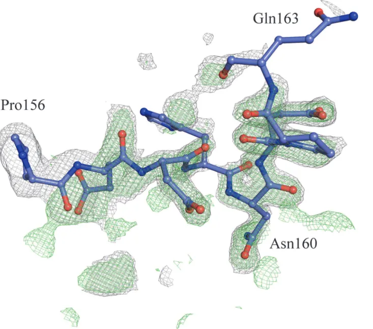
Met in the T190M mutant (34 Å2). Furthermore, the solvation energy contribution estimated Fig 4. SA-omitσ
A-weighted 2Fo-Fcmap (contoured at 1.0σand colored in grey) and Fo-Fcmap (contoured at 3.0σand colored in green) overlaid
with the loop region encompassing residues 156–160 as in the WT model (PDB id 1U2C).Maps have been calculated by using the T190M model where
residues (156–163) have been omitted from the calculation.
by PISA for each residue in the respective crystallographic models goes from -0.01 kcal/mole (Thr in WT) to 0.74 kcal/mole (Met in T190M), indicating a potentially perturbing effect of the methi-onine on the surrounding water molecules.
Fig 5. Superimposition of the WT and T190M mutant S6 domains.The models are represented as ribbons, with WT colored in pink-salmon and T190M colored in blue; Met 190 is highlighted in green and depicted as a ball-and-stick model. The“flexible loop”C-term residues Gln163 (T190M) and Asn160 (WT and T190M) are also depicted as ball-and-stick models (Asn160 residue is partially modeled in 1U2C). B1 and B5β-strands are white labeled for clarity. The superimposition of the two crystal structures emphasize the different conformations of the respective loops encompassing residues (157–163).
An analysis of the solvent structure in the crystallographic models shows that while several waters surrounding residue 190 are conserved in both WT and T190M, some differences in the solvent organization are actually evident (as shown in the four panels ofFig 6). Namely, WAT304 (WT, PDB id: 1U2C) interacts with Thr190 OH in WT but it is absent in T190M (Fig 6A and 6B). The lack of WAT304 in T190M is rather interesting, as this water molecule bridges together the B1 and B5β-strands by interacting with the backbone of residues Val188 (O) and His296 (O), and with the side chains of residues Thr190 (Oγ1) and His296 (Nδ1).
WAT304 acts as hydrogen bonds donor towards residues Val188 (O) and His296 (O) and as hydrogen bonds acceptor from Thr190 (Oγ1) and His296 (Nδ1) (Fig 6A). Such hydrogen
bonds are not observed in T190M due to the lack of a bridging solvent molecule that would sta-bilize the interaction between the B1 and B5β-strands (Fig 6B). Moreover, solvent structure
changes were also found in a region occupied in T190M by Asn160, whereas the same space is populated by solvent waters in WT (Fig 6B). Indeed, Asn160, which is part of the flexible loop discussed above, protrudes from the loop towards Met190 acting, through Oδ1, as an acceptor of bifurcated hydrogen bonds fromWAT660 and WAT638 water molecules (T190M, PDB id: 4QWI); Met190 in turn interacts, still through hydrogen bonds, with Oγof Ser257, and with other solvent molecules (Fig 6C). In the WT crystal structure WAT660 is not present and WAT638 does not mediate any interaction between the flexible loop and Ser257 (Fig 6D).
It is quite apparent that the substitution of a hydrophilic residue such as threonine with a bulkier and apolar one like methionine results in a solvent re-organization around the residue itself. Changes in the structure solvation sphere may have significant consequences on the con-formation of the (159–179) loop. Altogether, the loop relocation and the modified solvation sphere may affect the protein stability and, eventually, also the mutual orientation of the two domains in solution.
Stability measurements
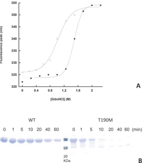
Chemical unfolding experiments have been performed in order to establish the relative stabili-ties of WT and T190M in solution. Fluorescence emission has been used as a mean to record the exposure of the aromatic residues (mainly Trp) to the solvent, with emission maxima that range from310 nm of the fully buried to350 nm of the fully exposed tryptophan residues
[33]. The four tryptophan residues in the WT primary sequence are good fluorescent probes of the unfolding behavior of the domain and its mutant upon addition of a chemical denatur-ant such as GdnHCl.Fig 7Ashows the unfolding curves of WT and T190M as a function of GdnHCl concentration. In the absence of denaturant, the emission spectra of both WT and T190M are those of native-like proteins, with peaks in the spectral region of buried tryptophan residues; the fact that the mutant’s peak is slightly red-shifted as compared to the wild-type might reflect an increased exposure of one or more of its tryptophan residues to the solvent. The plot of the emission spectra peaks as a function of GdnHCl reveals a cooperative unfolding behaviour for both WT and T190M, with transition midpoints (Gdn50%, i.e. the denaturant
concentration inducing 50% of the total peak shift) of 1.45M and 1M, respectively, indicating that WT is more stable than T190M.
Fig 6. Details of the hydrogen bond networks involving selected water molecules and residues.The structural elements of the N-terminal domain ofα -DG are represented as ribbon; residues involved in the interaction networks are represented as sticks. Interatomic distances are reported inÅ. The color scheme for WT and T190M models is the same as inFig 5. A) hydrogen bond network involving WAT304, Thr190 and Val188 in WT, with the B1 and B5β -strands labeled for clarity. B) The T190M model is shown in the same orientation as WT. Water molecules less than 8Åapart from a putative WAT304 are also shown. C) WT hydrogen bond network involving Ser257 and the“flexible loop”. Water molecules less than 8Åapart from a putative WAT660 (water numbering as in PDB id: 1U2C) are also shown. D) T190M hydrogen bond network involving Ser257, WAT660 and WAT638. The T190M model is shown in the same orientation as WT. The comparison of the mutated T190M with WT highlights the differences existing in their solvent structures and emphasizes their role in the interplay between protein structural elements.
variants is subjected to degradation in the absence of the protease in the same experimental conditions, T190M is clearly more susceptible to proteolysis than WT, confirming the latter to be more stable, in accordance with the results of the chemical denaturation experiments.
Super Resolution Microscopy
The Osteoclast cell line U2OS has been transfected both with the EGFP plasmids carrying the WT and T190M full-length DG variants [19], respectively and the cells have been analyzed by 3D structured illumination microscopy on an OMX v.4 system.
No relevant differences have been observed between WT and T190M, either following the EGFP signal reportingβ-DG localization (Fig 8A and 8B) or the anti-Myc reportingα-DG
lo-calization (Fig 8C). In both variants, DG undergoes its maturation cycle and it is extensively
Fig 7. Relative stabilities of WT and T190M.A. Equilibrium unfolding curves of WT and T190M. The red-shifts in the fluorescence peaks upon addition of GdnHCl indicate a progressive exposure of the tryptophan residues to the solvent: the data fit to a single cooperative transition for both WT, closed circles, and T190M, open circles, with a difference in midpoints (Gdn50%) of 0.45M, that reveals the mutant to be less stable than
the wild-type protein. Both experiments were performed in triplicate, and no significant deviation from the above curves was detected. B. Limited proteolysis of WT and T190M. The tryptic digestion time course of the two variants is shown. The faint band(s) below the main band of full length T190M are likely to represent some contaminants and/or the result of a minor degradation event taking place during the purification steps.
traffickedviathe endoplasmic reticulum to the plasma membrane where it is nicely localized to filopodia and also to podosomes located at the cell periphery. Notably,β-DG colocalizes with the plasma membrane stain while the anti-Mycα-DG signal appears slightly shifted at the
outside of the cell, suggesting no dramatic alterations as far as the distribution of the two DG subunits is concerned.
Discussion
According to the high resolution X-ray crystal structure here presented, the overall structure of the N-terminal domain ofα-DG is not significantly affected by the T190M mutation, nor re-markable local conformational changes of the residues close to position 190 have been de-tected. At large, this finding seems to be fully supported by the fluorescence microscopy analysis, which does not record dramatic differences in the phenotype of T190M transfected osteoclasts with respect to their WT counterpart. The outcome of the fluorescence microscopy experiments (showing that both DG subunits have a typical localization pattern in T190M) is definitely indicating that the mutation is acting in quite a subtle and intriguing molecular regu-latory fashion, or anyway that its effect would be much less observable in an isolated cellular context than in skeletal muscle fibres [18].
The major effect of the T190M mutation seems to be related to the disruption by the bulkier and apolar methionine side chain of the surrounding solvent network structure. Indeed, it is well known that solvation may play a critical role on the stability and the conformational equi-librium of a macromolecule [34], as well as on the protein-ligands interactions [35]. Further-more, it has been reported that even a single mutation at the surface of a protein, can
significantly affect the folding/unfolding free energies as a result of altering the solvent organi-zation [36]. In T190M the differences observed in the solvent organization around Met190 may account for the structural and thermodynamic differences observed with respect to WT. Indeed, T190M resolves in the formation of a new hydrogen bonding network involving Asn160 and the water molecule WAT660, which may influence the flexibility of the 159–179 loop. Moreover, the loss of stabilizing interactions between the B1 and B5β-strands due to the
absence of WT WAT304, may play a role in the T190M lower stability revealed by the chemical denaturation and limited proteolysis experiments (see below).
Indeed, a particularly interesting aspect seems to be that in this case part of the flexible loop encompassing residues 160–180 that in the WT domain was even less structured, due to its high mobility and flexibility [13], assumes in T190M a different conformation. Such a result leads to hypothesize that the Thr to Met mutation could have an impact on the structural dy-namics of the entire N-terminal domain. The enhanced rigidity of T190M may affect the over-all dynamics and orientation of the two subdomains within the L-shapedα-DG molecular
architecture, eventually influencing its global conformation and its function.
Fig 8. 3D Structured Illumination Microscopy images of U2OS cells expressing DG and DGT190Mfused
to EGFP.DG-EGFP (A) and DGT190M-EGFP (B) are both localized specifically at the plasma membrane and
accumulate in the cytoplasm. The columns show the individual images for DAPI (blue), EGFP (green), Cellmask Plasma Membrane Orange (red) and the resulting RGB overlay. The first row of images is the z stack maximum intensity projection of the 3D recording of the cell. The two subsequent rows of images are at a specific z position in the stack (XY) and X position slice through the stack (XZ). The white bar represents 10μm. Immunolabelling of Myc-tagged Dystroglycan (C) shows a specific signal at the plasma membrane. The individual maximum intensity z-stack projection images for the EGFP, Cellmask Orange and anti Myc channels are shown as well as an overlay. Four by fourμm magnifications of the corresponding white dashed boxes depict the Myc-tag signal at the outside of the plasma membrane (filopodia). The antibody stain appears nicely shifted toward the outside of the plasma membrane, while GFP and Cellmask orange are overlapping.
This possibility seems to be corroborated by the evidence that in solution T190M has a lower stability than the WT, as suggested by the different melting points calculated on their un-folding curves. Although the T190M denaturation curve has a melting point at about 1 M GdnHCl, denoting a rather stable protein, the decrease in stability with respect to WT is small but significant, as further confirmed by limited proteolysis experiments.
Potential consequences of T190M/T192M on the DG-LARGE
connection
The exact molecular mechanism leading to hypoglycosylation ofα-DG carrying the T190M
(T192M in human) mutation and causing a severe dystrophic phenotype, accompanied by some degree of intellectual impairment, is currently unknown [18].Via co-immunoprecipita-tion experiments, it was shown by Hara and colleagues that the mutated N-terminal domain of
α-DG is likely to exhibit a reduced affinity towards LARGE. This glycosyltransferase is
respon-sible for adding a repeating disaccharide unit [-α3-GlcA-β3-Xyl-], that is thought to be crucial for extracellular matrix protein binding, to O-mannosylatedα-DG [18]. Therefore, it is
reason-able to hypothesize that the degree of local perturbation and/or reduced stability measured in T190M in solution could affect the conformation of the N-terminal domain ofα-DG,
influenc-ing its bindinfluenc-ing to LARGE and provokinfluenc-ing, at least in some cell types or tissues, a significant hypoglycosylation ofα-DG.
Kanagawa and colleagues have shown that the N-terminal domain ofα-DG is shed by furin, in a process that is likely to take place into the intracellular Golgi compartments in which the final steps of the DG maturation process are completed [37]. An alternative chronological order dictating the chaotic molecular events taking place in the Golgi could imply that such furin-driven enzymatic step would even anticipate LARGE binding toα-DG. In which case, it is reasonable to postulate that such an autonomous domain ofα-DG [12] may represent not
only a recognition substrate but also an (allosteric?) activator for LARGE [38].
Interestingly, following the findings of a recent computational docking study, it was hypoth-esized that the T190M/T192M mutation might affect directly the binding ofα-DG to laminins [39]. However, we believe that such a mechanism would be highly unlikely. Although we previ-ously measured in the recombinant WT the presence of some residual binding activity towards laminin-1 [13], the estimated affinity was lower than that observed for full-length and glycosy-lated nativeα-DG binding to laminin and agrin [5,11,40]. In addition, such residual binding activity was apparently harbored by the first Ig-like domain ofα-DG and not by the S6-like
do-main where the T190M/T192M mutations resides [13].α-DG post-translationalO-glycans decorations along the endoplasmic reticulum and modifications accomplished in the Golgi, are crucial for its ability to function as an extracellular matrix (EM) receptor and namely to bind to EM proteins such as laminin, perlecan, agrin and neurexin in the brain and pikachurin in the retina [5]. Notably, the same are required for the binding ofα-DG to Old World arenaviruses [16]. Recently, aβ-1,4-glucuronyltransferase, designated B4GAT1, has been also identified to
be involved in the initiation of the LARGE-dependent enzymatic process [41,42].
Although a few glycosylation sites have been proposed and/or localized within the N-termi-nal and C-termiN-termi-nal domains ofα-DG, most of the O-linked oligosaccharide chains protrude from Thr and Ser residues placed within its central mucin-like domain [2,43,44]. A thorough biophysical characterization of the“naked”mucin-like domain ofα-DG clearly pointed to the dramatic role played by the glycosylation in its conformational stability [45].
peptide GPATPAP by the paired Ig-like type 2 receptorα[46] suggests that the Ig-like domain
within the N-terminalα-DG region could function as a protein“hub”for the recognition of the oligosaccharides protruding from the neighboringα-DG mucin-like domain. Furthermore,
the same domain could act as a direct docking site for one of the two domains of the glycosyl-transferase LARGE [47], belonging to the GT-8 and GT-49 families respectively [48]. Indeed, two mutations hitting the N-terminal Ig-like domain have been recently reported in a patient with hypoglycosylatedα-DG [49].
Possibly, in solution, the effect of the T190M mutation, which is next to the flexible hinge connecting the two subdomains, may“propagate”to the Ig-like domain by changing its relative orientation with respect to the S6-like domain also influencing its binding properties. It is wor-thy of note that the presence of a second Ig-like domain within the C-terminal region ofα-DG
has been confirmed by bioinformatics analysis and molecular modeling; the domain has been biochemically characterized and shown to include theβ-DG binding epitope [50].
Conclusions
Such interesting structural hypotheses necessarily await further rounds of experimental/ computational validations by means of techniques being able to explore the dynamical behav-ior ofα-DG (NMR, Molecular Dynamics). Furthermore, molecular modeling and docking tools will be exploited to investigate the likelihood ofα-DG binding to LARGE or to other
reg-ulatory factors important for DG maturation. The present structural analysis of the N-terminal T190M variant ofα-DG may pave the road to the elucidation of the molecular mechanism of a
specific case of dystroglycanopathy, with important consequences for clinicians and for design-ing possible therapies, but in addition it may be also relevant from a cellular and molecular bi-ology perspective, helping to improve the overall picture of the multiple structural aspects underlying the maturation and function of the DG complex.
Acknowledgments
This work was supported by a Wellcome Trust Career Re-entry fellowship to MGB. We thank Dr. Damir Bozic (University of Zurich) for thoughtful discussions and insights and Dr. Claudia Desiderio and Dr. Claudia Martelli (Catholic University of Rome) for the Orbitrap Mass Spectroscopy measurements.
Author Contributions
Conceived and designed the experiments: MB AC AB. Performed the experiments: MB AC SC MGB SB WH FS. Analyzed the data: MB AC MGB DL AB. Wrote the paper: MB AC DL AB.
References
1. Ibraghimov-Beskrovnaya O, Ervasti JM, Leveille CJ, Slaughter CA, Sernett SW, Campbell KP. Primary structure of dystrophin-associated glycoproteins linking dystrophin to the extracellular matrix. Nature. 1992; 355: 696–702. PMID:1741056
2. Barresi R, Campbell KP. Dystroglycan: from biosynthesis to pathogenesis of human disease. J Cell Sci. 2006; 119: 199–207. PMID:16410545
3. Ervasti JM, Campbell KP. Membrane organization of the dystrophin-glycoprotein complex. Cell. 1991; 66: 1121–1131. PMID:1913804
4. Ervasti JM, Campbell KP. A role for the dystrophin-glycoprotein complex as a transmembrane linker be-tween laminin and actin. J Cell Biol. 1993; 122: 809–823. PMID:8349731
6. Wells L. The O-mannosylation pathway: glycosyltransferases and proteins implicated in congenital muscular dystrophy. J Biol Chem. 2013; 288: 6930–6935. doi:10.1074/jbc.R112.438978PMID: 23329833
7. Michele DE, Barresi R, Kanagawa M, Saito F, Cohn RD, Satz JS, et al. Post-translational disruption of dystroglycan-ligand interactions in congenital muscular dystrophies. Nature. 2002; 418: 417–422. PMID:12140558
8. Sciandra F, Bozzi M, Morlacchi S, Galtieri A, Giardina B, Brancaccio A. Mutagenesis at theα-β inter-face impairs the cleavage of the dystroglycan precursor. FEBS J. 2009; 276: 4933–4945. doi:10.1111/ j.1742-4658.2009.07196.xPMID:19694806
9. Inamori K, Yoshida-Moriguchi T, Hara Y, Anderson ME, Yu L, Campbell KP. Dystroglycan function re-quires xylosyl- and glucuronyltransferase activities of LARGE. Science. 2012; 335: 93–96. doi:10. 1126/science.1214115PMID:22223806
10. Yoshida-Moriguchi T, Willer T, Anderson ME, Venzke D, Whyte T, Muntoni F, et al. SGK196 is a glyco-sylation-specific O-mannose kinase required for dystroglycan function. Science. 2013; 341: 896–899. doi:10.1126/science.1239951PMID:23929950
11. Brancaccio A, Schulthess T, Gesemann M, Engel J. Electron microscopic evidence for a mucin-like re-gion in chick muscleα-dystroglycan. FEBS Lett. 1995; 368: 139–142. PMID:7615068
12. Brancaccio A, Schulthess T, Gesemann M, Engel J. The N-terminal region ofα-dystroglycan is an au-tonomous globular domain. Eur J Biochem. 1997; 246: 166–172. PMID:9210479
13. Bozic D, Sciandra F, Lamba D, Brancaccio A. The structure of the N-terminal region of murine skeletal muscleα-dystroglycan discloses a modular architecture. J Biol Chem. 2004; 279: 44812–44816. PMID:15326183
14. Stalnaker SH, Hashmi S, Lim JM, Aoki K, Porterfield M, Gutierrez-Sanchez G, et al. Site mapping and characterization of O-glycan structures onα-dystroglycan isolated from rabbit skeletal muscle. J Biol Chem. 2010; 285: 24882–24891. doi:10.1074/jbc.M110.126474PMID:20507986
15. Tran DT, Lim JM, Liu M, Stalnaker SH, Wells L, Ten Hagen KG, et al. Glycosylation ofα-dystroglycan: O-mannosylation influences the subsequent addition of GalNAc by UDP-GalNAc polypeptide N-acetyl-galactosaminyltransferases. J Biol Chem. 2012; 287: 20967–20974. doi:10.1074/jbc.M112.370387 PMID:22549772
16. Hara Y, Kanagawa M, Kunz S, Yoshida-Moriguchi T, Satz JS, Kobayashi YM, et al. Like-acetylglucosa-minyltransferase (LARGE)-dependent modification of dystroglycan at Thr-317/319 is required for lami-nin binding and arenavirus infection. Proc Natl Acad Sci USA. 2011a; 108: 17426–17431. doi:10. 1073/pnas.1114836108PMID:21987822
17. Harrison R, Hitchen PG, Panico M, Morris HR, Mekhaiel D, Pleass RJ, et al. Glycoproteomic characteri-zation of recombinant mouseα-dystroglycan. Glycobiology. 2012; 22: 662–675. doi:10.1093/glycob/ cws002PMID:22241827
18. Hara Y Balci-Hayta B, Yoshida-Moriguchi T, Kanagawa M, Beltrán-Valero de Bernabé D, Gündeşli H,
et al. A dystroglycan mutation associated with limb-girdle muscular dystrophy. N Engl J Med. 2011b; 364: 939–946. doi:10.1056/NEJMoa1006939PMID:21388311
19. Morlacchi S, Sciandra F, Bigotti MG, Bozzi M, Hübner W, Galtieri A, et al. Insertion of a myc-tag within
α-dystroglycan domains improves its biochemical and microscopic detection. BMC Biochem. 2012; 13:14. doi:10.1186/1471-2091-13-14PMID:22835149
20. Schagger H, von Jagow G. Tricine-sodium dodecyl sulfate-polyacrylamide gel electrophoresis for the separation of proteins in the range from 1 to 100 kDa. Anal Biochem. 1987; 166: 368–379. PMID: 2449095
21. Kabsch W. XDS. Acta Crystallogr D Biol Crystallogr. 2010; 66: 125–132. doi:10.1107/ S0907444909047337PMID:20124692
22. Collaborative Computational Project, N. 4. The CCP4 suite: programs for protein crystallography. Acta Crystallogr D Biol Crystallogr. 1994; 50: 760–763. PMID:15299374
23. McCoy A J, Grosse-Kunstleve RW, Adams PD, Winn MD, Storoni LC, Read RJ. Phaser crystallograph-ic software. J Appl Crystallogr. 2007; 40: 658–674. PMID:19461840
24. Afonine PV, Grosse-Kunstleve RW, Echols N, Headd JJ, Moriarty NW, Mustyakimov M, et al. Towards automated crystallographic structure refinement with phenix.refine. Acta Crystallogr D Biol Crystallogr. 2012; 68: 352–367. doi:10.1107/S0907444912001308PMID:22505256
25. Emsley P, Lohkamp B, Scott WG, Cowtan K. Features and development of Coot. Acta Crystallogr D Biol Crystallogr. 2010; 66: 486–501. doi:10.1107/S0907444910007493PMID:20383002 26. Chen VB, Arendall WB, Headd JJ, Keedy DA, Immormino RM, Kapral GJ, et al. MolProbity: all-atom
27. Heinig M, Frishman D. STRIDE: a web server for secondary structure assignment from known atomic coordinates of proteins. Nucleic Acids Res. 2004; 32: W500–502. PMID:15215436
28. Krissinel E, Henrick K. Inference of Macromolecular Assemblies from Crystalline State. J Mol Biol. 2007; 372: 774–797. PMID:17681537
29. Praveen S, Ramesh J, Sivasankari P, Sowmiya G, Sekar K.WAP(version 2.0): an updated computing and visualization server for water molecules. J Appl Crystallogr. 2008; 41: 952–954.
30. Schrödinger L. The PyMOL Molecular Graphics System, Version 1.3r1. 2010
31. Di Stasio E, Bizzarri P, Misiti F, Pavoni E, Brancaccio A. A fast and accurate procedure to collect and analyze unfolding fluorescence signal: the case of dystroglycan domains. Biophys Chem. 2004; 107: 197–211. PMID:14962600
32. Schindelin J, Arganda-Carreras I, Frise E, Kaynig V, Longair M, Pietzsch T, et al. Fiji: an open-source platform for biological-image analysis. Nat Methods. 2012; 9: 676–682. doi:10.1038/nmeth.2019 PMID:22743772
33. Lakowicz JR. Principles of fluorescence spectroscopy. 3rded. Springer; 2006 ISBN
978-0-387-46312-4.
34. Levy Y, Onuchic JN. Water Mediation in Protein Folding and Molecular Recognition. Annu Rev Biophys Biomol Struct. 2006; 35: 389–415. PMID:16689642
35. Ladbury JE. Just add water! The effect of water on the specificity of protein-ligand binding sites and its potential application to drug design. Chem Biol. 1996; 3: 973–980. PMID:9000013
36. Covalt JC Jr, Roy M, Jennings PA. Core and surface mutations affect folding kinetics, stability and cooperativity in IL-1β: does alteration in buried water play a role? J Mol Biol. 2001; 307: 657–669. PMID:11254388
37. Kanagawa M, Saito F, Kunz S, Yoshida-Moriguchi T, Barresi R, Kobayashi YM, et al. Molecular recog-nition by LARGE is essential for expression of functional dystroglycan. Cell. 2004; 117: 953–964. PMID:15210115
38. Brancaccio A. Increased levels of expression of dystroglycan may protect the heart. Neuromuscul Dis-ord. 2013; 23: 867–870. doi:10.1016/j.nmd.2013.06.371PMID:23911074
39. Bhattacharya S, Das A, Ghosh S, Dasgupta R, Bagchi A. Hypoglycosylation of dystroglycan due to T192M mutation: a molecular insight behind the fact. Gene. 2014; 537: 108–114. doi:10.1016/j.gene. 2013.11.071PMID:24361964
40. Gesemann M, Brancaccio A, Schumacher B, Ruegg MA. Agrin is a high-affinity binding protein of dys-troglycan in non-muscle tissue. J Biol Chem. 1998; 273: 600–605. PMID:9417121
41. Willer T, Inamori KI, Venzke D, Harvey C, Morgensen G, Hara Y, et al. The glucuronyltransferase B4GAT1 is required for initiation of LARGE-mediatedα-dystroglycan functional glycosylation. 2014; 3: e03941.
42. Praissman JL, Live DH, Wang S, Ramiah A, Chinoy ZS, Boons GJ, et al. B4GAT1 is the priming en-zyme for the LARGE-dependent functional glycosylation ofα-dystroglycan. 2014; 3: e03943.
43. Liu M, Borgert A, Barany G, Live D. Conformational consequences of protein glycosylation: preparation of O-mannosyl serine and threonine building blocks, and their incorporation into glycopeptide se-quences derived fromα-dystroglycan. Biopolymers. 2008; 90: 358–368. PMID:17868094
44. Live D, Wells L, Boons GJ. Dissecting the molecular basis of the role of O-mannosylation pathway in disease:α-distroglycan and forms of muscular dystrophy. ChemBioChem. 2013; 14: 2392–2402. doi: 10.1002/cbic.201300417PMID:24318691
45. Bozzi M, Di Stasio E, Scaglione GL, Desiderio C, Martelli C, Giardina B, et al. Probing the stability of the
“naked”mucin-like domain of humanα-dystroglycan. BMC Biochemistry. 2013; 14: 15. doi:10.1186/ 1471-2091-14-15PMID:23815856
46. Kuroki K, Wang J, Ose T, Yamaguchi M, Tabata S, Maita N, et al. Structural basis for simultaneous rec-ognition of an O-glycan and its attached peptide of mucin family by immune receptor PILRα. Proc Natl Acad Sci USA. 2014; 111: 8877–8882. doi:10.1073/pnas.1324105111PMID:24889612
47. Stanley P. Golgi glycosylation. Cold Spring Harb Perspect Biol. 2011; 3: a005199. doi:10.1101/ cshperspect.a005199PMID:21441588